BACKGROUND & AIMS:Predicting outcomes of primary sclerosing cholangitis (PSC) by reliable and simple tools is an unmet need. Retrospective studies have suggested that liver stiffness measurement (LSM) by vibration-controlled transient elastography (FibroScan, Echosens) can predict outcomes. We aimed to validate the prognostic value of LSM statics and determine the relevance of LSM dynamics in a large, prospective, cohort study. METHODS:Clinical, biological, and LSM data of adult patients with uncomplicated PSC were prospectively recorded annually for 5 years. LSM was assessed continuously or according to 3 Baveno VII classes (<10 kPa, ≥10 to <15 kPa, and ≥15 kPa). The primary end point was transplant-free survival. Adjusted hazard ratios and 95% confidence intervals were determined using time-dependent multivariable Cox regression analyses. Effect of LSM change was evaluated using joint modeling. Progression was defined by a significantly positive individual LSM slope. RESULTS:The study analyzed 538 patients with at least 1 reliable LSM and follow-up available (median, 60.7 months; interquartile range [IQR], 48.6-66.0 months). Median baseline LSM was 7.6 kPa (IQR, 5.8-11.7 kPa). Nineteen patients died, and 72 received a transplant. Baseline LSM was strongly and independently linked to the risk of death or transplantation. For the groups 2.5 to <10 kPa, 10 to <15 kPa, and ≥15 kPa, the 5-year transplant-free survival was 93.9% (IQR, 90.3%-96.2%), 78.1% (IQR, 66.2%-86.3%), and 46.0% (IQR, 34.6%-56.7%), respectively. Progressors experienced worse transplant-free survival vs nonprogressors: adjusted hazard ratio of 3.12 (95% confidence interval, 1.55-6.24; P = .001). Each 1-kPa/y increment was associated with 18% increase in risk of death or transplantation. CONCLUSIONS:This observational study validates the strong prognostic value of both static and dynamic LSM in PSC, as assessed by FibroScan. These results support the use of LSM as a risk stratification tool and potential surrogate end point in clinical trials.
Background & Aims: Gli1+ cells were previously described as myofibroblast precursors. We aimed to delineate the identity of Gli1+ cells and their contribution to wound healing in liver. Methods: Gli1+ cells were analyzed in normal mouse liver by single-cell RNA sequencing (n = 3). Genetic cell fate tracing and depletion of Gli1+ cells were achieved in male mice, undergoing cholestatic injury induced by 3,5-diethoxycarbonyl-1,4-dihydrocollidine (DDC) or bile duct ligation (BDL), and metabolic dysfunction-associated steatohepatitis (MASH). Gli1 expression was examined by fluorescence in-situ hybridization in human liver (n = 16). Cell interactions were assessed using organoids. Results: In normal liver, Gli1 demarcated a periductal subpopulation (11.8%) of portal fibroblasts, which transcriptome pertains to extracellular matrix and structure organization. In liver injury, Gli1+ fibroblasts accumulated with fibrosis as multilayers around bile ducts and discontinuously along the inner border of the ductular reaction, predominantly in biliary models and less markedly in MASH. In humans, periductal Gli1+ cells were present in normal liver and accumulated in primary sclerosing cholangitis and, to a lesser extent, in MASH, notably alongside the ductular reaction in late-stage diseases. Co-culture of cholangiocyte organoids with Gli1+ fibroblasts showed bidirectional interactions fostering proliferation of both cell types. Genetic depletion of Gli1+ cells (n ≥4/group) caused a marked reduction of fibrosis, assessed by Sirius Red area, in DDC-fed and BDL mice after 1 week (p <0.01) and of ductular reaction, assessed by cytokeratin-19+ cell count in DDC (p <0.01), although cholestatic injury persisted or even increased. Conclusions: Gli1 demarcates a subpopulation of fibroblasts that play a key role in the wound healing process combining portal fibrosis and ductular reaction notably in biliary diseases. Impact and implications: The heterogeneity and the functions of hepatic fibroblasts in liver fibrosis remain poorly defined. Gli1+ fibroblasts are a transcriptionally distinct subset of hepatic fibroblasts, mainly located around bile ducts. Gli1+ fibroblasts develop bidirectional interactions with cholangiocytes, underlying portal fibrosis and ductular reaction formation, as they typically occur in biliary diseases. The therapeutic targeting of Gli1+ hepatic fibroblasts is of potential interest in biliary diseases.
Portal fibrosis, a determinant of progression in virtually all chronic liver diseases, prototypically develops in biliary diseases. Using single-cell RNA sequencing and genetic cell fate tracing in mouse models, we identify Clec3b⁺ fibroblasts as a distinct subset of portal fibroblasts, which rapidly expand after biliary injury and give rise to the bulk of portal myofibroblasts. Mechanistic analyses reveal that Clec3b⁺ portal fibroblasts activation is governed by a Krüppel-like factor 4 (KLF4)/periostin (POSTN) axis, i.e., KLF4 directly binds to the Postn promoter and represses its transcription in quiescent fibroblasts, whereas after injury, KLF4 is downregulated, which allows POSTN, acting via αvβ5 integrin, to drive portal fibroblast activation and portal fibrosis. Our findings identify Clec3b+ portal fibroblasts as the primary effectors of portal fibrosis and demonstrate that the KLF4/POSTN signaling axis regulates their activation, offering potential therapeutic targets for inhibiting fibrosis in biliary diseases.
BACKGROUND:Ex situ perfusion has been shown to improve the preservation of marginal liver grafts. Normothermic conditions allow graft viability evaluation and metabolic modifications, such as drugs injection to modify lipid content. In a previous study, we demonstrated that a pharmacologic defatting cocktail with rapamycin (DFAT) significantly reduced the triglyceride content of steatotic hepatocytes in vitro. The objective of the present study was to test the defatting cocktail in discarded human livers during ex situ perfusion. METHODS:Discarded livers were perfused according to an uninterrupted cold-to-warm protocol. The defatting cocktail was injected after 30 minutes under normothermic conditions (37 °C), whereas no defatting cocktail was administered to controls. The objective of this study was to evaluate the effect of the defatting cocktail on lipid metabolism and reduction in histologic steatosis. Liver viability was evaluated using UK viability criteria. RESULTS:Twelve discarded highly marginal livers (median Donor Risk Index 2.02 [1.6-3.47], with a median macrosteatosis of 55% [10%-90%]) underwent cold-to-warm perfusion for a median of 765 minutes (450-1,320). Defatting cocktail injection was performed in 9 livers (vs 3 controls), triggering a marked increase in triglyceride into the perfusate (at 90 minutes +164% vs +23% in controls; P < .001); a reduction of triglyceride content in liver tissue with a peak effect at 30 minutes (-46.6% vs +14.8% in controls, P = .0032); an increase in the hepatic expressions of genes related to lipid export, β-oxidation, and autophagy; and a decrease in that of lipogenesis-related genes. Five livers (41.6%) reached viability criteria, including 3 (33%) in the defatting cocktail group and 2 (67%) in the control group. A 10% reduction of histologic macrosteatosis was observed in the defatting cocktail group vs 0% in controls. CONCLUSION:Continuous cold-to-warm ex situ perfusion combined with defatting cocktail injection triggers the release of triglycerides and the activation of lipid export, β-oxidation, and autophagy pathways, together with a trend toward steatosis reduction.
Primary sclerosing cholangitis (PSC) is a chronic cholestatic liver disease with no effective curative therapies. Necroptosis, a regulated necrotic cell death pathway controlled by receptor-interacting protein kinase 1 (RIPK1), has emerged as a potential driver of inflammation and fibrosis in chronic liver disorders. We investigated the role of necroptosis in PSC and whether RIPK1 inhibition could modify disease course and progression. Spatial profiling of human PSC biopsies revealed that necroptosis primarily affects cholangiocytes, while apoptosis was more frequent in hepatocytes and nonbiliary cells. In vitro, necroptosis inhibition protected cholangiocytes, and RIPK1 deletion conferred resistance to TNF-mediated cytotoxicity. In a murine model of PSC, pharmacological RIPK1 inhibition reduced cholestatic injury, hepatic inflammation, and biliary fibrosis. Multiomic analyses comprehensively demonstrated reprogramming toward wild-type-like profiles following treatment. These findings identify necroptosis as a critical effector in PSC and highlight RIPK1 inhibition as a promising disease-modifying approach, opening the door to targeted necroptosis-based therapies for this otherwise untreatable disease.
Obesity is a global epidemic characterized by chronic low-grade inflammation and metabolic dysfunction, with adipose tissue playing a pivotal role in these processes. The mixed lineage kinase domain-like pseudokinase (MLKL) is a critical mediator of necroptosis but also exhibits noncanonical roles in metabolic regulation. This study aimed to investigate the adipocyte-specific functions of MLKL in obesity. Using adipocyte-specific Mlkl knockout (MlklAdi-KO) mice, we observed reduced susceptibility to high-fat diet (HFD)-induced obesity, enhanced glucose tolerance, and improved insulin sensitivity. MlklAdi-KO mice showed elevated energy expenditure independent of changes in food intake or locomotor activity, correlating with increased mitochondrial function and reduced lipid accumulation in white adipose tissue (WAT). Transcriptomic analyses of WAT revealed significant modulation of pathways linked to oxidative phosphorylation, inflammation, and lipid metabolism. Furthermore, metabolomic profiling highlighted reductions in TCA cycle intermediates, acylcarnitines, and pro-inflammatory amino acids in MlklAdi-KO mice under HFD conditions. These findings were accompanied by improved hepatic lipid profiles and decreased steatosis, underscoring systemic benefits of adipocyte-specific Mlkl deletion. Mechanistically, Mlkl deficiency altered adipocyte differentiation. These results position MLKL as a promising therapeutic target for obesity and related metabolic disorders, emphasizing the need for future studies using conditional knockout and overexpression models to explore its cell-specific and noncanonical functions in metabolic regulation.
Cholecystectomy is considered as a safe procedure to treat patients with gallstones. However, epidemiological studies highlighted an association between cholecystectomy and metabolic disorders, such as type 2 diabetes mellitus and metabolic dysfunction-associated steatotic liver disease (MASLD), independently of the gallstone disease. Following cholecystectomy, bile acids flow directly from the liver into the intestine, leading to changes in the entero-hepatic circulation of bile acids and their metabolism. The changes in bile acids metabolism impact the gut microbiota. Therefore, cholecystectomized patients display gut dysbiosis characterized by a reduced diversity, a loss of bacteria producing short-chain fatty acids and an increase in pro-inflammatory bacteria. Alterations of both bile acids metabolism and gut microbiota occurring after cholecystectomy can promote the development of metabolic disorders. In this review, we discuss the impact of cholecystectomy on bile acids and gut microbiota and its consequences on metabolic functions.
Sorbonne Université, INSERM, Centre de Recherche Saint-Antoine (CRSA), Paris, France Correspondence to Chantal Housset, Faculté de Santé Sorbonne Université, Centre de Recherche Saint-Antoine, 27 rue Chaligny, 75012 Paris, France. E-mail: [email protected]
ABCB4 (ATP-binding cassette subfamily B member 4) is a hepatocanalicular floppase involved in biliary phosphatidylcholine (PC) secretion. Variations in the ABCB4 gene give rise to several biliary diseases, including progressive familial intrahepatic cholestasis type 3 (PFIC3), an autosomal recessive disease that can be lethal in the absence of liver transplantation. In this study, we investigated the effect and potential rescue of ten ABCB4 missense variations in NBD1:NBD2 homologous positions (Y403H/Y1043H, K435M/K1075M, E558K/E1200A, D564G/D1206G and H589Y/H1231Y) all localized at the conserved and functionally critical motifs of ABC transporters, six of which are mutated in patients. By combining structure analysis and in vitro studies, we found that all ten mutants were normally processed and localized at the canalicular membrane of HepG2 cells, but showed dramatically impaired PC transport activity that was significantly rescued by treatment with the clinically approved CFTR potentiator ivacaftor. Our results provide evidence that functional ABCB4 mutations are rescued by ivacaftor, paving the way for the repositioning of this potentiator for the treatment of selected patients with PFIC3 caused by mutations in the ATP-binding sites of ABCB4.
<p>Supplementary Tables S1-S5 - PDF file 94K, Clinicopathologic characteristics of patients with HCC (S1); Pharmacological inhibitors (S2); Antibodies for Western blot and immunofluorescence (S3); Primer pairs for qualitative and quantitative PCR (S4); Correlations between IR-A and splicing factor mRNA levels in human HCC tumours (S5)</p>
Supplementary Figure S4 - PDF file 227K, CUGBP1, hnRNPH, hnRNPA2B1 and S2/ASF mRNA levels are controlled by EGFR in HuH7 cells
Supplementary Figure S3 - PDF file 53K, Basal and EGFR-activated ERK levels in HCC cell lines
Sorbonne Université, INSERM, Centre de Recherche Saint-Antoine (CRSA), Assistance Publique-Hôpitaux de Paris (AP-HP). Sorbonne Université, Department of Hepatology, Reference Center for Inflammatory Biliary Diseases and Autoimmune Hepatitis (CRMR MIVB-H), ERN RARE-LIVER, Saint-Antoine Hospital, Paris, France Correspondence to Chantal Housset, MD, PhD, Faculté de Médecine Sorbonne Université, 75012 Paris, France. E-mail: [email protected]
Supplementary Data from Insulin-Like Growth Factor-1 Receptor Inhibition Induces a Resistance Mechanism via the Epidermal Growth Factor Receptor/HER3/AKT Signaling Pathway: Rational Basis for Cotargeting Insulin-Like Growth Factor-1 Receptor and Epidermal Growth Factor Receptor in Hepatocellular Carcinoma
Supplementary Figure S5 - PDF file 190K, The blockage of EGFR signalling does not alter the subcellular localization of splicing factors in HCC cells
Supplementary Figure S2 - PDF file 640K, EGFR-dependent signalling increases IR-A mRNA splicing in HCC cells
 Dominique Wendum合作论文数Sorbonne Université71
 Laura Fouassier合作论文数Centre de Recherche Saint-Antoine, INSERM60
 Raoul Poupon合作论文数Sorbonne Université59
 Nicolas Chignard合作论文数Public Health Sciences Division, Fred Hutchinson Cancer Research Center46M Mergey合作论文数45
 Vlad Ratziu合作论文数Sorbonne University39
 Sara Lemoinne合作论文数Sorbonne Universités, UPMC Université Paris 06;UMR_S 938, INSERM;UPMC Université Paris 06, Sorbonne Universités37Olivier Rosmorduc合作论文数Centre Hépato-Biliaire, Assistance Publique-Hôpitaux de Paris37V Barbu合作论文数37Colette Rey合作论文数35Veronique Barbu合作论文数33
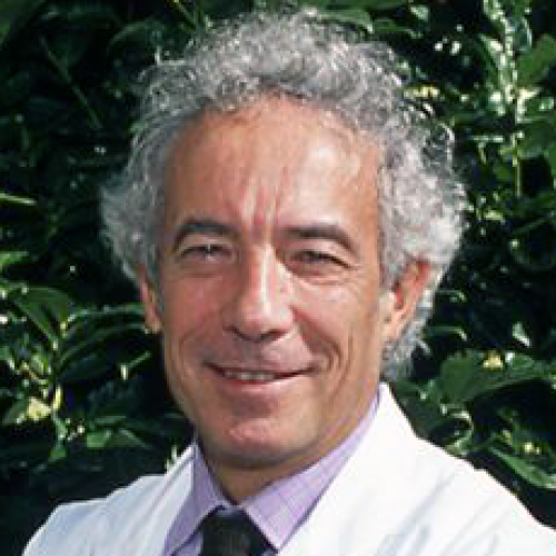 Thierry Poynard合作论文数Centre de Recherche Saint-Antoine - Paris, Hospital Saint-Antoine;Sorbonne University, INSERM;Pitié-Salpêtrière Hospital33Christele Desbois-Mouthon合作论文数INSERM U.402, Faculté de Médecine Saint-Antoine, Paris, France32Axelle Cadoret合作论文数32
 Christophe Corpechot合作论文数INSERMUnité 402, HôpitalSaint-Antoine31Y Chretien合作论文数29Clapéron Audrey合作论文数28
 Dominique Damais-Thabut合作论文数Hôpital Pitié-Salpêtrière;Hôpital Saint-Antoine;Sorbonne-Université28
 Christian Brechot合作论文数Center for Global Health and Inter, College of Public Health, University of South Florida;College of Medicine Internal Medicine, University of South Florida;Morsani College of Medicine, University of South Florida27
 Lynda Aoudjehane合作论文数25Olivier Deckmyn合作论文数24El Mourabit Haquima合作论文数23
 Olivier Chazouillères合作论文数Hôpital Saint-Antoine;Assistance Publique-Hopitaux De Paris23Yen Ngo合作论文数22Jacqueline Capeau合作论文数Laboratoire de Biochimie, I.N.S.E.R.M. U181, Faculté de Médecine Saint-Antoine, 75571 Paris Cedex 12, France21Filomena Conti合作论文数20
 Olivier Scatton合作论文数20Mona Munteanu合作论文数20
 Laetitia Fartoux合作论文数Department of Oncology, Hôpital Paris Saint Joseph;Groupe GPSanté19
 Valentina Peta合作论文数18Jean-Louis Delaunay合作论文数18Tounsia Aït-Slimane合作论文数17Elisabeth Lasnier合作论文数17Delphine Firrincieli合作论文数16
 Jeremie Gautheron合作论文数INSERM U781, Hôpital Necker-Enfants Malades and Université Paris-Descartes15Lawrence Serfaty合作论文数15Dominique Debray合作论文数14Hamza Chettouh合作论文数14Pierre Berthelot合作论文数14
 Dominique Rainteau合作论文数14Gonzalez-Sanchez E合作论文数13A. Paul合作论文数12Michel Vaubourdolle合作论文数12
 Francoise Praz合作论文数INSERM U762, Paris 6 University12
 Thomas Falguieres合作论文数11
 Lemoine Maud合作论文数Hôpital Saint-Antoine, AP-HP11
 Olivier Soubrane合作论文数Service de chirurgie hépato-bilio-pancréatique et transplantation hépatique, Hôpital Beaujon, AP-HP et Université de Paris11Anne-Marie Durand-Schneider合作论文数11




