Could HAMET increase pregnancy rates in predicted and unexpected difficult transfer patients? Data of this study show a strong correlation between HAMET and pregnancy rates. Mock embryo transfer is regularly performed to adequately plan the correct strategy for positioning embryos inside the uterus of patients predicted to have difficult access to uterine cavity procedure. This is a single-center, observational, retrospective study conducted at S.I.S.Me.R. center in Bologna, Italy. 1184 consecutive diagnostic hysteroscopies were performed for patients suffering for primary or secondary infertility before ART. All patients from 18 to 49 years old underwent a mock embryo transfer performed with 10 to 15 μl of methylene blue dye. Diagnostic hysteroscopy was performed immediately after mock embryo transfer to assess if the dye has entered the uterine cavity. Out of the 1184 patients who underwent hysteroscopy, in 617 patients 1118 transfers were executed after IVF-ICSI cycles. Patients were subdivided in: group A where whole cavity including fundus was filled by blue; group B where cavity but not fundus was filled by methylene blue; group C patients with partial captation or with no captation at all of dye. The difficulty of mock embryo-transfer (scale 1 to 3) was also recorded by the physician. the results of 1118 transfers are summarized in the table below. While patients with whole cavity plus fundal captation (group A) had the highest implantantion rate (IR) compared to group B and C, reflecting the same difference in the clinical pregnancy rate (CPR), the live birth rate (LBR) was statistically superior only for group A when compared to group B. Main limitation of these findings is the absence of control group. HAMET is able to predict which patients have the higher chances to achieve a pregnancy and, even more important, a term pregnancy. The use of HAMET it is also useful to assess the capability of physicians to enter the uterine cavity when the cervical canal route is not regular. not applicable
Journal Article Reply: Failure to detect DNA in blastocoel fluids after whole genome amplification—is it the next add-on? Get access L Gianaroli, L Gianaroli SISMER, Reproductive Medicine Unit, Bologna, Italy Search for other works by this author on: Oxford Academic PubMed Google Scholar D Perruzza, D Perruzza SISMER, Reproductive Medicine Unit, Bologna, Italy Search for other works by this author on: Oxford Academic PubMed Google Scholar C Albanese, C Albanese SISMER, Reproductive Medicine Unit, Bologna, Italy Search for other works by this author on: Oxford Academic PubMed Google Scholar S Azzena, S Azzena SISMER, Reproductive Medicine Unit, Bologna, Italy Search for other works by this author on: Oxford Academic PubMed Google Scholar C Tabanelli, C Tabanelli SISMER, Reproductive Medicine Unit, Bologna, Italy Search for other works by this author on: Oxford Academic PubMed Google Scholar A P Ferraretti, A P Ferraretti SISMER, Reproductive Medicine Unit, Bologna, Italy Search for other works by this author on: Oxford Academic PubMed Google Scholar M C Magli M C Magli SISMER, Reproductive Medicine Unit, Bologna, Italy Correspondence address. SISMER, Reproductive Medicine Unit, Bologna, Italy. E-mail: cristina.magli@sismer.it https://orcid.org/0000-0002-2889-8479 Search for other works by this author on: Oxford Academic PubMed Google Scholar Human Reproduction, dead141, https://doi.org/10.1093/humrep/dead141 Published: 19 July 2023
STUDY QUESTION Is the presence of DNA in the blastocoel fluid (BF) of expanded blastocysts, assessed by whole genome amplification (WGA), associated with the clinical outcome at the first transfer? SUMMARY ANSWER At the first transfer, blastocysts with negative BF-WGA have more chance to implant and to develop to term than those with positive BF-WGA results, both in preimplantation genetic testing for aneuploidies (PGT-A) cycles (where only euploid blastocysts resulting from the chromosomal analysis of trophectoderm (TE) biopsies were transferred) and in IVF/ICSI conventional cycles. WHAT IS KNOWN ALREADY Retrospective studies conducted in patients undergoing PGT-A have shown that the incidence of negative BF-WGA was significantly higher in TE-euploid blastocysts than in TE-aneuploid blastocysts. In addition, after the transfer of TE-euploid blastocysts, the ongoing clinical pregnancy rate was significantly higher in the group with negative BF-WGA compared with those with positive BF-WGA. STUDY DESIGN, SIZE, DURATION A prospective cohort study including 102 consecutive PGT-A patients (Group 1) and 88 consecutive conventional IVF/ICSI patients (Group 2), was conducted between January 2019 and December 2021. PARTICIPANTS/MATERIALS, SETTING, METHODS In both groups, BFs were collected from expanded blastocysts of high grade and processed for WGA. DNA amplification was evaluated by agarose gel electrophoresis for the presence (positive BF-WGA) or absence (negative BF-WGA) of a band. Directly after the BF retrieval, blastocysts from Group 1 underwent TE biopsy and vitrification. In Group 2, blastocysts were vitrified immediately after BF collection. In Group 1, only euploid blastocysts were considered for transfer according to the results of TE biopsies. In both groups, the selection of the blastocyst to be transferred was based on BF-WGA results giving priority, if available, to those with negative amplification. The primary outcome investigated was the live birth rate (LBR) at the first transfer. The main variable under investigation was the negative BF-WGA and results were corrected for confounders (maternal and paternal age, number of retrieved oocytes, male factor) by multiple logistic regression analysis. MAIN RESULTS AND THE ROLE OF CHANCE In Group 1, 60 patients transferred negative BF-WGA blastocysts and 42 positive BF-WGA blastocysts, and the LBR at the first transfer was 53.3% and 26.2%, respectively (P = 0.0081). After testing for selected confounders in a multiple logistic analysis, the transfer of blastocysts with negative BF-WGA resulted in an odds ratio of (OR) 3.52 (95% CI: 1.48-8.88, P = 0.0057) compared to transfer of positive BF-WGA blastocysts. In Group 2, at the first transfer 30 deliveries resulted from blastocysts with negative BF-WGA (48.4%) and three from the transfer of positive BF-WGA blastocysts in 26 patients (11.5%; P = 0.0014). Multiple logistic analysis indicated that the transfer of blastocysts with negative BF-WGA resulted in an OR 6.89 (95% CI: 1.98-32.95, P = 0.0056) compared to transfer of positive BF-WGA blastocysts. The LBR per transfer and the cumulative LBR per patient showed the same trend. LIMITATIONS, REASONS FOR CAUTION The study was performed in a single center. WIDER IMPLICATIONS OF THE FINDINGS The data from this study highlight the heterogeneity of blastocysts of similar morphology, even in those classified as euploid by TE analysis. Failure to detect DNA in BFs after WGA is associated with a significantly higher LBR at the first embryo transfer as well as per transfer and per patient. The processing of the BF by WGA is an easy and cost-effective tool that could become a valuable option to offer patients the highest chances of term pregnancy in the shortest time possible. STUDY FUNDING/COMPETING INTEREST(S) The study received no funding from external sources. There are no conflicts of interest to declare. TRIAL REGISTRATION NUMBER N/A.
Abstract Study question Is the presence or absence of DNA in the blastocoelic fluid detected by whole genomic amplification (WGA) a valid method to prioritize embryos for transfer? Summary answer Blastocysts with DNA in the BF have lower ongoing pregnancy rates compared with blastocysts without DNA, both in PGT-A and in conventional IVF cycles What is known already The detection of DNA in BFs from expanded blastocysts has been reported in different studies. After amplification, this DNA can be analyzed to provide information on the blastocyst chromosome condition, but the degree to which it is representative of the corresponding embryo ploidy is still controversial. The reason of this divergence could reside in several factors, including the different status of the studied embryos. A recent study comparing euploid and aneuploid blastocysts reported a significantly higher incidence of failed BF-DNA amplification in euploid blastocysts compared with aneuploid blastocysts suggesting an effect of the embryo ploidy condition on BF content Study design, size, duration This prospective study included 142 cycles with PGT-A (Group-1; 24-chromosome analysis was performed on trophectoderm (TE) biopsies) and 121 conventional IVF consecutive cycles (Group-2) treated in the last three years. In both groups, the BF was collected from expanded blastocysts before vitrification, and submitted to WGA. Single blastocyst transfers were performed by selecting blastocysts based on BF-WGA results giving priority to those with failed amplification. In Group-1, only TE-euploid blastocysts were transferred Participants/materials, setting, methods Patients in Group-1 had a maternal age higher than in Group-2 (36.8±3 vs 34.1±3.5 years). The same protocol of vitrification was used for all patients, and only expanded blastocysts of high grade were included in the study. Amplification after WGA was evaluated by loading an aliquot of the amplified product onto a 1.5% agarose gel. An ongoing pregnancy was defined as a pregnancy regularly ongoing beyond the 16th week of gestation Main results and the role of chance In Group-1, a total of 622 blastocysts underwent trophectoderm (TE) biopsy and 261 were euploid. The BF was retrieved from 237 euploid blastocysts and submitted to WGA. Amplification failure resulted in 98 BFs, whereas 139 BF gave a positive amplification. In all, 57 clinical pregnancies resulted, 53 of which were regularly ongoing. 61 transfers were performed with euploid blastocysts with failed BF-WGA, and 81 with positive BF-WGA. When looking at the transfer outcome, the ongoing pregnancy rate was significantly higher for euploid blastocysts with failed BF-WGA (31/61, 50.8%) when compared to those with positive BF-WGA results (22/81, 27.2%, p<0.01). In Group-2, there were 62 clinical pregnancies, 52 of which were ongoing. In relation to the BF-WGA results, the ongoing pregnancy rate showed the same trend of Group-1, and was 20% (52/121) for blastocysts with failed BF-WGA and 59.1% (42/71, p<0.001) for blastocysts with positive BF-WGA Limitations, reasons for caution This is a prospective cohort study. The results should be confirmed by a prospectively randomized study Wider implications of the findings The presence of DNA in the BF could be indicative of an abnormal embryos that is trying to reach a viable condition. Therefore, failure to detect DNA after BF amplification could represent an additional criterion to select viable embryos for transfer both in PGT-A and in conventional IVF cycles Trial registration number Not applicable
Abstract Study question Do RIF patients have the preimplantation genetic testing for aneuploidy (PGT-A) overcome their infertility condition? Summary answer PGT-A positively impact on implantation rate in RIF patients What is known already The most common definition of RIF is failure to achieve a pregnancy after three consecutive transfers of good quality embryos. This term possibly represents a heterogeneous category of infertile couples as the causes of repeated failures can be diverse. Especially intriguing is the case of patients with an age lower than 39 years for which the oocyte quality is expected not to be compromised by the well known age effect on female fertility. The chromosome analysis of the resulting embryos has been proposed as a valid method to improve implantation in the great majority of RIF patients Study design, size, duration This retrospective study included 49 patients with at least three previous consecutive implantation failures, which underwent PGT-A from January 2016 to April 2020. Both partners had a normal karyotype. Only patients with a female age below 39 years were included, who presented with a normal uterine cavity. Couples with a severe male factor were excluded. Single frozen blastocysts were transferred according to chromosomal results Participants/materials, setting, methods Maternal age was 35.5 ± 3.1 years. All blastocysts were vitrified after trophectoderm biopsy. Whole genome amplification and array comparative genomic hybridization were performed on biopsies. Only euploid embryos were transferred. The primary outcome was the live-birth delivery rate after the first transfer Main results and the role of chance Before starting a PGT-A cycle, these patients underwent 213 embryo transfers with 251 embryos replaced. A total of 264 blastocysts were analyzed, 140 of which were aneuploid (53%). Monosomy or trisomy was reported in 67 of the diagnosed samples (67/140, 48%) whereas the remaining 73 carried complex aneuploidies (73/140, 52%). The remaining 124 blastocysts (47%) were diagnosed as euploid. All patients performed an embryo transfer resulting in 28 clinical pregnancies (57%). There were 5 spontaneous abortions and the live-birth delivery rate per patient was 47% Limitations, reasons for caution This study suffers from the weakness related to retrospectivity. In addition, as euploid embryos are still cryopreserved, the delivery rate could change at completion of the cycles Wider implications of the findings: A RIF condition can be attributed, at least in a good proportion of cases, to the generation of high percentages of aneuploid embryos. In this case, the transfer of euploid blastocysts has high chances to classify this category of RIF patients has having an embryonic cause of infertilit. Trial registration number Not applicable
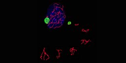
STUDY QUESTION: Is de novo segmental aneuploidy (SA) a biological event or an artifact that is erroneously interpreted as partial chromosome imbalance? SUMMARY ANSWER: The detection of de novo SA in sequential biopsies of preimplantation embryos supports the biological nature of SA. WHAT IS KNOWN ALREADY: Although some SAs are detected in oocytes and in blastocysts, the highest incidence is observed in cleavage-stage embryos. Based on these findings, we can postulate that the majority of cells affected by SAs are eliminated by apoptosis or that affected embryos mainly undergo developmental arrest. STUDY DESIGN, SIZE, DURATION: This retrospective study includes 342 preimplantation genetic testing for aneuploidy (PGT-A) cycles performed between January 2014 and December 2018. Chromosome analysis was performed on 331 oocytes, 886 cleavage-stage embryos and 570 blastocysts (n = 1787). From 268 expanded blastocysts, the blastocoelic fluid (BF) was also analyzed (resulting in 2025 samples in total). In cases of SAs involving loss or gain in excess of 15 Mb, embryos were not considered for transfer and sequential biopsies were performed at following stages. This resulted in 66 sets where the initial diagnosis of SAs (4 made in polar bodies, 25 in blastomeres and 37 in trophectoderm (TE) cells) was followed up. PARTICIPANTS/MATERIALS, SETTING, METHODS: A total of 2082 samples (2025 + 27 whole embryos) were processed by whole genome amplification followed by array comparative genomic hybridization. MAIN RESULTS AND THE ROLE OF CHANCE: The incidence of SAs was 6.3% in oocytes, increased to 16.6% in cleavage-stage embryos (P < 0.001) and decreased to 11.2% in blastocysts (P < 0.025 versus oocytes; P < 0.01 versus cleavage-stage embryos). The highest incidence of SAs was found in BFs (26.1%, P < 0.001). The analysis of 66 sets of sequential biopsies revealed that the initial finding was confirmed in all following samples from 39 sets (59.1% full concordance). In 12 additional sets, SAs were detected in some samples while in others the interested chromosome had full aneuploidy (18.2%). In three more sets, there was a partial concordance with the initial diagnosis in some samples, but in all TE samples the interested chromosome was clearly euploid (4.5%). In the remaining 12 sets, the initial SA was not confirmed at any stage and the corresponding chromosomes were euploid (18.2% no concordance). The pattern of concordance was not affected by the number of SAs in the original biopsy (single, double or complex) or by the absence or presence of concomitant aneuploidies for full chromosomes. LIMITATIONS, REASONS FOR CAUTION: Chromosome analyses were performed on biopsies that might not be representative of the true constitution of the embryo itself due to the occurrence of mosaicism. WIDER IMPLICATIONS OF THE FINDINGS: The permanence of SAs throughout the following stages of embryo development in more than half of the analyzed sets suggests for this dataset a very early origin of this type of chromosome imbalance, either at meiosis or at the first mitotic divisions. Since SAs remained in full concordance with the initial diagnosis until the blastocyst stage, a corrective mechanism seems not to be in place. In the remaining cases, it is likely that, as for full chromosome aneuploidy, mosaicism derived from mitotic errors could have occurred. In following cell divisions, euploid cell lines could prevail preserving the embryo chances of implantation. Due to the scarcity of data available, the transfer of embryos with SAs should be strictly followed up to establish possible clinical consequences related to this condition.
Introduction Preimplantation genetic testing for monogenic diseases (PGT-M) is a clinical method developed to prevent the transmission of monogenic inherited disorders to the future offspring. The reproductive risk of carriers of single gene disorders depends on the typology of the disorder, with the probability of affected conceptions ranging from 25% in recessive or X-linked diseases to 50% in dominant diseases. Therefore, the possibility of having embryos suitable for transfer and the consequent chances of ongoing pregnancy are strictly related to the number of embryos available for the genetic analysis. The aim of this study was to evaluate how many embryos need to be analyzed for having one or more genetically transferrable embryos. Material and Methods 86 carriers of genetic disorders performed 99 cycles of genetic analysis between January 2010 and September 2018. They were divided into two groups according to the typology of the disorder transmission. Group 1 included recessive and X-linked diseases (58 patients, 70 cycles of analysis), while with dominant disorders were in Group 2 (28 patients, 29 cycles of analysis). Maternal age was comparable in the two groups. Following blastocyst biopsy, embryos were vitrified to complete the genetic analysis. Results Clinical results were the following. For both groups, the mean number of analyzed embryos to have genetically transferrable embryos are reported in the following table: Conclusions Based on the reported data, we define for each case the minimum number of analyzable embryos needed to have one or more transferrable embryo depending on the typology of genetic disorder. Therefore, we eventually advise patients to undergo further oocyte retrievals for the best clinical outcome.
 Luca Gianaroli合作论文数International Institutes for Advanced Reproduction and Genetics;SISMeR29
 M. Cristina Magli合作论文数27
 Anna Pia Ferraretti合作论文数Reproductive Medicine Unit, SISMeR24Elisabetta Feliciani合作论文数9C. Albanese合作论文数7S. Azzena合作论文数4
 Davide Perruzza合作论文数4
 Marco Gergolet合作论文数Slovene Institute for Fertility and Endoscopic Surgery4B. Balicchia合作论文数4A. Pomante合作论文数3Daniela Fortini合作论文数3Andor Crippa合作论文数3G. Terzuoli合作论文数2N. Kenda Suster合作论文数2G Mascaretti合作论文数2Vincenzo Trengia合作论文数2
 Valeria Farfalli合作论文数2J L Marino合作论文数1G. Cafueri合作论文数1I. Afaneh合作论文数1R. Roopnarinesingh合作论文数1M. Croitor合作论文数1P. Mora-Guanche合作论文数1E. Rasohin合作论文数1Z. Ciorap合作论文数1A. Hotineanu合作论文数1M. Martinez-Moya合作论文数1E. Iammarrone合作论文数1S. Masouridou合作论文数1s crespi合作论文数1E. Boudjema合作论文数1R. Fadini合作论文数1A. Mitsoli合作论文数1C. Brigante合作论文数1H. Benli合作论文数1R Garcia Guzman合作论文数1E Erdei合作论文数1Serkan Aydoğdu合作论文数1Giulia Pirastu合作论文数1P. Naval Diaz合作论文数1Magdaliz Gorritz-Kindu合作论文数1M. Toschi合作论文数1N. Grieco合作论文数1N. Mishieva合作论文数1G. Quaranta合作论文数1I. Korneeva合作论文数1h hu合作论文数1M. Valerio合作论文数1
 Gina La Sala合作论文数CNR, Italian Natl Res Council1




