Mural cells are essential for maintaining the proper functions of microvasculatures. However, a key challenge of microvascular tissue engineering is identifying a cellular source for mural cells. We showed that in vitro , circulating fibrocytes (CFs) can (1) shear and stabilize the microvasculatures formed by vascular endothelial cells (VECs) in a collagen gel, (2) form gap junctions with VECs and (3) induce basement membrane formation. CFs transplanted into nude mice along with VECs in either collagen gel or Matrigel exhibited activities similar to those mentioned above, that is, sheathing microvasculatures formed by VECs, inducing basement membrane formation and facilitating the connection of the engineered microvasculatures with the host circulation. Interestingly, the behaviour of CFs also differs from that of human brain vascular pericytes (HBVPs) in vitro , which often infiltrate the lumen of capillary-like structures in a mosaic pattern, actively proliferate and exhibit lower endocytosis and migration capacities. We concluded that CFs are a suitable cellular source for mural cells in the construction of tissue-engineered microvasculatures.
Adeno-associated virus (AAV) vectors have been successfully used in patients with bleeding disorders and blindness. For human liver targeting, two major factors restrict effective AAV transduction after systemic administration of AAV vectors: human hepatocyte tropism and neutralizing antibodies (Nabs). In this study, we attempted to isolate AAV variants with the ability to transduce human hepatocytes and escape Nabs using a directed evolution approach in vivo. After four cycles of selection, 14 AAV capsid mutants were identified from a capsid shuffling library selected in the presence of human Intravenous Immunoglobulin (IVIG) and isolated from human hepatocytes xenografted into chimeric mice. AAV neutralization assays using IVIG showed that most of the mutants showed the Nab escape pattern in a manner similar to that of AAV8 or AAV9 and better than that of other AAV serotypes. Different mutants displayed varying capacities to escape Nab activity from individual serum samples collected from healthy subjects or hemophilia patients. The mutant AAV LP2-10 was found in 12 colonies out of 25, which was composed of capsids from AAV serotypes 2, 6, 8, and 9, with VP3 subunits derived from AAV8 swapped with AAV6 from residues 261 to 272. The mutant AAV LP2-10 manifested a higher ability than that of other serotypes to escape Nabs in IVIG and most human serum samples. After injection of AAV vectors encoding a self-complementary GFP cassette into chimeric mice, LP2-10 transduced human hepatocytes with efficiency similar to that of AAV8. In summary, AAV mutants can be isolated in humanized mice with both human hepatocyte tropism and the ability to evade Nab activity.
Although therapeutic outcomes have been achieved in hemophilia patients after delivery of clotting factor genes to the liver using adeno-associated virus (AAV) vectors, it is well known that the preclinical results generated from hemophilia animal models have not been directly predictive of successful translation in humans. To address this discrepancy humanized mouse models have recently been used to predict AAV transduction efficiency for human hepatocytes. In this study we evaluated AAV vector transduction from several serotypes in human liver hepatocytes xenografted into chimeric mice. After systemic administration of AAV vectors encoding a GFP transgene in humanized mice, the liver was harvested for either immunohistochemistry staining or flow cytometry assay for AAV human hepatocyte transduction analysis. We observed that AAV7 consistently transduced human hepatocytes more efficiently than other serotypes in both immunohistochemistry assay and flow cytometry analysis. To better assess the future application of AAV7 for systemic administration in the treatment of hemophilia or other liver diseases, we analyzed the prevalence of neutralizing antibodies (NAbs) to AAV7 in sera from healthy subjects and patients with hemophilia. In the general population, the prevalence of NAbs to AAV7 was lower than that of AAV2 or AAV3B. However, a higher prevalence of AAV7 NAbs was found in patients with hemophilia. In summary, results from this study suggest that AAV7 vectors should be considered as an effective vehicle for human liver targeting in future clinical trials.
Isolation and long-term maintenance of hepatic progenitor cells (HPCs) from healthy, non-injured adult livers remains challenging due to the lack of specific surface markers for selection and a limited understanding of the mechanisms for maintaining self-renewal. Previously, we identified a Sca-1 positive, bipotent HPC population in the peri-portal region of adult liver, and found MAPK/ERK and Wnt/β-Catenin pathways to be synergistically involved in their proliferation. In this study, we report the long-term culture of Sca-1 positive HPCs with epidermal growth factor (EGF) and CHIR99021, a small molecule inhibitor of glycogen synthase kinase 3 (GSK-3). Sca-1+ HPCs remain non-tumorigenic when passaged 35 times in vitro over 1 year. Flow cytometric analysis indicates that HPCs are positive for Sca-1 and putative liver progenitor cell markers, including CD13, CD24 and Prominin-1, but negative for hematopoietic/endothelial cell markers CD31, CD34, CD45, CD90 and CD117. Immunocyto-chemistry and RT-PCR indicate Sca-1+ HPCs express albumin (ALB), α-fetoprotein (AFP), cytokeratin19 (CK19), Sox9 and a panel of special hepatic progenitor transcriptional factors. Moreover, Sca-1+ HPCs are able to differentiate into hepatocyte-like and cholangiocyte-like cells under appropriate culture conditions in vitro and can take part in liver repopulation in an acetaminophen (APAP) induced liver injury mouse model. This study provides a paradigm to capture and maintain HPCs from naive liver tissue and offers a valuable cell model for investigating the molecular mechanisms underlying the cell lineage relationship in normal liver.
Accumulating evidence supports that circulating fibrocytes play important roles in angiogenesis. However, the specific role of fibrocytes in angiogenesis and the underlying mechanisms remain unclear. In this study, we found that fibrocytes stabilized newly formed blood vessels in a mouse wound-healing model by inhibiting angiogenesis during the proliferative phase and inhibiting blood vessel regression during the remodeling phase. Fibrocytes also inhibited angiogenesis in a Matrigel mouse model. In vitro study showed that fibrocytes inhibited both the apoptosis and proliferation of vascular endothelial cells (VECs) in a permeable support (Transwell) co-culture system. In a three-dimensional collagen gel, fibrocytes stabilized the VEC tubes by decreasing VEC tube density on stimulation with growth factors and preventing VEC tube regression on withdrawal of growth factors. Further mechanistic investigation revealed that fibrocytes expressed many prosurvival factors that are responsible for the prosurvival effect of fibrocytes on VECs and blood vessels. Fibrocytes also expressed angiogenesis inhibitors, including thrombospondin-1 (THBS1). THBS1 knockdown partially blocked the fibrocyte-induced inhibition of VEC proliferation in the Transwell co-culture system and recovered the fibrocyte-induced decrease of VEC tube density in collagen gel. Purified fibrocytes transfected with THBS1 siRNA partially recovered the fibrocyte-induced inhibition of angiogenesis in both the wound-healing and Matrigel models. In conclusion, our findings reveal that fibrocytes stabilize blood vessels via prosurvival factors and anti-angiogenic factors, including THBS1.
Skin cancer cells with donor genotype have been identified in allogeneic transplant patients; however, the donor contribution to the recipient's epithelial malignancy remains to be established. In this issue of the JCI, Verneuil et al. provide the first evidence for donor contribution to the malignant epithelium of skin squamous cell carcinoma in a kidney transplant recipient. This case report may have important implications for cancer research and clinical care of long-surviving kidney transplant patients.
Regenerative medicine is transitioning into clinical programs utilizing stem/progenitor cell therapies for repair of damaged organs. We summarize those for liver and pancreas, organs that share endodermal stem cell populations, biliary tree stem cells (hBTSCs), located in peribiliary glands: they are precursors to hepatic stem/progenitors in canals of Hering and to committed progenitors in pancreatic duct glands. They give rise to maturational lineages along a radial axis within bile duct walls and a proximal-to-distal axis starting at the duodenum and ending with mature cells in the liver or pancreas.
Regenerative medicine is transitioning into clinical programs using stem/progenitor cell therapies for repair of damaged organs. We summarize those for liver and pancreas, organs that share endodermal stem cell populations, biliary tree stem cells (hBTSCs), located in peribiliary glands. They are precursors to hepatic stem/progenitors in canals of Hering and to committed progenitors in pancreatic duct glands. They give rise to maturational lineages along a radial axis within bile duct walls and a proximal-to-distal axis starting at the duodenum and ending with mature cells in the liver or pancreas. Clinical trials have been ongoing for years assessing effects of determined stem cells (fetal-liver-derived hepatic stem/progenitors) transplanted into the hepatic artery of patients with various liver diseases. Immunosuppression was not required. Control subjects, those given standard of care for a given condition, all died within a year or deteriorated in their liver functions. Subjects transplanted with 100-150 million hepatic stem/progenitor cells had improved liver functions and survival extending for several years. Full evaluations of safety and efficacy of transplants are still in progress. Determined stem cell therapies for diabetes using hBTSCs remain to be explored but are likely to occur following ongoing preclinical studies. In addition, mesenchymal stem cells (MSCs) and hematopoietic stem cells (HSCs) are being used for patients with chronic liver conditions or with diabetes. MSCs have demonstrated significant effects through paracrine signaling of trophic and immunomodulatory factors, and there is limited evidence for inefficient lineage restriction into mature parenchymal or islet cells. HSCs' effects are primarily via modulation of immune mechanisms.
Peribiliary glands (PBGs) in bile duct walls, and pancreatic duct glands (PDGs) associated with pancreatic ducts, in humans of all ages, contain a continuous, ramifying network of cells in overlapping maturational lineages. We show that proximal (PBGs)-to-distal (PDGs) maturational lineages start near the duodenum with cells expressing markers of pluripotency (NANOG, OCT4, and SOX2), proliferation (Ki67), self-replication (SALL4), and early hepato-pancreatic commitment (SOX9, SOX17, PDX1, and LGR5), transitioning to PDG cells with no expression of pluripotency or self-replication markers, maintenance of pancreatic genes (PDX1), and expression of markers of pancreatic endocrine maturation (NGN3, MUC6, and insulin). Radial-axis lineages start in PBGs near the ducts' fibromuscular layers with stem cells and end at the ducts' lumens with cells devoid of stem cell traits and positive for pancreatic endocrine genes. Biliary tree-derived cells behaved as stem cells in culture under expansion conditions, culture plastic and serum-free Kubota's Medium, proliferating for months as undifferentiated cells, whereas pancreas-derived cells underwent only approximately 8-10 divisions, then partially differentiated towards an islet fate. Biliary tree-derived cells proved precursors of pancreas' committed progenitors. Both could be driven by three-dimensional conditions, islet-derived matrix components and a serum-free, hormonally defined medium for an islet fate (HDM-P), to form spheroids with ultrastructural, electrophysiological and functional characteristics of neoislets, including glucose regulatability. Implantation of these neoislets into epididymal fat pads of immunocompromised mice, chemically rendered diabetic, resulted in secretion of human C-peptide, regulatable by glucose, and able to alleviate hyperglycemia in hosts. The biliary tree-derived stem cells and their connections to pancreatic committed progenitors constitute a biological framework for life-long pancreatic organogenesis.
Current protocols for differentiation of stem cells make use of multiple treatments of soluble signals and/or matrix factors and result typically in partial differentiation to mature cells with under- or overexpression of adult tissue-specific genes. We developed a strategy for rapid and efficient differentiation of stem cells using substrata of biomatrix scaffolds, tissue-specific extracts enriched in extracellular matrix, and associated growth factors and cytokines, in combination with a serum-free, hormonally defined medium (HDM) tailored for the adult cell type of interest. Biomatrix scaffolds were prepared by a novel, four-step perfusion decellularization protocol using conditions designed to keep all collagen types insoluble. The scaffolds maintained native histology, patent vasculatures, and ≈1% of the tissue's proteins but >95% of its collagens, most of the tissue's collagen-associated matrix components, and physiological levels of matrix-bound growth factors and cytokines. Collagens increased from almost undetectable levels to >15% of the scaffold's proteins with the remainder including laminins, fibronectins, elastin, nidogen/entactin, proteoglycans, and matrix-bound cytokines and growth factors in patterns that correlate with histology. Human hepatic stem cells (hHpSCs), seeded onto liver biomatrix scaffolds and in an HDM tailored for adult liver cells, lost stem cell markers and differentiated to mature, functional parenchymal cells in ≈1 week, remaining viable and with stable mature cell phenotypes for more than 8 weeks. Conclusion: Biomatrix scaffolds can be used for biological and pharmaceutical studies of lineage-restricted stem cells, for maintenance of mature cells, and, in the future, for implantable, vascularized engineered tissues or organs. (HEPATOLOGY 2011.)
Multipotent stem/progenitors are present in peribiliary glands of extrahepatic biliary trees from humans of all ages and in high numbers in hepato-pancreatic common duct, cystic duct, and hilum. They express endodermal transcription factors (e.g., Sox9, SOX17, FOXA2, PDX1, HES1, NGN3, PROX1) intranuclearly, stem/progenitor surface markers (EpCAM, NCAM, CD133, CXCR4), and sometimes weakly adult liver, bile duct, and pancreatic genes (albumin, cystic fibrosis transmembrane conductance regulator [CFTR], and insulin). They clonogenically expand on plastic and in serum-free medium, tailored for endodermal progenitors, remaining phenotypically stable as undifferentiated cells for months with a cell division initially every approximate to 36 hours and slowing to one every 2-3 days. Transfer into distinct culture conditions, each comprised of a specific mix of hormones and matrix components, yields either cords of hepatocytes (express albumin, CYP3A4, and transferrin), branching ducts of cholangiocytes (expressing anion exchanger-2-AE2 and CFTR), or regulatable C-peptide secreting neoislet-like clusters (expressing glucagon, insulin) and accompanied by changes in gene expression correlating with the adult fate. Transplantation into quiescent livers of immunocompromised mice results in functional human hepatocytes and cholangiocytes, whereas if into fat pads of streptozocin-induced diabetic mice, results in functional islets secreting glucose-regulatable human C-peptide. Conclusion: The phenotypes and availability from all age donors suggest that these stem/progenitors have considerable potential for regenerative therapies of liver, bile duct, and pancreatic diseases including diabetes. (HEPATOLOGY 2011; 54: 2159-2172)
During embryogenesis, hEHBDs emerges from midgut endoderm with endodermal proliferation resulting in formation of peribiliary glands (PBGs).To investigate whether MPS exist in hEHBDs, common hepatic duct, cystic duct, gallbladder, bile duct were obtained from normal human adult livers while hepato-pancreatic ampulla from pancreas.Tissues or isolated cells were characterized by histology, immunohistochemistry (IH), immunofluorescence and by endpoint and quantitative RT-PCR for stem/progenitor and mature cell markers.Freshly isolated cells were plated onto plastic and in Kubota's Medium for self-replication and successively transferred to each of several, distinct 2-and 3-dimensional (2D, 3D) conditions.Parallel, in vivo studies were conducted to evaluate the adult fates possible for PBG cells.The branching points along the EHBDs (e.g.hepato-pancreatic ampulla) contained higher densities of PBGs.The in situ IH demonstrated that PBGs contain cells positive for CK7, CK19, NCAM, CD133/1, insulin, EpCAM, SOX9, SOX17 and PDX1 and weakly positive for albumin.With transition of cells from the PBGs towards the luminal surface, a gradual loss of endodermal stem cell markers was observed.Tissue PCR assays indicated expression of a broad repertoire of primitive endoderm genes.Gallbladder does not contain PBGs, but it showed cells related to those in PBGs.Cultures in KM yielded several distinct, colony types and were phenotypically stable for more than 8 weeks; throughout they expressed transcription factors and surface markers of primitive endoderm (PDX1, SOX17, CXCR4) and remained negative for markers of mature hepatocytes, cholangiocytes, mesenchymal, endothelial and hematopoietic cells.Transfer into 2D/3D differentiation conditions resulted in distinct fates that, depending on the conditions, yielded cords of hepatocytes positive for albumin/CK18, branching ducts of cholangiocytes positive for CK19/secretin-receptor, or functional pancreatic islets with glucose regulatable c-peptide secretion.In vivo there was lineage restriction to mature liver parenchymal cells with transplantation to the livers of SCID mice.Transplantation of the stem/progenitors into the fat pads of streptozocin-induced diabetic SCID mice resulted in lineage restriction of cells to pancreatic islet fate accompanied by detectable human c-peptide levels in mouse serum, levels that were regulatable by glucose administration.We have identified candidate endodermal stem cells in PBGs of normal adult hEHBDs.
Hepatic progenitor cells (HPCs) persist in adulthood and have the potential to play a major role in regenerating diseased liver. However, the signaling pathways that both directly and indirectly regulate HPCs' self-renewal and differentiation remain elusive. Previously, we identified a bipotent, stem cell antigen-1 (Sca-1) positive HPC population from naïve adult liver tissue. In the present study, we aimed to investigate the involvement of various signaling pathways in Sca-1(+) HPC proliferation. Epidermal growth factor (EGF) supplementation shows a significant increase in Sca-1(+) HPC proliferation and colony formation while stimulating phosphorylation of ERK1/2 and activating the induction of Cyclin D1. There were no demonstrable effects of EGF on Akt. The MEK inhibitor, PD0325901, inhibits proliferation and ERK1/2 phosphorylation while also suppressing the expression of Cyclin D1. In addition, activation of either IL-6/STAT3 or Wnt/β-Catenin pathway did not independently support cell proliferation and colony formation of HPCs. The Wnt/β-Catenin pathway can cooperate with EGF to significantly promote HPC colony formation ratio and maintain long-term HPC in vitro. The data indicates that the MAPK/ERK pathway is both essential and critical for HPC proliferation, and the Wnt signaling pathway is not sufficient, while it works synergistically with the MAPK/ERK signaling pathway to promote HPC proliferation.
Osteogenesis associated with persistent inflammation or infection exists in a broad range of conditions including rheumatoid arthritis and traumatic bone fracture. The poor outcomes of these conditions will benefit from more effective treatments. Here we investigated the molecular mechanisms and tested NEMO-binding domain peptide as a new approach of circumventing TNF-α inhibition of osteoblast differentiation. Our results showed: TNF-α markedly decreased BMP-2-induced alkaline phosphatase activity in the multipotent myoblast C2C12 cells in a dose dependent manner; stepwise experiments demonstrated that BMP-2-induced Smad1 activity was abrogated by addition of exogenous TNF-α or overexpression of NF-κB, and it was significantly elevated by overexpression of IκBα, an inhibitor of NF-κB; Western blotting showed that TNF-α markedly decreased the amount of phospho-Smad1 in BMP-2-activated C2C12 cells, but it did not alter Smad1 mRNA abundance as measured by real-time PCR; addition of a functional cell-permeable NEMO-binding domain (NBD) peptide antagonized NF-κB activity and ameliorated TNF-α inhibition of osteoblast differentiation. Taken together, our study reveals for the first time that NF-κB activation inhibits osteoblast differentiation by attenuating Smad1 activity and application of NBD peptide ameliorates this inhibitory effect. This could lead to new therapeutic drugs that circumvent the inflammatory inhibition of osteogenesis for treatment of traumatic open fractures with infection, rheumatoid arthritis and other bone loss disorders.
Abstract Since its discovery, osteoclast-associated receptor (OSCAR) gene has been studied as a receptor protein. Our research is the first to identify and characterize a non-receptor form of OSCAR. While searching for proteins differentially expressed during bone remodeling, a clone of a non-receptor form of OSCAR gene (3D10) was isolated using representational difference analysis. 3D10 was found to be one of a non-receptor group of OSCAR. This group was found to have a non-spliced intron resulting in larger transcripts and proteins lacking a transmembrane domain. Comparisons were made in tissue distribution between the non-receptor and the membrane receptor forms of OSCAR using specific PCR primers and a rabbit polyclonal antibody that we synthesized. The non-receptor forms were found to be expressed in the lung, pancreas, spleen, kidneys and bone marrow, as well as in multinucleated cells and peripheral blood leukocytes. To determine potential binding partners, yeast two-hybrid screening was performed, which showed several potential binding proteins with multiple hits like URP2, PFAAP5 and BAT3. Interestingly, while 3D10 does not have a nuclear localization signal, two of its binding partners were nuclear proteins one of which is an apoptotic regulator (BAT3). 3D10 and some of its binding partners were studied using transfected cell lines with various constructs. Transfection studies using luciferase reporter plasmid for NF-κB binding sites and HEK 293 cells suggest that 3D10 down-regulates NF-κB-dependant inflammatory responses. In conclusion, 3D10 represents a novel non-receptor form of OSCAR which may be involved in the regulation of NF-κB-mediated inflammatory responses and apoptosis regulation.
The alternatively spliced type III extradomain B (EIIIB) of fibronectin (FN) is expressed only during embryogenesis, wound healing and tumorigenesis. The biological function of this domain is unclear. We describe here the first crystal structure of the interface between alternatively spliced EIIIB and its adjacent FN type III domain 8 (FN B-8). The opened CC' loop of EIIIB, and the rotation and tilt of EIIIB allow good access to the FG loop of FN-8, which is normally hindered by the CC' loop of FN-7. In addition, the AGEGIP sequence of the CC'' loop of EIIIB replaces the NGQQGN sequence of the CC' loop of FN-7. Finally, the CC'' loop of EIIIB forms an acidic groove with FN-8. These structural findings warrant future studies directed at identifying potential binding partners for FN B-8 interface, linking EIIIB to skeletal and cartilaginous development, wound healing, and tumorigenesis, respectively.
 David A. Gerber合作论文数9
 Yunfang Wang (王韫芳)合作论文数School of Clinical Medicine, Tsinghua University;Beijing Tsinghua Changgung Hospital8Domenico Alvaro合作论文数7Vincenzo Cardinale合作论文数7
 Eugenio Gaudio合作论文数Dipartimento di Scienze Anatomiche, Istologiche, Medico Legali e dell'Apparato Locomotore, Sapienza Università di Roma6Guido Carpino合作论文数Department of Health Sciences, University of Rome “Foro Italico”6
 Juan Dominguezbendala合作论文数University of Miami School of Medicine5Luca Inverardi合作论文数5Eliane Wauthier合作论文数4
 Mark Furth合作论文数4
 Lola Reid合作论文数Department of Cell Biology and Physiology, School of Medicine, The University of North Carolina at Chapel Hil4Tsunekazu Oikawa合作论文数4David J. Gerber合作论文数3Reid Lola M合作论文数3
 Giacomo Lanzoni合作论文数Interuniversity Consortium I.N.B.B.3Lisa Samuelson合作论文数3Yasmina L Abajas合作论文数2yangzhong sun合作论文数2Caixia Jin合作论文数2
 Xiaolei Pei (裴晓磊)合作论文数2Xiaojing Chen合作论文数2
 Charles Askew合作论文数Gene Therapy Ctr, Univ North Carolina Chapel Hill2
 Jinqing Li (李金清)合作论文数The Second Affiliated Hospital of Xi'an Jiaotong University2Aukhil Ikramuddin合作论文数2
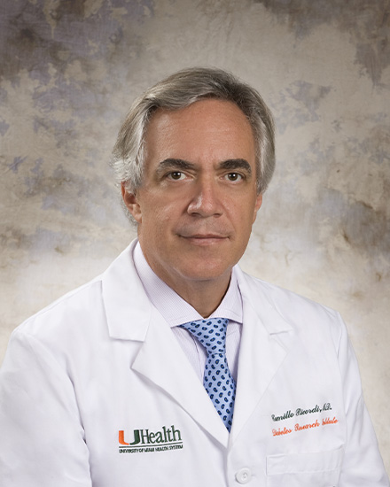 Camillo Ricordi合作论文数Division of Cellular Transplantation, DeWitt Daughtry Family Department of Surgery, Miller School of Medicine, University of Miami2
 Hong Tan (谭泓)合作论文数School of Medicine, Northwest University2Xueyong Li (李学拥)合作论文数2
 R. Jude Samulski合作论文数Gene Therapy Center, University of North Carolina;Department of Pharmacology, School of Medicine, University of North Carolina;Asklepios BioPharmaceuticals, Inc.2
 Tim Nichols合作论文数Division of Cardiology, Department of Medicine, School of Medicine, The University of North Carolina at Chapel Hill;Francis Owen Blood Research Laboratory, The University of North Carolina at Chapel Hill2P. B. Berloco合作论文数2
 Alfredo Cantafora合作论文数2Chengwen Li合作论文数2Manuela Gatto合作论文数2Amanda Dobbins合作论文数1Massimo Rossi合作论文数Umberto I Polyclinic of Rome, Sapienza University of Rome1
 Xiaolin Wang (王晓林)合作论文数School of Integrated Circuits, Shanghai Jiao Tong University1Wuping Li (李武平)合作论文数Chinese Academy of Medical Sciences(Chinese Academy of Medical Sciences),Beijing,China1
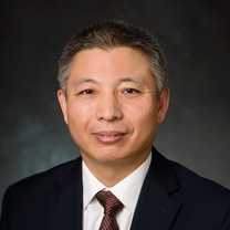 Hongjun Wang合作论文数Department of Biomedical Engineering, Charles V. Schaefer, Jr. School of Engineering and Science, Stevens Institute of Technology;Center for Healthcare Innovation, Stevens Institute of Technology1Hu Yanjun合作论文数1Malavarca Richard合作论文数1
 Wenwei Shao (邵文威)合作论文数Medical School of Tianjin University1Wenfeng Li合作论文数1
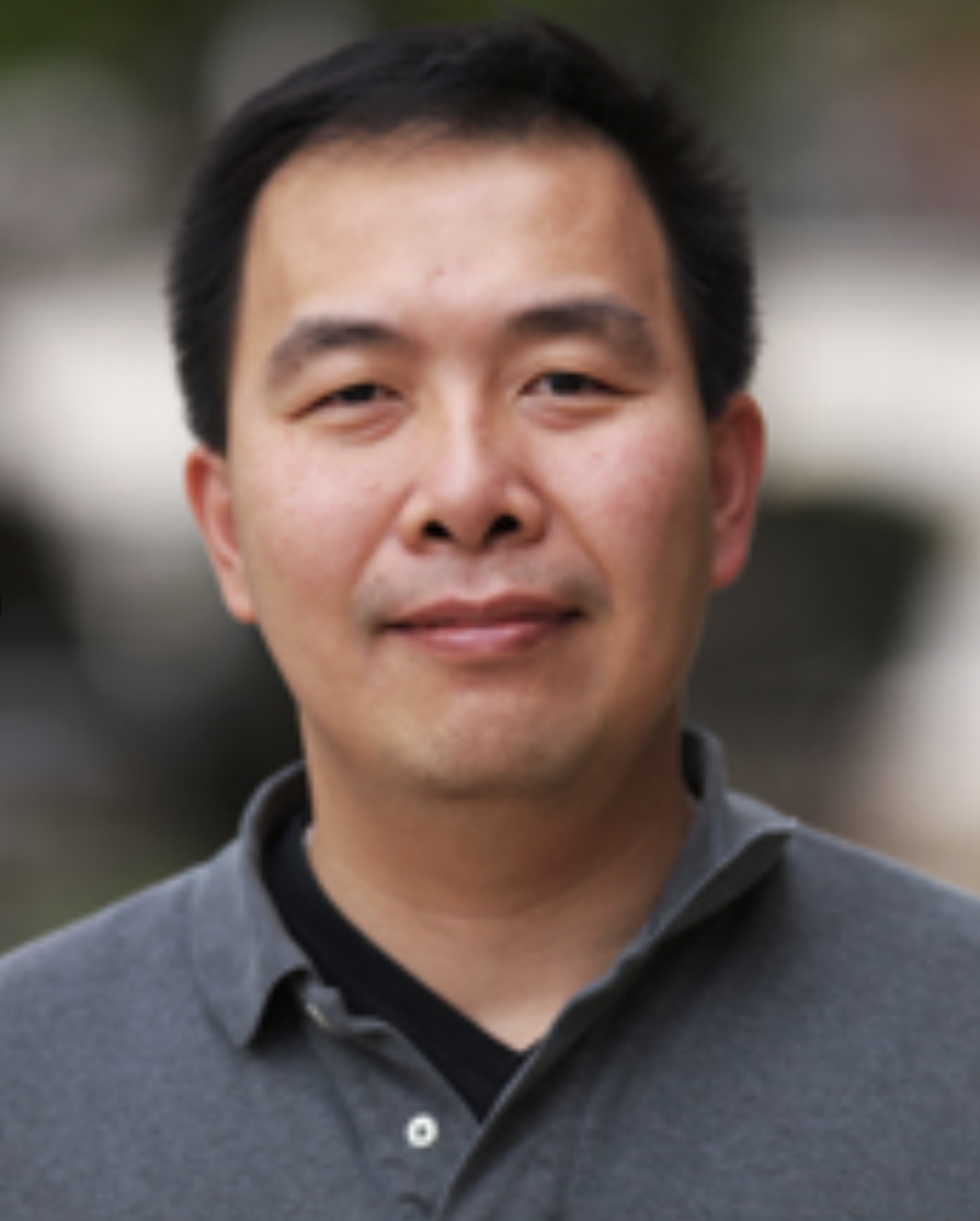 Mitsuo Yamauchi合作论文数Department of Oral and Craniofacial Health Sciences, University of North Carolina1Yuejun li (李曰俊)合作论文数lnslitute of Geology and Geophysics, Chinese Academy of Sciences1
 Shuxun Hou (侯树勋)合作论文数First Affiliated Hospital of People's Liberation Army General Hospital1
 Hongbing Deng (邓红兵)合作论文数School of Resource and Environmental Sciences, Wuhan University1
 Bin Yu (余斌)合作论文数Nanfang Hospital, Southern Medical University1
 John Sondek合作论文数Department of Biochemistry and Biophysics, School of Medicine, The University of North Carolina at Chapel Hil1




