The scope of fetal therapy has evolved over the last several decades to include interventions intended to treat or mitigate morbidities of complex fetal disorders. As a result, maternal-fetal surgery centers have been established across the country to better meet the needs of this patient population. Centers offering fetal interventions need to utilize a multidisciplinary approach to optimally balance the pregnant patient's autonomy and safety while striving to optimize fetal health. Although there is literature highlighting the components that an experienced maternal-fetal surgery center should contain, there are limited publications illustrating the process of creating a maternal-fetal surgery center. The journey of building a maternal-fetal surgery center is often as complex as the care delivered. The convergence of resources from both adult and pediatric medicine along with extensive hospital executive support are necessary. As a group of centers with diverse experience and geographic locations, we present a staged approach to building a comprehensive maternal-fetal surgery center and the lessons learned along the way.
Spina bifida aperta (SBA) is a serious neural tube defect that can lead to a range of disabilities and health complications in affected individuals. In recent years, fetoscopic surgical repair has emerged as a promising new approach to treat spina bifida prenatally, offering the potential for improved outcomes compared with traditional open surgery. As one of the few centres in Europe to offer this innovative technique, the Departments of Obstetrics and Gynaecology, Neurosurgery, and Anaesthesiology and Intensive Care Medicine at the University Medical Centre of Marburg (UKGM Marburg) have faced unique challenges in developing and establishing standards of care for the pregnant patients undergoing this complex procedure. In this publication, we aim to present details of our initial experience with the first 15 patients and propose a clinical concept for the rather complex perioperative management of these patients.
Spina bifida aperta (SBA) is a serious neural tube defect that can lead to a range of disabilities and health complications in affected individuals. In recent years, fetoscopic surgical repair has emerged as a promising new approach to treat spina bifida prenatally, offering the potential for improved outcomes compared with traditional open surgery. As one of the few centres in Europe to offer this innovative technique, the Departments of Obstetrics and Gynaecology, Neurosurgery, and Anaesthesiology and Intensive Care Medicine at the University Medical Centre of Marburg (UKGM Marburg) have faced unique challenges in developing and establishing standards of care for the pregnant patients undergoing this complex procedure. In this publication, we aim to present details of our initial experience with the first 15 patients and propose a clinical concept for the rather complex perioperative management of these patients.
Prenatal repair of myelomeningocele (MMC) is associated with lower rates of hydrocephalus requiring ventriculoperitoneal shunt and improved motor function when compared with postnatal repair. Efforts aiming to develop less invasive surgical techniques to decrease the risk for the pregnant patient while achieving similar benefits for the fetus have led to the implementation of fetoscopic surgical techniques. While no ideal anesthetic technique for fetoscopic MMC repair has been demonstrated, we present our anesthetic approach for these repairs, including considerations for both the pregnant patient and the fetus. We emphasize the importance of the preoperative consultation to optimize any medical conditions and to set expectations for the perioperative course. Our preferred anesthetic technique for the pregnant patient includes general anesthesia with an epidural for postoperative analgesia. Intraoperative anesthetic considerations for patients undergoing fetoscopic surgery include tocolysis, meticulous control of hemodynamics, judicious fluid administration, and maternal temperature regulation. We also avoid long-acting neuromuscular blocking agents due to significant weakness observed when given in combination with magnesium sulfate. While the maternal anesthetic crosses the placenta, direct administration of anesthesia to the fetus is required to reliably blunt the stress response. Additional considerations for the fetus include monitoring, fetal resuscitation strategies, and the theoretical risk of anesthetic neurotoxicity. Postoperatively, we use a multi-modal, opioid sparing regimen for analgesia. As advances in fetal surgery aiming to minimize risk to the pregnant patient alter the surgical approach, maternal-fetal anesthesiologists must adapt and incorporate the unique considerations of fetoscopy into their anesthetic management.
OBJECTIVE:This study reports the infant to preschool outcomes of a laparotomy-assisted, two-port fetoscopic myelomeningocele (MMC) repair and compares the results with those of a contemporary, same-center cohort that underwent either fetal MMC surgery via hysterotomy or postnatal MMC repair. METHODS:All MMC closures between December 2011 and July 2021 were screened. Singleton pregnancies with hindbrain herniation and MMC between T1 and S1 were included. Fetuses were excluded for genetic abnormalities, severe kyphosis, and other congenital anomalies. The pregnant woman determined the method of MMC repair (fetoscopic, hysterotomy, or postnatal repair). RESULTS:Two hundred MMC closures met the study criteria (100 fetoscopic, 41 hysterotomy, and 59 postnatal). The median length of follow-up was beyond 46 months for all groups. The median gestational age at delivery was 38.1 weeks (IQR 35.1, 39.1 weeks) for the fetoscopic group, 35.7 weeks (IQR 33.6, 37.0 weeks) for the hysterotomy group, and 38.6 weeks (IQR 37.7, 39.0 weeks) for the postnatal group. Vaginal delivery occurred in 51% of the fetoscopic cases, and there were no instances of uterine dehiscence or rupture. Treatment for hydrocephalus in the 1st year occurred in 35% (95% CI 27%-50%) of fetoscopic, 33% (95% CI 20%-50%) of hysterotomy, and 81% (95% CI 70%-90%) of postnatal repair cases. At 30 months, patients who underwent fetal intervention were twice as likely to be community ambulators (with or without devices) as those who underwent postnatal repair (52% [95% CI 42%-62%] of fetoscopic, 54% [95% CI 39%-68%] of hysterotomy, and 24% [95% CI 14%-36%] of postnatal cases). Surgery for symptomatic tethered cord occurred in 12% (95% CI 7%-19%) of fetoscopic, 17% (95% CI 8%-31%) of hysterotomy, and 2% (95% CI 1%-8%) of postnatal repair cases. Surgery for symptomatic spinal inclusion cysts was required in 4% (95% CI 1%-9%) of fetoscopic, 7% (95% CI 2%-18%) of hysterotomy, and none (95% CI 0%-8%) of the postnatal cases. CONCLUSIONS:Laparotomy-assisted, two-port fetoscopic repair provides significant benefits for maternal health. It negates the risk of uterine rupture for the index pregnancy and subsequent pregnancies and allows for vaginal delivery. The benefits to the fetus are the same as those of hysterotomy repairs, with a lower risk of prematurity. There was no difference in the rate of surgery for tethered cord or spinal inclusion cysts between fetoscopic and hysterotomy procedures. Overall, laparotomy-assisted, two-port fetoscopic repair is safer for the fetus and the mother than fetal MMC surgery via hysterotomy.
Open spina bifida (OSB) is a congenital, non-lethal malformation with multifactorial etiology. Fetal therapy can be offered under certain conditions to parents after accurate prenatal diagnostic and interdisciplinary counseling. Since the advent of prenatal OSB surgery, various modifications of the original surgical techniques have evolved, including laparotomy-assisted fetoscopic repair. After a two-year preparation time, the team at the University of Giessen and Marburg (UKGM) became the first center to provide a three-port, three-layer fetoscopic repair of OSB via a laparotomy-assisted approach in the German-speaking area. We point out that under the guidance of experienced centers and by intensive multidisciplinary preparation and training, a previously described and applied technique could be transferred to a different setting.
Anesthesiologists are critical members of the multidisciplinary team managing patients with suspected placenta accreta spectrum (PAS). Preoperatively, anesthesiologists provide predelivery consultation for patients with suspected PAS where anesthetic modality and invasive monitor placement is discussed. Additionally, anesthesiologists carefully assess patient and surgical risk factors to choose an anesthetic plan and to prepare for massive intraoperative hemorrhage. Postoperatively, the obstetric anesthesiologist hold unique skills to assist with postoperative pain management for cesarean hysterectomy. We review the unique aspects of peripartum care for patients with PAS who undergo cesarean hysterectomy and explain why these responsibilities are critical for achieving successful outcomes for patients with PAS. KEY POINTS: · Anesthesiologists are critical members of the multidisciplinary team planning for patients with suspected placenta accreta spectrum.. · Intraoperative preparation for massive hemorrhage is a key component of anesthetic care for patients with PAS.. · Obstetric anesthesiologists have a unique skill set to manage postpartum pain and postoperative disposition for patients with PAS who undergo cesarean hysterectomy..
OBJECTIVE: To evaluate the performance characteristics of existing screening tools for the prediction of sepsis during antepartum and postpartum readmissions. METHODS: This was a case–control study using electronic health record data obtained between 2016 and 2021 from 67 hospitals for antepartum sepsis admissions and 71 hospitals for postpartum readmissions up to 42 days. Patients in the sepsis case group were matched in a 1:4 ratio to a comparison cohort of patients without sepsis admitted antepartum or postpartum. The following screening criteria were evaluated: the CMQCC (California Maternal Quality Care Collaborative) initial sepsis screen, the non–pregnancy-adjusted SIRS (Systemic Inflammatory Response Syndrome), the MEWC (Maternal Early Warning Criteria), UKOSS (United Kingdom Obstetric Surveillance System) obstetric SIRS, and the MEWT (Maternal Early Warning Trigger Tool). Time periods were divided into early pregnancy (less than 20 weeks of gestation), more than 20 weeks of gestation, early postpartum (less than 3 days postpartum), and late postpartum through 42 days. False-positive screening rates, C-statistics, sensitivity, and specificity were reported for each overall screening tool and each individual criterion. RESULTS: We identified 525 patients with sepsis during an antepartum hospitalization and 728 patients with sepsis during a postpartum readmission. For early pregnancy and more than 3 days postpartum, non–pregnancy-adjusted SIRS had the highest C-statistics (0.78 and 0.83, respectively). For more than 20 weeks of gestation and less than 3 days postpartum, the pregnancy-adjusted sepsis screening tools (CMQCC and UKOSS) had the highest C-statistics (0.87–0.94). The MEWC maintained the highest sensitivity rates during all time periods (81.9–94.4%) but also had the highest false-positive rates (30.4–63.9%). The pregnancy-adjusted sepsis screening tools (CMQCC, UKOSS) had the lowest false-positive rates in all time periods (3.9–10.1%). All tools had the lowest C-statistics in the periods of less than 20 weeks of gestation and more than 3 days postpartum. CONCLUSION: For admissions early in pregnancy and more than 3 days postpartum, non–pregnancy-adjusted sepsis screening tools performed better than pregnancy-adjusted tools. From 20 weeks of gestation through up to 3 days postpartum, using a pregnancy-adjusted sepsis screening tool increased sensitivity and minimized false-positive rates. The overall false-positive rate remained high.
OBJECTIVE:To evaluate the screening performance characteristics of existing tools for the diagnosis of sepsis during delivery admissions. METHODS:This was a case-control study using electronic health record data, including vital signs and laboratory results, for all delivery admissions of patients with sepsis from 59 nationally distributed hospitals. Patients with sepsis were matched by gestational age at delivery in a 1:4 ratio with patients without sepsis to create a comparison group. Patients with chorioamnionitis and sepsis were compared with a complete cohort of patients with chorioamnionitis without sepsis. Multiple screening criteria for sepsis were evaluated: the CMQCC (California Maternal Quality Care Collaborative), SIRS (Systemic Inflammatory Response Syndrome), the MEWC (the Maternal Early Warning Criteria), UKOSS (United Kingdom Obstetric Surveillance System), and the MEWT (Maternal Early Warning Trigger Tool). Sensitivity, false-positive rates, and C-statistics were reported for each screening tool. Analyses were stratified into cohort 1, which excluded patients with chorioamnionitis-endometritis, and cohort 2, which included those patients. RESULTS:Delivery admissions at 59 hospitals were extracted for patients with sepsis. Cohort 1 comprised 647 patients with sepsis, including 228 with end-organ injury, matched with a control group of 2,588 patients without sepsis. Cohort 2 comprised 14,591 patients with chorioamnionitis-endometritis, of whom 1,049 had sepsis and 238 had end-organ injury. In cohort 1, the CMQCC and the UKOSS pregnancy-adjusted criteria had the lowest false-positive rates (6.9% and 9.6%, respectively) and the highest C-statistics (0.92 and 0.91, respectively). Although other screening criteria, such as SIRS and the MEWC, had similar sensitivities, it was at the cost of much higher false-positive rates (21.3% and 38.3%, respectively). In cohort 2, including all patients with chorioamnionitis-endometritis, the highest C-statistics were again for the CMQCC (0.67) and UKOSS (0.64). All screening tools had high false-positive rates, but the false-positive rates for the CMQCC and UKOSS were substantially lower than those for SIRS and the MEWC. CONCLUSION:During delivery admissions, the CMQCC and UKOSS pregnancy-adjusted screening criteria have the lowest false-positive results while maintaining greater than 90% sensitivity rates. Performance of all screening tools was degraded in the setting of chorioamnionitis-endometritis.
The effect of red blood cell (RBC) transfusion on mortality after lung transplantation (LT) was assessed in some retrospective studies, with contradictory results. The first aim of this study was to assess the 90-day survival of LT recipients according to massive intraoperative transfusion (MIOT).This prospective, observational, single-centre study analysed the intraoperative transfusion (IOT) of all consecutive LT recipients between January 2016 and February 2019. MIOT was defined as transfusion of 5 RBC units or more. The results are presented as the median [IQR] and absolute numbers (proportions) and were analysed using χ2, Fisher, and Mann–Whitney tests (p < 0.05 as significance). Multivariate analyses were performed to identify independent risk factors for MIOT, 90-day and one-year mortality and grade 3 PGD at day 3. Ninety-day and one-year survivals were studied (Kaplan–Meier curves, log rank test). The Paris-North-Hospitals Institutional Review Board approved the study.Overall, 147 patients were included in the analysis, 27 (18%) of them received MIOT. In multivariate analysis, predictive factors of MIOT included preoperative ECMO support (p = 0.017), and bilateral LT (p = 0.023). The SOFA score on ICU admission after LT was higher in cases with MIOT (p < 0.001). MIOT was an independent risk factor for 90-days and one-year mortality (p = 0.002 and 0.008 respectively). The number of RBCs unit transfused during surgery was an independent risk factor for grade 3 PGD at day 3 (OR 1.14, 95% CI [1.00–1.29], p = 0.040).Increased preoperative severity of recipients predicts MIOT. MIOT is associated with increased early postoperative morbidity and mortality rates.
Obstetric anesthesiologists can use the tenets of principlism to identify ethically supportable decisions. This approach begins with recognizing and upholding the fiduciary obligations that every physician has to their patient. For the pregnant patient, these obligations are both autonomy- and beneficence-based. Additional beneficence-based obligations arise in cases where the fetus is also considered a patient. Ethical dilemmas result from conflict between any of these obligations. A shared decision-making model that incorporates the foundational principles of ethics can be used to resolve these conflicts, leaving clinicians and patients confident that a sound decision-making process has taken place and an ethically supportable decision has been reached.
BACKGROUND: Opioid overprescription has the potential to lead to harmful medications remaining in homes and to a rise in accidental or deliberate ingestion by children and adolescents. Although methods for opioid disposal are available, many are costly or require greater than minimal effort for the patient. In this study, we used a mail-back return envelope to retrieve unused opioids after ambulatory pediatric surgery. METHODS: This feasibility study was performed to assess the rate of opioid return by using a mail-back envelope for children ages 0 to 18 prescribed opioids after outpatient surgery. Participants were provided a return envelope as well as instruction on the dangers of opioids in the home. Our primary outcome was to assess the absolute percent return rate through the use of a mail-back envelope. RESULTS: Between November 2017 and October 2018, we identified 355 patients, of whom 331 were included in the analysis. In total, 64 (19.3%) returned opioids. In total,.2000 mL of liquid opioids and >250 tablets or nearly 3000 mg of oral morphine equivalents were removed from the homes of the 64 participants. Of those patients returning unused medications, the median rate of return was 58% (interquartile range = 34.7%-86.1%) of the written prescription. CONCLUSIONS: The findings suggest that providing a free mail-back return envelope is a suitable way to remove unused opioids from the home after pediatric surgery. Additional research is needed to identify barriers to return of unused medications.
There are many situations that the pediatric anesthesiologist encounters on a daily basis. However, caring for the patient with do-not-resuscitate (DNR) orders requires attention to a variety of requests that may not be applicable in all scenarios. End-of-life care for pediatric patients is complex and presents many challenges for all physicians. The care of a patient with a DNR order who presents for surgery requires thoughtful, open communication between all stakeholders including the primary team, the surgeon, the anesthesiologist, the patient (when mental status and development allows), and the patient’s family. To best align care with goals, DNR orders can be rescinded, continued, or—most often—modified, for the perioperative period.
Study Objective: We examined the characteristics of women who choose nitrous oxide for labor analgesia and identified factors that predict conversion from nitrous oxide to labor neuraxial analgesia.Design: Retrospective descriptive study.Setting: Labor and Delivery Ward.Patients: 146 pregnant women who used nitrous oxide for analgesia during labor and delivery between September 2014 and September 2015.Interventions: Chart review only.Measurements: Demographic, obstetric, and intrapartum characteristics of women using nitrous oxide were examined. Multivariable logistic regression was performed to identify factors associated with conversion from nitrous oxide to neuraxial analgesia. Data are presented as n (%), median [IQR], adjusted relative risk (aRR), and 95% confidence intervals (CI) as appropriate.Results: During the study period, 146 women used nitrous oxide for labor analgesia (accounting for 3% of the total deliveries). The majority (71.9%) of women who used nitrous oxide were nulliparous, and over half (51.9%) had expressed an initial preference for "nonmedical birth." The conversion rate to neuraxial blockade was 63.2%, compared to a concurrent institutional rate of 85.1% in women who did not use nitrous oxide. Factors associated with conversion from nitrous oxide to neuraxial blockade were labor induction (aRR = 2.0, CI 1.2-33) and labor augmentation (aRR = 1.7, CI 1.0-2.9).Conclusion: Only a small number of women opted to use nitrous oxide during labor, analgesia was minimal, and most converted to neuraxial analgesia. Women with induced and augmented labors should be counseled about the increased likelihood that they will convert to neuraxial analgesia. (C) 2017 Elsevier Inc. All rights reserved.
We read with interest the article titled “What is trending now? An analysis of trends in internet searches for labor epidural” by Sutton et al. 1 Sutton C.D. Carvalho B. What's trending now? An analysis of trends in internet searches for labor epidurals. Int J Obstet Anesth. 2017; 30: 52-57 Abstract Full Text Full Text PDF PubMed Scopus (11) Google Scholar We agree with the authors regarding the importance of targeting how and what type of information patients are seeking. Over a year ago, my research team and I performed a survey addressing similar questions as part of a resident-driven quality assurance project. Once institutional review board approval was obtained, we conducted a survey to gauge the impact of an educational pamphlet, printed both in Spanish and English, that was given to every parturient at the antenatal clinic and upon admission to the labor and delivery suite over a period of two months. Our primary outcome was to inform parturients regarding the various labor pain management options. The secondary outcome was to evaluate how and what women are seeking when considering labor pain management options. As a quality assurance project, we evaluated 100 surveys.
 Michael A Belfort合作论文数Texas Children's Pavilion for Women;Department of Obstetrics and Gynecology, Baylor College of Medicine4
 Gerald Kalmus合作论文数3David G Mann合作论文数3Corinna Keil合作论文数3
 Ivonne Bedei合作论文数3
 William E. Whitehead合作论文数Division of Gastroenterology and Hepatology, Department of Medicine, School of Medicine, University of North Carolina at Chapel Hill;Center for Functional Gastrointestinal Disorders, University of North Carolina at Chapel Hill3Mindy Foster合作论文数2Kurt Wharton合作论文数2Magdalena S. Cortes合作论文数2Matt Fuller合作论文数2
 Alex Butwick合作论文数Department of Anesthesiology, Perioperative and Pain Medicin, School of Medicine, Stanford University2
 Alyssa Burgart合作论文数Department of Anesthesiology, Perioperative and Pain Medicine, Stanford University2Ann-Kristin Schubert合作论文数2
 Sylvia E K Sudat合作论文数Corresponding author at: 2121 N. California Blvd, Suite 310, Walnut Creek, CA 94596, United States.2Rebecca Johnson合作论文数Texas Children's Hospital Pavilion for Women, Baylor College of Medicine2
 Luc Joyeux合作论文数Texas Children's Hospital, Baylor College of Medicine2Melissa G Rosenstein合作论文数2
 Brenna L Hughes合作论文数Department of Obstetrics and Gynecology, School of Medicine, Duke University2
 Melissa E. Bauer合作论文数Department of Anesthesiology, School of Medicine, Duke University2
 Paloma Toledo合作论文数Department of Anesthesiology, Feinberg School of Medicine, Northwestern University2
 Elliott Main合作论文数Maternal-Fetal Medicine and Obstetrics Division, School of Medicine, Stanford University;California Maternal Quality Care Collaborative2Laurence E. Shields合作论文数2
 Brendan Carvalho合作论文数Department of Anesthesiology, Perioperative and Pain Medicine, School of Medicine, Stanford University2King Alice合作论文数Laboratory for Regenerative Wound Healing, Cincinnati Children's Hospital Medical Center2
 Azar Kristen M J合作论文数Palo Alto Medical Foundation Research Institute2
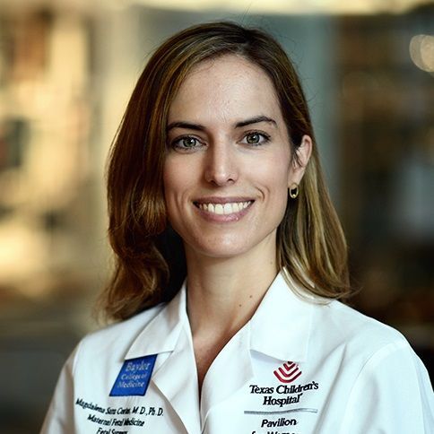 Magdalena Sanz Cortes合作论文数Baylor College of Medicine;Texas Children's Hospital Pavilion for Women2
 Ronald Gibbs合作论文数Department of Obstetrics & Gynecology, Stanford University2
 Hinnerk Wulf合作论文数Universitätsklinikum Gießen und Marburg;Klinik für Anästhesie und Intensivtherapie, Universität Marburg2
 Vesela P Kovacheva合作论文数2
 Ramesha Papanna合作论文数1Amy Hermesch合作论文数1Mirjam Wege合作论文数1Stephanie Dukhovny合作论文数1Siegmund Köhler合作论文数1
 Christine M. Warrick合作论文数1Danielle Guffey合作论文数1
 Pamela Flood合作论文数Department of Anesthesiology, Perioperative and Pain Medicine, School of Medicine, Stanford University1Nilesh K Desai合作论文数1P. Hess合作论文数1Arvind Chandrakantan合作论文数1
 Raphael Sun合作论文数Div Pediat Surg, Texas Childrens Hosp1Guillermo Aldave合作论文数Texas Children's Hospital, Baylor College of Medicine1Amanda Kim合作论文数1
 Benjamin Saß合作论文数1Mubeen Jafri合作论文数Doernbecher Childrens Hosp, Oregon Hlth & Sci Univ1
 Erin Madriago合作论文数1Azarow Kenneth S合作论文数1
 Michaela Kristina Farber合作论文数Department of Anesthesiology, Perioperative, and Pain Medicine, Brigham and Women's Hospital, Boston, MA, USA.1
 Dean B. Andropoulos合作论文数Department of Anesthesiology, Perioperative & Pain Medicine, Texas Children's Hospital;Department of Anesthesiology, Baylor College of Medicine;Pediatric Heart Network Core Clinical Center, Baylor College of Medicine1




