Because many tumors possess blood vessels permeable to particles with diameters of 200 nm, it is possible that submicron perfluorocarbon droplets could constitute a novel extravascular ultrasound contrast agent capable of selectively enhancing tumors. Under exposure to bursts of ultrasound of sufficient rarefactional pressure, droplets can undergo vaporization to form echogenic microbubbles. In this study, phase-change thresholds of 220-nm-diameter droplets composed of perfluoropentane were studied in polyacrylamide gel phantoms maintained at temperatures of 21-37 degrees C, exposed to high-pressure bursts of ultrasound with frequencies ranging from 5-15 MHz and durations of 1 mu s to 1 ms. The thresholds were found to depend inversely and significantly (p < 0.001) on ultrasound frequency, pulse duration, and droplet temperature, ranging from 9.4 +/- 0.8 MPa at 29 degrees C for a 1-mu s burst at 5 MHz to 3.2 +/- 0.5 MPa at 37 degrees C for a 1-ms burst at 15 MHz. The diameters of microbubbles formed from droplets decreased significantly as phantom stiffness increased (p < 0.0001), and were independent of pulse duration, although substantially more droplets were converted to microbubbles for 1-ms pulse durations compared with briefer exposures. In vivo experiments in a mouse tumor model demonstrated that intravenously injected droplets can be converted into highly echogenic microbubbles 1 h after administration. (E-mail: williams@sri.utoronto.ca) (C) 2013 World Federation for Ultrasound in Medicine & Biology.
Objectives: To characterize the ability of high-intensity focused ultrasound to achieve thrombolysis in vitro and investigate the feasibility of this approach as a means of restoring blood flow in thrombus-occluded arteries in vivo. Materials and Methods: All experiments were approved by the Institutional Animal Care Committee. Thrombolysis was performed with a 1.51-MHz focused ultrasound transducer with pulse lengths of 0.1 to 10 milliseconds and acoustic powers up to 300 W. In vitro experiments were performed with blood clots formed from rabbit arterial blood and situated in 2-mm diameter tubing. Both single location and flow bypass recanalization experiments were conducted. In vitro clot erosion was assessed with 30-MHz ultrasound, with debris size measured with filters and a Coulter counter. In vivo clots were initiated in the femoral arteries of rabbits (n = 26). Cavitation signals from bubbles formed during exposure were monitored. In vivo flow restoration was assessed with 23-MHz Doppler ultrasound. Results: At a single location, in vitro clot erosion volumes increased with exposure power and pulse length, with debris size reducing with increasing pulse length. Flow bypass experiments achieved 99.2% clot erosion with 1.1% of debris above 0.5 mm in size. In vivo, 10 milliseconds pulses were associated with bleeding, but at 1 millisecond, it was feasible to achieve partial flow restoration in 6of the 10 clots with only 1of the 10 showing evidence of bleeding. In all cases, thrombolysis occurred only in the presence of cavitation. Conclusion: High-intensity focused ultrasound thrombolysis is feasible as a means of restoring partial blood flow in thrombus-occluded arteries in the absence of thrombolytic agents. The potential for bleeding with this approach requires further investigation.
Ultrasound has been shown to potentiate the effects of tissue plasminogen activator to improve clot lysis in a range of in vitro and in vivo studies as well as in clinical trials. One possible mechanism of action is acoustic radiation force-induced clot displacements. In this study, we investigate the temporal and spatial dynamics of clot displacements and strain initiated by focused ultrasound pulses. Displacements were produced by a 1.51 MHz f-number 1 transducer over a range of acoustic powers (1-85 W) in clots constrained within an agar vessel phantom channel. Displacements were tracked during and after a 5.45 ms therapy pulse using a 20 MHz high-frequency ultrasound imaging probe. Peak thrombus displacements were found to be linear as a function of acoustic power up to 60 W before leveling off near 128 μm for the highest transmit powers. The time to peak displacement and recovery time of blood clots was largely independent of acoustic powers with measured values near 2 ms. A linear relationship between peak axial strain and transmit power was observed, reaching a peak value of 11% at 35 W. The peak strain occurred ~0.75 mm from the focal zone for all powers investigated in both lateral and axial directions. These results indicate that substantial displacements can be induced by focused ultrasound in confined blood clots, and that the spatial and temporal displacement patterns are complex and highly dependent on exposure conditions, which has implications for future work investigating their link to clot lysis and for developing approaches to exploit these effects.
The objective of this study was to investigate the kinetics of contrast agent concentration within the brain when it is subjected to pulsed focused ultrasound. An ultrasound imaging array was mounted within the aperture of a 1.68 MHz focused therapy transducer. Experiments were performed in the brains of rabbits that had undergone craniotomies, with the therapy beam focus localized within one hemisphere. Pulse lengths of 10 ms were employed at repetition rates of 0.1-2 Hz and acoustic powers of 0.1-1 watts. Contrast imaging was performed for up to 9 minutes following the bolus injection of Definity. Contrast signal power was then quantified in regions of interest within the acoustic focus of the therapy beam, as well as in the contra-lateral hemisphere. Following the injection of the contrast agent, signals peaked within 10 seconds and then underwent a period of slow decay. Individual therapy pulses resulted in microbubble depletion within the focus for powers >=0.25 W, with full recovery of signal occurring within 1-3 seconds, depending on transmit power. As the pulse repetition rates increased, agent reperfusion between pulses was incomplete and the concentration within the beam was progressively diminished, to a degree dependent upon both power and repetition rates. These results demonstrate that microbubble concentration can be substantially influenced by destruction induced by therapeutic ultrasound pulses. The kinetics of this effect may therefore be a significant factor influencing the efficiency of BBB breakdown.
Despite a significant body of work establishing the feasibility of ultrasound mediated thrombolysis in vitro, in vivo, and in clinical settings, there remains considerable uncertainty about the specific mechanisms involved in this process. This motivates further work to elucidate these mechanisms, which will be central to optimizing safe and effective operating conditions, and to guide the development of novel approaches and instrumentation. In this study, we investigate the use of high frequency ultrasound as a means of gaining mechanistic insight into sonothrombolysis. A high frequency ultrasound (20-50 MHz) instrument is employed which provides the ability to conduct volumetric clot imaging as well as pulsed-wave Doppler to monitor hemodynamics within vessels and clots. With modifications, it is enabled to perform the acquisition of RF data to assess the displacement of clots and vessel walls subjected to therapeutic pulses. Additional modifications were made to perform nonlinear imaging of micron to submicron sized bubbles, which are of interest in enhancing clot lysis. Experiments were performed on in vitro clots, and in vivo using a rabbit femoral artery clot model initiated by the injection of thrombin. Therapeutic pulses are provided by a single element spherically focused air backed transducer with transmit frequencies of 1.68 MHz. Clear visualization of the clots, displacements, and presence or absence of flow within these vessels is shown to be feasible, indicating the potential of this approach as a tool for providing insight into sonothrombolysis.
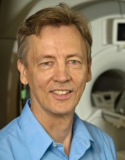 Kullervo Hynynen合作论文数Sunnybrook Research Institute;Department of Medical Biophysics, Temerty Faculty of Medicine, University of Toronto4
 David Goertz合作论文数Department of Medical Biophysics, Temerty Faculty of Medicine, University of Toronto4
 Mike Lee合作论文数Department of Medical Biophysics, Sunnybrook Health Sciences Centre1
 Naomi Matsuura合作论文数Department of Materials Science and Engineering, Faculty of Applied Science and Engineering, University of Toronto1
 Peter N Burns合作论文数Department of Medical Biophysics, University of Toronto/Sunnybrook Research Institute1
 Williams Ross合作论文数Sunnybrook Research Institute, University of Toronto1
 Stuart Foster合作论文数Sunnybrook Health Sciences Centre, Sunnybrook Research Institute, Temerty Faculty of Medicine, University of Toronto;Department of Medical Biophysics, Temerty Faculty of Medicine, University of Toronto1
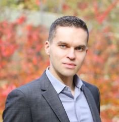 Reznik Nikita合作论文数Department of Medical Biophysics, University of Toronto1
 Ivan Gorelikov合作论文数1
 Emmanuel Cherin合作论文数Imaging Research, Sunnybrook Health Sciences Centre1




