The growing use of biomarker testing for personalized oncologic care has increased the demand for complex and repeated percutaneous biopsies in abdominal and interventional radiology practices. Challenging biopsies can be daunting, requiring a careful assessment of feasibility, safety, and risk tolerance. Several strategies can help optimize the chances of achieving success. Such strategies include leveraging the advantages of the modality chosen for image guidance, ensuring patient comfort, and the use of special techniques, such as IV contrast, navigational tols, and biopsy needle modifications. This review outlines practical tips and tricks for radiologists performing challenging percutaneous biopsies.
Ultrasound (US) has many known advantages, including lack of ionizing radiation, low operating cost, and an unmatched safety profile. The development of contrast-enhanced US (CEUS) has proven to be a useful alternative or adjunct in the assessment of multiple organ system pathology, including genitourinary (GU), gastrointestinal, and pulmonary applications. CEUS offers several advantages over other contrast-enhanced modalities, including real-time enhancement kinetics, decreased contrast reactions, and lack of renal toxicity. CEUS is especially useful in assessment of GU pathology, where it can be used intravascularly as well as intraluminally within the collecting system to augment the evaluation of multiple GU pathologies.
Tissue changes and the enlargement of the prostate, whether benign or malignant, are among the most common groups of diseases that affect men and can have significant impacts on length and quality of life. The prevalence of benign prostatic hyperplasia (BPH) increases significantly with age and affects nearly all men as they grow older. Other than skin cancers, prostate cancer is the most common cancer among men in the United States. Imaging is an essential component in the diagnosis and management of these conditions. Multiple modalities are available for prostate imaging, including several novel imaging modalities that have changed the landscape of prostate imaging in recent years. This review will cover the data relating to commonly used standard-of-care prostate imaging modalities, advances in newer technologies, and newer standards that impact prostate gland imaging.
With PET/MRI, the strengths of PET and MRI are combined to allow simultaneous image acquisition and near-perfect image coregistration. MRI is increasingly being used for staging and restaging of abdominopelvic oncologic lesions, including prostate, hepatobiliary, pancreatic, neuroendocrine, cervical, and rectal cancers. Fluorine 18-fluorodeoxyglucose PET/CT has long been considered a cornerstone of oncologic imaging, and the development of multiple targeted radiotracers has led to increased research on and use of these agents in clinical practice. Thus, simultaneously performed PET/MRI enables the acquisition of complementary imaging information, with distinct advantages over PET/CT and MR image acquisitions. The authors provide an overview of PET/MRI, including descriptions of the major differences between PET/MRI and PET/CT, as well as case examples and treatment protocols for patients with commonly encountered malignancies in the abdomen and pelvis. Online supplemental material is available for this article.©RSNA, 2021.
INTRODUCTION Despite advances in standard-of-care treatment for prostate cancer, including pelvic radiotherapy and radical prostatectomy with lymph node dissection, biochemical recurrence remains a problem with some studies reporting up to 30% of patients with biochemical recurrence at 10-years following treatment. Upon detection of a rise in serum prostate-specific antigen following treatment, clinicians are left with the dilemma of how best to treat these patients. Frequently, these patients are restaged with CT of the abdomen and pelvis or MRI of the pelvis in addition to a nuclear medicine bone scan. However, detection of recurrent disease both within the prostate gland/prostatectomy bed and in the region of pelvic lymph nodes remains challenging and is limited by size criteria per RECIST 1.1. [F]Fluciclovine PET imaging has improved detection of these previously occult metastases and helped guide treatment planning. In some select patients, a targeted lymphadenectomy may be the treatment of choice if the PET-avid disease is limited to a solitary lymph node site of spread. However, this concept has not been well described in the literature and here we provide 3 cases and short-term outcomes of patients that underwent [F]fluciclovine PET guided surgical excision of their biochemically recurrent prostate cancer.
Despite advances in standard-of-care treatment for prostate cancer, including pelvic radiotherapy and radical prostatectomy with lymph node dissection, biochemical recurrence remains a problem with some studies reporting up to 30% of patients with biochemical recurrence at 10-years following treatment. Upon detection of a rise in serum prostate-specific antigen following treatment, clinicians are left with the dilemma of how best to treat these patients. Frequently, these patients are restaged with CT of the abdomen and pelvis or MRI of the pelvis in addition to a nuclear medicine bone scan. However, detection of recurrent disease both within the prostate gland/prostatectomy bed and in the region of pelvic lymph nodes remains challenging and is limited by size criteria per RECIST 1.1. [18F]Fluciclovine PET imaging has improved detection of these previously occult metastases and helped guide treatment planning. In some select patients, a targeted lymphadenectomy may be the treatment of choice if the PET-avid disease is limited to a solitary lymph node site of spread. However, this concept has not been well described in the literature and here we provide 3 cases and short-term outcomes of patients that underwent [18F]fluciclovine PET guided surgical excision of their biochemically recurrent prostate cancer.
Magnetic resonance imaging (MRI)/ultrasound fusion-targeted biopsy (TB) has been shown to more accurately identify higher-grade prostate cancers compared with standard-of-care systematic sextant prostate biopsy (SB). However, occasional false-positive imaging findings occur. We investigated the histologic findings associated with false-positive prostate MRI findings. A retrospective review was performed on our surgical pathology database from 2014 to 2017 selecting patients with no cancer detected on TB with concurrent SB after at least 1 prior benign SB session. Histologic features evaluated included percentage of core involvement by chronic inflammation, percentage of core composed of stroma, percentage of glands involved by atrophy, and presence of the following features: acute or granulomatous inflammation, stromal nodular hyperplasia, adenosis, squamous metaplasia, basal cell hyperplasia, and presence of skeletal muscle. Histologic findings were compared between TB and concurrent SB. We identified 544 patients who underwent TB. Of these, 41 patients, including 62 targeted lesions, met criteria. Compared with SB tissue, the mean percentage of stroma was increased in TB (P = .02). Basal cell hyperplasia was also found to be more common on TB (P = .02). Both high percentage of stroma (P = .046) and presence of basal cell hyperplasia (P = .038) were independent predictors on multivariate analysis. The combination of high chronic inflammation, high stroma, acute inflammation, and basal cell hyperplasia was associated with TB (P = .001). Atrophic glands and chronic inflammation showed a positive correlation (r = 0.67, P = .003), which was especially seen in high prostate imaging reporting and data system lesions. Specific benign histologic entities are associated with false-positive findings on prostate MRI. (C) 2018 Elsevier Inc. All rights reserved.
Prostate cancer is the most common noncutaneous malignancy in men in the United States with an estimated 164,690 new cases diagnosed in 2018 [ 1 American Cancer Society. Facts and figures 2018. Available at https://www.cancer.org/research/cancer-facts-statistics/all-cancer-facts-figures/cancer-facts-figures-2018.html. Accessed June 3, 2018. Google Scholar ]. Approximately one in nine men will be diagnosed with prostate cancer in their lifetime, but many do not die from the disease [ 1 American Cancer Society. Facts and figures 2018. Available at https://www.cancer.org/research/cancer-facts-statistics/all-cancer-facts-figures/cancer-facts-figures-2018.html. Accessed June 3, 2018. Google Scholar ]. There are an estimated 2.9 million men living in the United States diagnosed with prostate cancer at some point in their lives either on surveillance or after treatment [ 1 American Cancer Society. Facts and figures 2018. Available at https://www.cancer.org/research/cancer-facts-statistics/all-cancer-facts-figures/cancer-facts-figures-2018.html. Accessed June 3, 2018. Google Scholar ]. However, approximately 29,430 deaths from prostate cancer occurred in 2018 (1 in 41 men), making it the second leading cause of cancer-related death in American men [ 1 American Cancer Society. Facts and figures 2018. Available at https://www.cancer.org/research/cancer-facts-statistics/all-cancer-facts-figures/cancer-facts-figures-2018.html. Accessed June 3, 2018. Google Scholar ].
We reviewed the role of transesophageal echocardiography (TEE) in the management of renal cell carcinoma (RCC) with associated tumor thrombus. Many consider intraoperative TEE as imperative in cases of Level 4 thrombi with atrial involvement, as well as in cases that require the use of cardiopulmonary bypass (CPB). However, the role of TEE in the surgical management of RCC with associated inferior vena cava (IVC) thrombus may expand beyond this subset. When performed after induction, TEE provides updated information regarding tumor thrombus staging, which is essential for optimal surgical planning. Furthermore, TEE provides feedback regarding properties of the thrombus, such as fragility and adherence, which may alter surgical technique. TEE can also be used intraoperatively for central venous line placement, to monitor cardiovascular and fluid status, to guide vascular clamp placement, and to ensure complete removal of the tumor thrombus. In some cases, the use of TEE allows for less morbid procedures and safe avoidance of CPB. We therefore recommend the use of preoperative TEE in all cases with a known tumor thrombus with discretion as to what extent TEE is used throughout the remainder of the case. Further investigation is necessary to elucidate the effect of TEE on patient outcomes, including surgical complication rates, morbidity and mortality of procedures, and cancer control.
Radical cystectomy is considered the standard of care for muscle invasive bladder cancer (MIBC), as well as some high-grade or recurrent non-muscle invasive bladder cancers. There are a number of reasons that urologic surgeons may find themselves considering a salvage cystectomy (SC) for a patient. Understanding the risks associated with performing SC is important for patient counseling when considering primary management of MIBC. Awareness of the unique complications that can occur can alter surgical technique and approach and can equip clinical staff with knowledge to properly monitor the patient during post-operative surveillance to identify and treat these situations. In this review, we characterize the outcomes of patients who underwent SC, elucidate the special considerations that must be made when performing this surgery, and discuss these outcomes in comparison to primary surgery in the absence of radiation.
 Soroush Rais-Bahrami合作论文数Department of Urology, Heersink School of Medicine, University of Alabama;Department of Radiology, Heersink School of Medicine, University of Alabama7
 Nix Jeffrey W合作论文数5
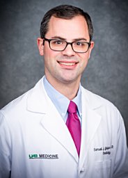 Samuel Galgano合作论文数5Kristin Kelly Porter合作论文数Center for Cognitive Neuroscience, Dartmouth College4David Ullman合作论文数2
 Luciana Schultz合作论文数2
 Jonathan Mcconathy合作论文数2Jennifer B Gordetsky合作论文数Univ Alabama Birmingham2Stephen H. Gray合作论文数1Janelle T West合作论文数1
 Elainea N Smith合作论文数Department of Radiology, University of Alabama at Birmingham1David Summerlin合作论文数1
 Jennifer Taylor合作论文数Scripps Institution of Oceanography, University of California, San Diego1Maria Del Carmen Rodriguez Pena合作论文数Dept Pathol, Univ Alabama Birmingham1deShazo Mollie合作论文数1Tuck Benjamin C合作论文数1
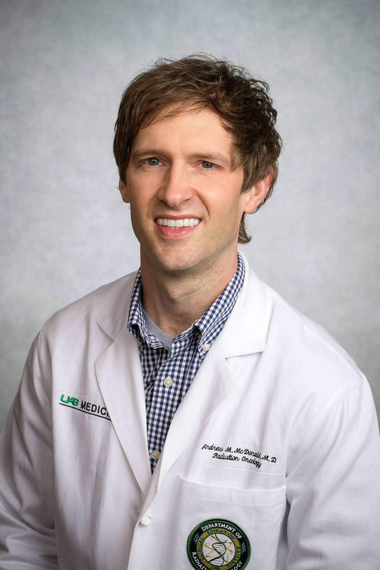 Andrew McDonald合作论文数Department of Radiation Oncology, Heersink School of Medicine, The University of Alabama at Birmingham1
 Seth Paul Lerner合作论文数Medical Center, Baylor College of Medicine;Scott Department of Urology, Baylor College of Medicine1
 Sejong Bae合作论文数Center for Outcomes and Effectiveness Research and Education, School of Medicine, The University of Alabama at Birmingham;Integrative Center for Aging Research, School of Medicine, The University of Alabama at Birmingham;Biostatistics and Bioinformatics O'Neal Comprehensive Cancer Center, University of Alabama at Birmingham1Mark E. Lockhart合作论文数1David T. Fetzer合作论文数1
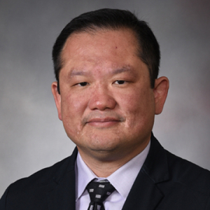 Eddy S Yang合作论文数Department of Radiation Medicine, College of Medicine, University of Kentucky1




