OBJECTIVEThe aim of this study was to evaluate the safety and clinical outcomes associated with stereotactic surgical implantation of modified bone marrow–derived mesenchymal stem cells (SB623) in patients with stable chronic ischemic stroke.METHODSThis was a 2-year, open-label, single-arm, phase 1/2a study; the selected patients had chronic motor deficits between 6 and 60 months after nonhemorrhagic stroke. SB623 cells were administered to the target sites surrounding the subcortical stroke region using MRI stereotactic image guidance.RESULTSA total of 18 patients were treated with SB623 cells. All experienced at least 1 treatment-emergent adverse event (TEAE). No patients withdrew due to adverse events, and there were no dose-limiting toxicities or deaths. The most frequent TEAE was headache related to the surgical procedure (88.9%). Seven patients experienced 9 serious adverse events, which resolved without sequelae. In 16 patients who completed 24 months of treatment, statistically significant improvements from baseline (mean) at 24 months were reported for the European Stroke Scale (ESS) score, 5.7 (95% CI 1.4–10.1, p < 0.05); National Institutes of Health Stroke Scale (NIHSS) score, −2.1 (95% CI −3.3 to −1.0, p < 0.01), Fugl-Meyer (F-M) total score, 19.4 (95% CI 9.9–29.0, p < 0.01); and F-M motor scale score, 10.4 (95% CI 4.0–16.7, p < 0.01). Measures of efficacy reached plateau by 12 months with no decline thereafter. There were no statistically significant changes in the modified Rankin Scale score. The size of transient lesions detected by T2-weighted FLAIR imaging in the ipsilateral cortex at weeks 1–2 postimplantation significantly correlated with improvement in ESS (0.619, p < 0.05) and NIHSS (−0.735, p < 0.01) scores at 24 months.CONCLUSIONSIn this completed 2-year phase 1/2a study, implantation of SB623 cells in patients with stable chronic stroke was safe and was accompanied by improvements in clinical outcomes.Clinical trial registration no.: NCT01287936 (clinicaltrials.gov)
BACKGROUND Telomerase activity in leukemic blasts frequently is increased among patients with high‐risk acute myeloid leukemia (AML). In the current study, the authors evaluated the feasibility, safety, immunogenicity, and therapeutic potential of human telomerase reverse transcriptase (hTERT)‐expressing autologous dendritic cells (hTERT‐DCs) in adult patients with AML. METHODS hTERT‐DCs were produced from patient‐specific leukapheresis, electroporated with an mRNA‐encoding hTERT and a lysosomal‐targeting sequence, and cryopreserved. A total of 22 patients with a median age of 58 years (range, 30‐75 years) with intermediate‐risk or high‐risk AML in first or second complete remission (CR) were enrolled. hTERT‐DCs were generated for 24 patients (73%). A median of 17 intradermal vaccinations (range, 6‐32 intradermal vaccinations) containing 1×10 7 cells were administered as 6 weekly injections followed by 6 biweekly injections. A total of 21 patients (16 in first CR, 3 in second CR, and 2 with early disease recurrence) received hTERT‐DCs. RESULTS hTERT‐DCs were well tolerated with no severe toxicities reported, with the exception of 1 patient who developed idiopathic thrombocytopenic purpura. Of the 19 patients receiving hTERT‐DCs in CR, 11 patients (58%) developed hTERT‐specific T‐cell responses that primarily were targeted toward hTERT peptides with predicted low human leukocyte antigen (HLA)‐binding affinities. With a median follow‐up of 52 months, 58% of patients in CR (11 of 19 patients) were free of disease recurrence at the time of their last follow‐up visit; 57% of the patients who were aged ≥60 years (4 of 7 patients) also were found to be free of disease recurrence at a median follow‐up of 54 months. CONCLUSIONS The generation of hTERT‐DCs is feasible and vaccination with hTERT‐DCs appears to be safe and may be associated with favorable recurrence‐free survival. Cancer 2017;123:3061–72. © 2017 American Cancer Society .
Regenerative medicine for the treatment of motor features in Parkinson's disease (PD) is a promising therapeutic option. Donor cells can simultaneously address multiple pathological mechanisms while responding to the needs of the host tissue. Previous studies have demonstrated that mesenchymal stromal cells (MSCs) promote recovery using various animal models of PD. SanBio Inc. has developed a novel cell type designated SB623, which are adult bone marrow-derived MSCs transfected with Notch intracellular domain. In this preclinical study, SB623 cells protected against 1-methyl-4-phenyl-1,2,3,6-tetrahydropyridine (MPTP)-induced nigrostriatal injury when transplanted unilaterally into C57BL/6 mouse striatum 3days prior to toxin exposure. Specifically, mice with the SB623 cell transplants revealed significantly higher levels of striatal dopamine, tyrosine hydroxylase immunoreactivity and stereological nigral cell counts in the ipsilateral hemisphere vs vehicle-treated mice following MPTP administration. Interestingly, improvement in markers of striatal dopaminergic integrity was also noted in the contralateral hemisphere. These data indicate that MSCs transplantation, specifically SB623 cells, may represent a novel therapeutic option to ameliorate damage related to PD, not only at the level of striatal terminals (i.e. the site of implantation) but also at the level of the nigral cell body. Copyright (c) 2015 John Wiley & Sons, Ltd.
Background and Purpose— Preclinical data suggest that cell-based therapies have the potential to improve stroke outcomes. Methods— Eighteen patients with stable, chronic stroke were enrolled in a 2-year, open-label, single-arm study to evaluate the safety and clinical outcomes of surgical transplantation of modified bone marrow–derived mesenchymal stem cells (SB623). Results— All patients in the safety population (N=18) experienced at least 1 treatment-emergent adverse event. Six patients experienced 6 serious treatment-emergent adverse events; 2 were probably or definitely related to surgical procedure; none were related to cell treatment. All serious treatment-emergent adverse events resolved without sequelae. There were no dose-limiting toxicities or deaths. Sixteen patients completed 12 months of follow-up at the time of this analysis. Significant improvement from baseline (mean) was reported for: (1) European Stroke Scale: mean increase 6.88 (95% confidence interval, 3.5–10.3; P <0.001), (2) National Institutes of Health Stroke Scale: mean decrease 2.00 (95% confidence interval, −2.7 to −1.3; P <0.001), (3) Fugl-Meyer total score: mean increase 19.20 (95% confidence interval, 11.4–27.0; P <0.001), and (4) Fugl-Meyer motor function total score: mean increase 11.40 (95% confidence interval, 4.6–18.2; P <0.001). No changes were observed in modified Rankin Scale. The area of magnetic resonance T2 fluid-attenuated inversion recovery signal change in the ipsilateral cortex 1 week after implantation significantly correlated with clinical improvement at 12 months ( P <0.001 for European Stroke Scale). Conclusions— In this interim report, SB623 cells were safe and associated with improvement in clinical outcome end points at 12 months. Clinical Trial Registration— URL: https://www.clinicaltrials.gov . Unique identifier: NCT01287936.
AST-VAC2 is a cancer immunotherapy product comprising embryonic stem cell-derived dendritic cells electroporated with an mRNA encoding a telomerase/lysosome-associated membrane protein 1 (LAMP hTERT) chimeric tumor antigen. Previous studies have shown that AST-VAC2 can elicit a telomerase-specific T-cell response from partially HLA-matched donor peripheral blood mononuclear cells. A related product (AST-VAC1), an autologous dendritic cell/LAMP hTERT cancer immunotherapy, has been tested in clinical trials on patients with prostate cancer or acute myeloid leukemia. The AST-VAC1 treatment was well tolerated and generated anti-telomerase immune responses. AST-VAC2 is being developed as an off-the-shelf allogeneic cancer vaccine for clinical testing in partially HLA matched patients with non-small cell lung carcinoma. The AST-VAC2 product is derived from the H1 human embryonic stem cell line. The stem cells are differentiated into mature dendritic cells in a multi-step process via embryoid bodies, prepared growth surfaces and specific growth factors. The differentiation process requires 34 days in culture. The cells are then electroporated with the LAMP hTERT mRNA and irradiated prior to cryopreservation. In preparation for clinical testing the manufacturing process has been scaled up from T-flasks to large surface area cell stacks. This has required a number of novel process modifications including large scale embryoid body filtration and flow-through electroporation. The final product will be rendered incapable of replication by gamma irradiation. The radiation dose required for halting replication while still preserving antigen presentation was determined using a combination colony formation/potency assay. It is anticipated that the proposed Phase 1/2a clinical trial will enroll approximately 30 patients and will include both a dose escalation and a broadening of inclusion criteria to permit an assessment of safety, toxicity and immunogenicity in patients with advanced disease. Asterias has recently partnered with Cancer Research United Kingdom (CRUK) and the University of Southampton for the GMP manufacturing and initial clinical testing of AST-VAC2 in the UK.
Large animal and primate models of spinal cord injury (SCI) are being increasingly utilized for the testing of novel therapies. While these represent intermediary animal species between rodents and humans and offer the opportunity to pose unique research questions prior to clinical trials, the role that such large animal and primate models should play in the translational pipeline is unclear. In this initiative we engaged members of the SCI research community in a questionnaire and round-table focus group discussion around the use of such models. Forty-one SCI researchers from academia, industry, and granting agencies were asked to complete a questionnaire about their opinion regarding the use of large animal and primate models in the context of testing novel therapeutics. The questions centered around how large animal and primate models of SCI would be best utilized in the spectrum of preclinical testing, and how much testing in rodent models was warranted before employing these models. Further questions were posed at a focus group meeting attended by the respondents. The group generally felt that large animal and primate models of SCI serve a potentially useful role in the translational pipeline for novel therapies, and that the rational use of these models would depend on the type of therapy and specific research question being addressed. While testing within these models should not be mandatory, the detection of beneficial effects using these models lends additional support for translating a therapy to humans. These models provides an opportunity to evaluate and refine surgical procedures prior to use in humans, and safety and bio-distribution in a spinal cord more similar in size and anatomy to that of humans. Our results reveal that while many feel that these models are valuable in the testing of novel therapies, important questions remain unanswered about how they should be used and how data derived from them should be interpreted.
Introduction: No treatment exists to restore lost brain function after stroke. Animal studies demonstrate that brain transplantation of SB623, a human bone marrow derived stromal cell with transient transfection of Notch-1 gene, after experimental stroke can improve neurologic outcome. This clinical study is the first North American trial of intraparenchymal transplantation of bone marrow derived cell therapy for chronic stroke patients. METHODS: This is a two center (Stanford University and the University of Pittsburgh) open label safety and dose escalation feasibility study. Stereotactic transplantation is targeted to the subcortical peri-infarct area. Inclusion criteria include 18-75 yo, 6-60 mos post subcortical MCA ischemic stroke, mRS 3-4 and NIHSS > 7. Safety endpoints include WHO toxicity scale, MRIs and clinical follow-up to 2 years. The primary efficacy endpoint is European Stroke Scale (ESS) at 6 mos; secondary efficacy measures are ESS, NIHSS, Fugl-Meyer, mRS, cognitive scores up to 2 years, and FDG-PET at 6 months. RESULTS: Seventeen patients have been treated (6 with 2.5M cells, 6 with 5M and 5 with 10M). Follow-up is currently 6 mos in 12 pts, 9 mos in 9 pts and 12 mos in 6 pts. There were 3 serious adverse events related to the surgery, but not to the cells (seizure, subdural hematoma, pneumonia). Cytokine levels, HLA antibody levels, and PBMC function did not change from baseline. Three measures of efficacy (NIHSS, ESS, Fugl-Meyer) all show a trend toward improvement. Since the sample size is small, no statistical analysis has been done. Two patients showed remarkable improvement in their motor (2) and language function (1) within 24 h of surgery, effects which have been sustained during follow-up (12 and 3 mos). These were the only 2 patients with new FLAIR lesions (DWI neg) in the motor cortex that resolved at 2 mos. CONCLUSIONS: Intraparenchymal transplantation of human modified bone marrow derived stromal cells in chronic stroke patients is safe, feasible, and shows a trend toward neurologic improvement. Larger studies will be initiated to further assess clinical efficacy.
Cell-based therapy continues to grow as a new field to explore investigational treatments for stroke. Leaders from academia and industry convened an inaugural meeting in 2007 with members of the National Institutes of Health and Food and Drug Administration (FDA) to generate consensus-based guidelines on the development of cell therapies for stroke, entitled “Stem Cells as an Emerging Paradigm in Stroke” (STEPS).1 These guidelines focused on preclinical studies that are considered important as part of a development program to support clinical testing of cell therapies. The STEPS meeting also provided recommendations on the conduct of early-stage clinical trials. Given the rapid advances in the field, a second meeting was held in 2009 to update and expand these guidelines, which were published as STEPS 2.2 In December 2011, investigators in academia, industry leaders, and members of the National Institutes of Health and FDA gathered at a third meeting, STEPS 3, to discuss emerging data on the mechanisms of action of cell therapy, the barriers to successful translation from animal models to patients, and the design of current clinical trials for acute and chronic stroke. Since the prior STEPS meeting, there are now several active cell therapy platforms for stroke and other neurological disorders, in stages that range from preclinical to clinical trials, and with sponsors that include industry, the National Institutes of Health, and the California Institute of Regenerative Medicine. As the field continues to progress and as pilot clinical studies are starting to show safety for some cell types, it has become necessary to formulate a new set of guidelines that address topics not covered in prior STEPS publications. Specifically, the current document reflects a compilation of recommendations that focus on more advanced stages of clinical testing, as well as the testing of cell therapies in a broader …
C ell-based therapy continues to grow as a new field to explore investigational treatments for stroke.Leaders from academia and industry convened an inaugural meeting in 2007 with members of the National Institutes of Health and Food and Drug Administration (FDA) to generate consensus-based guidelines on the development of cell therapies for stroke, entitled "Stem Cells as an Emerging Paradigm in Stroke" (STEPS). 1 These guidelines focused on preclinical studies that are considered important as part of a development program to support clinical testing of cell therapies.The STEPS meeting also provided recommendations on the conduct of early-stage clinical trials.Given the rapid advances in the field, a second meeting was held in 2009 to update and expand these guidelines, which were published as STEPS 2. 2 In December 2011, investigators in academia, industry leaders, and members of the National Institutes of Health and FDA gathered at a third meeting, STEPS 3, to discuss emerging data on the mechanisms of action of cell therapy, the barriers to successful translation from animal models to patients, and the design of current clinical trials for acute and chronic stroke.Since the prior STEPS meeting, there are now several active cell therapy platforms for stroke and other neurological disorders, in stages that range from preclinical to clinical trials, and with sponsors that include industry, the National Institutes of Health, and the California Institute of Regenerative Medicine.As the field continues to progress and as pilot clinical studies are starting to show safety for some cell types, it has become necessary to formulate a new set of guidelines that address topics not covered in prior STEPS publications.Specifically, the current document reflects a compilation of recommendations that focus on more advanced stages of clinical testing, as well as the testing of cell therapies in a broader stroke population that includes chronic stroke.
INTRODUCTION:Transplanting mesenchymal stromal cells (MSCs) or their derivatives into a neurodegenerative environment is believed to be beneficial because of the trophic support, migratory guidance, immunosuppression, and neurogenic stimuli they provide. SB623, a cell therapy for the treatment of chronic stroke, currently in a clinical trial, is derived from bone marrow MSCs by using transient transfection with a vector encoding the human Notch1 intracellular domain. This creates a new phenotype, which is effective in experimental stroke, exhibits immunosuppressive and angiogenic activity equal or superior to parental MSCs in vitro, and produces extracellular matrix (ECM) that is exceptionally supportive for neural cell growth. The neuropoietic activity of SB623 and parental MSCs has not been compared, and the SB623-derived neuropoietic mediators have not been identified.METHODS:SB623 or parental MSCs were cocultured with rat embryonic brain cortex cells on cell-derived ECM in a previously characterized quantitative neuropoiesis assay. Changes in expression of rat neural differentiation markers were quantified by using rat-specific qRT-PCR. Human mediators were identified by using expression profiling, an enzymatic crosslinking activity, and functional interference studies by means of blocking antibodies, biologic inhibitors, and siRNA. Cocultures were immunolabeled for presynaptic vesicular transporters to assess neuronal specialization.RESULTS:Among six MSC/SB623 pairs, SB623 induced expression of rat neural precursor, oligodendrocyte, and astrocyte markers on average 2.6 to 3 times stronger than did their parental MSCs. SB623 expressed significantly higher FGF2, FGF1, and BMP4, and lower FGFR1 and FGFR2 levels; and human FGF1, FGF2, BMPs, and HGF were implicated as neuropoietic mediators. Neural precursors grew faster on SB623- than on MSC-derived ECM. SB623 exhibited higher expression levels and crosslinking activity of tissue transglutaminase (TGM2). TGM2 silencing reduced neural precursor growth on SB623-ECM. SB623 also promoted the induction of GABA-ergic, but not glutamatergic, neurons more effectively than did MSCs.CONCLUSIONS:These data demonstrate that SB623 cells tend to support neural cell growth more effectively than their parental MSCs and identify both soluble and insoluble mediators responsible, at least in part, for enhanced neuropoietic potency of SB623. The neuropoiesis assay is a useful tool for identifying beneficial factors produced by MSCs and their derivatives.
Modified mesenchymal stromal cells (MSCs) display a unique mechanism of action during the repair phase of traumatic brain injury by exhibiting the ability to build a biobridge between the neurogenic niche and the site of injury. Immunohistochemistry and laser capture assay have visualized this biobridge in the area between the neurogenic subventricular zone and the injured cortex. This biobridge expresses high levels of extracellular matrix metalloproteinases (MMPs), which are initially co-localized with a stream of transplanted MSCs, but later this region contains only few to non-detectable grafts and becomes overgrown by newly recruited host cells. We have reported that long-distance migration of host cells from the neurogenic niche to the injured brain site can be attained via these transplanted stem cell-paved biobridges, which serve as a key regenerative process for the initiation of endogenous repair mechanisms. Thus, far the two major schools of discipline in stem cell repair mechanisms support the idea of "cell replacement" and the bystander effects of "trophic factor secretion." Our novel observation of stem cell-paved biobridges as pathways for directed migration of host cells from neurogenic niche toward the injured brain site adds another mode of action underlying stem cell therapy. More in-depth investigations on graft-host interaction will likely aid translational research focused on advancing this stem cell-paved biobridge from its current place, as an equally potent repair mechanism as cell replacement and trophic factor secretion, into a new treatment strategy for traumatic brain injury and other neurological disorders.
Transplanting mesenchymal stromal cells (MSCs) or their derivatives in a neurodegenerative environment is believed to be beneficial because of the trophic support, migratory guidance, and neurogenic stimuli they provide. There is a growing need for in vitro models of mesenchymal-neural cell interactions to enable identification of mediators of the MSC activity and quantitative assessment of neuropoietic potency of MSC preparations. Here, we characterize a microplate-format coculture system in which primary embryonic rat cortex cells are directly cocultured with human MSCs on cell-derived extracellular matrix (ECM) in the absence of exogenous growth factors. In this system, expression levels of the rat neural stem/early progenitor marker nestin, as well as neuronal and astrocytic markers, directly depended on MSC dose, whereas an oligodendrogenic marker exhibited a biphasic MSC-dose response, as measured using species-specific quantitative reverse transcription-polymerase chain reaction in total cell lysates and confirmed using immunostaining. Both neural cell proliferation and differentiation contributed to the MSC-mediated neuropoiesis. ECM's heparan sulfate proteoglycans were essential for the growth of the nestin-positive cell population. Neutralization studies showed that MSC-derived fibroblast growth factor 2 was a major and diffusible inducer of rat nestin, whereas MSC-derived bone morphogenetic proteins (BMPs), particularly, BMP4, were astrogenesis mediators, predominantly acting in a coculture setting. This system enables analysis of multifactorial MSC-neural cell interactions and can be used for elucidating the neuropoietic potency of MSCs and their derivative preparations.
Background: Angiogenesis is a critical part of the endogenous repair process in brain injury and disease, and requires at least two sequential steps. First, angiogenic sprouting of endothelial cells occurs, which entails the initial proliferation of endothelial cells and remodeling of the surrounding extracellular matrix. Second, vessel stabilization is necessary to prevent vascular regression, which relies on vascular smooth muscle recruitment to surround the young vessels. Marrow stromal cells (MSCs) have been shown to promote revascularization after hindlimb ischemia, cardiac ischemia, and stroke. SB623 cells are derived from marrow stromal cells by transfection with a Notch1 intracellular domain (NICD)-expressing plasmid and are known to elicit functional improvement in experimental stroke. These cells are currently used in human clinical testing for treatment of chronic stroke. In the current study, the angiogenic property of SB623 cells was investigated using cell-based assays.Methods: Angiogenic paracrine factors secreted by SB623 cells and the parental MSCs were identified using the Qantibody Human Angiogenesis Array. To measure the angiogenic activity of conditioned medium from SB623 cells and MSCs, endothelial tube formation in the human umbilical vein endothelial cell (HUVEC) assay and endothelial cell sprouting and branching in the rodent aortic ring assay were quantified. To validate the angiogenic contribution of VEGF in conditioned medium, endothelial cells and aortic rings were treated with SU5416, which inhibits VEGFR2 at low dose.Results: Conditioned medium from SB623 cells promoted survival and proliferation of endothelial cells under serum-deprived conditions and supports HUVEC vascular tube formation. In a rodent aortic ring assay, there was enhanced endothelial sprouting and branching in response to SB623-derived conditioned medium. SU5416 treatment partially reversed the effect of conditioned medium on endothelial cell survival and proliferation while completely abrogate HUVEC tube formation and endothelial cell sprouting and branching in aortic ring assays.Conclusions: These data indicate that SB623 cell-secreted angiogenic factors promoted several aspects of angiogenesis, which likely contribute to promoting recovery in the injured brain.
Here, we report that a unique mechanism of action exerted by stem cells in the repair of the traumatically injured brain involves their ability to harness a biobridge between neurogenic niche and injured brain site. This biobridge, visualized immunohistochemically and laser captured, corresponded to an area between the neurogenic subventricular zone and the injured cortex. That the biobridge expressed high levels of extracellular matrix metalloproteinases characterized initially by a stream of transplanted stem cells, but subsequently contained only few to non-detectable grafts and overgrown by newly formed host cells, implicates a novel property of stem cells. The transplanted stem cells manifest themselves as pathways for trafficking the migration of host neurogenic cells, but once this biobridge is formed between the neurogenic site and the injured brain site, the grafted cells disappear and relinquish their task to the host neurogenic cells. Our findings reveal that long-distance migration of host cells from the neurogenic niche to the injured brain site can be achieved through transplanted stem cells serving as biobridges for initiation of endogenous repair mechanisms. This is the first report of a stem cell-paved “biobridge”. Indeed, to date the two major schools of discipline in stem cell repair mechanism primarily support the concept of “cell replacement” and bystander effects of “trophic factor secretion”. The present novel observations of a stem cell seducing a host cell to engage in brain repair advances basic science concepts on stem cell biology and extracellular matrix, as well as provokes translational research on propagating this stem cell-paved biobridge beyond cell replacement and trophic factor secretion for the treatment of traumatic brain injury and other neurological disorders.
 Edward Rebar合作论文数Sana Biotechnology, Inc.21
 Michael Mcgrogan合作论文数14Lei Zhang合作论文数13Ciara C. Tate合作论文数9Philip D. Gregory合作论文数8Yankee Ernest合作论文数8
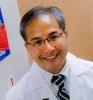 Cesario Borlongan合作论文数Center of Excellence for Aging & Brain Repair, Department of Neurosurgery and Brain Repair, University of South Florida Morsani College of Medicine7Irina Aizman合作论文数7Linda Hinh合作论文数7S Kaye Spratt合作论文数6Alan P. Wolffe合作论文数Sangamo BioSciences6Sharon Kaye Spratt合作论文数6C Pabo合作论文数5stephen p eisenberg合作论文数5K Venstrom合作论文数5YX Liang合作论文数5Yuxin Liang合作论文数5
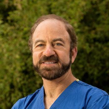 Gary K. Steinberg合作论文数Stanford Stroke Center, Department of Neurology and Neurological Sciences, School of Medicine, Stanford University;Moyamoya Center, School of Medicine, Stanford University5
 Douglas Kondziolka合作论文数Center for Advanced Radiosurgery, NYU Langone Health;Department of Neurosurgery, Grossman School of Medicine, New York University5George N. Cox合作论文数5
 Dmitry Guschin合作论文数Joint Human Genome Program between Argonne National Laboratory5Andrew C. Jamieson合作论文数4DF Xia合作论文数4PIELER CHRISTIAN合作论文数4steve zhang合作论文数4J Olin合作论文数4Xiao-Yong Li合作论文数4Reed Hickey合作论文数4
 Lawrence Wechsler合作论文数University of Pennsylvania School of Medicine Philadelphia Pennsylvania4
 John R. Stephenson合作论文数Viral Carcinogenesis Branch, National Cancer Institute4Brian Johnstone合作论文数4J. Gordon Foulkes合作论文数4eric edward jarvis合作论文数3Mike Kunis合作论文数3J Rokovich合作论文数3
 S. Thomas Carmichael合作论文数Department of Neurology, David Geffen School of Medicine, University of California, Los Angeles3
 Yan Huang合作论文数Cardiovascular Medicine, Internal Medicine, Yale School of Medicine, Yale University3Frank Giordano合作论文数Cardiovascular Medicine, Internal Medicine, Yale School of Medicine, Yale University3DAO M合作论文数3Tan Siyuan合作论文数3Qiang Liu合作论文数3Yuanyue Zhou合作论文数3
 Andrew G. Jamieson合作论文数Department of Chemistry, University of Glasgow3
 L. Dade Lunsford合作论文数Department of Neurological Surgery, School of Medicine, University of Pittsburgh;Center for Image-Guided Neurosurgery, University of Pittsburgh Medical Center3Victor V. Bartsevich合作论文数3
 Neil Schwartz合作论文数Department of Neurology and Neurological Sciences, School of Medicine, Stanford University;Department of Neurosurgery, School of Medicine, Stanford University3Jane S. Lebkowski合作论文数3Fyodor D. Urnov合作论文数3S. Kaye Spratt合作论文数3




