Clinical activity of ponatinib in a patient with FGFR1 -rearranged mixed-phenotype acute leukemia
The chromosomal translocation t(9;14)(p13;q32) has been reported in association with lymphoplasmacytic lymphoma (LPL). Although this translocation involving the paired homeobox-5 (PAX5) gene at chromosome band 9p13 and the immunoglobulin heavy chain (IgH) gene at 14q32 has been described in approximately 50% of LPL cases, the actual number of cases studied is quite small. Many of the initial cases associated with t(9;14)(p13;q32) were actually low-grade B-cell lymphomas with plasmacytic differentiation other than LPL. Thus, we analyzed a series of low-grade B-cell lymphomas for PAX5 gene rearrangements. We searched records from the Department of Pathology, Stanford University Medical Center for low-grade B-cell lymphomas, with an emphasis on plasmacytic differentiation, that had available paraffin blocks or frozen tissue. We identified 37 cases, including 13 LPL, 18 marginal zone lymphomas (nodal, extranodal, splenic, and alpha-heavy chain disease), and 6 small lymphocytic lymphomas. A novel dual-color break-apart bacterial artificial chromosome probe was designed to flank the PAX5 gene, spanning previously described PAX5 breakpoints, and samples were analyzed by interphase fluorescence in situ hybridization. All cases failed to demonstrate a PAX5 translocation, indicating that t(9;14)(p13;q32) and other PAX5 translocations are uncommon events in low-grade B-cell lymphomas with plasmacytic differentiation. This study also confirms recent reports that found an absence of PAX5 rearrangements in LPL, suggesting the reassessment of PAX5 rearrangements in LPL.
BACKGROUND:The diagnosis and classification of lymphoma require correlation of morphologic, immunophenotypic, and molecular-cytogenetic studies. Fine-needle aspiration biopsy (FNAB) is a valuable diagnostic technique that allows material to be collected for these ancillary studies, and for morphologic evaluation.METHODS:The authors report a series of seven cases clinically or morphologically suspicious for Burkitt lymphoma. Fluorescence in situ hybridization studies (FISH) for c-myc were performed on FNAB material and correlated with cytologic and immunophenotypic data.RESULTS:Six of seven specimens were positive for c-myc rearrangement by FISH. However, only three of these cases represented Burkitt lymphoma, with one additional case of atypical Burkitt lymphoma. The other cases included diffuse large B-cell lymphoma, monomorphic posttransplant B-cell lymphoma, and an aggressive B-cell lymphoma, with the latter case negative for c-myc rearrangement by FISH. Of 2 non-Burkitt lymphoma specimens tested, 1 was positive for the immunoglobulin H/bcl-2 rearrangement, in addition to the c-myc rearrangement, suggesting transformation from a lower grade lymphoma.CONCLUSIONS:These cases illustrated the value of FNAB in the diagnosis of Burkitt lymphoma, as well as the importance of obtaining material for, and integrating results of, ancillary studies for the final diagnosis.
The AML1 (CBFA2) gene is the most frequent target of chromosomal rearrangements observed in human acute leukemia. These rearrangements include the commonly reported t(8;21)(q22;q22) or AML1/ETO fusion in AML-M2, the t(3;21)(q26;q22) or AML1 fusion with one of three genes, MDS1, EAP or EVI1, in therapy-related AML and MDS, as well as in blast crisis in CML and the t(12;21)(p13;q22) or TEL/AML1 fusion in B-cell ALL. In addition to the t(3;21), other AML1 translocations have also been reported in therapy-related MDS and AML, particularly after treatment with topoisomerase II inhibitors. AML1 gene rearrangements have also been observed less frequently with numerous other chromosomal partners. Here, we describe a patient with AML-M4 and a previously unreported rearrangement involving the AML1 locus and an unknown locus on the short arm of chromosome 1 at 1p32.
Purpose The authors report the use of a cyclophosphamide, hydroxydaunorubicin, vincristine, and prednisone (CHOP)-based chemotherapy regimen in treating six children with posttransplantation lymphoproliferative disorder (PTLD) that developed after solid organ transplantation. Materials and Methods The chemotherapy regimen consisted of a 29-day induction with CHOP and then as many as 15 cycles of maintenance therapy using methotrexate and cytarabine alternating with vincristine, adriamycin, mercaptopurine, and prednisone. Results All patients attained remission. One patient died of sepsis while in remission. Four of the five remaining patients have been followed-up in remission for as long as 8 years without losing the graft. One of the patients experienced relapse after completing therapy and subsequently died with disease. Conclusions The authors conclude that pediatric patients with PTLD after solid organ transplantation that fails conservative management can be treated successfully with CHOP-based chemotherapy.
Spindle cell lipoma demonstrates a distinctive histologic appearance and characteristic clinical presentation. We recently observed two cases of solitary subcutaneous neoplasm of the foot with histologic features of spindle cell lipoma that in one case includes a minor component of the overlapping tumor, pleomorphic lipoma. Because the foot is an unusual location for these neoplasms, immunoperoxidase and cytogenetic studies were performed. In both cases, staining was strongly positive for CD34 and negative for smooth muscle actin. Cytogenetic studies from the tumor with a pleomorphic component revealed features consistent with a lipomatous neoplasm, but are otherwise diagnostically nonspecific. An analysis of the literature reveals that although CD34 immunoreactivity is characteristic of spindle cell lipoma and helps exclude nonlipomatous neoplasms, it does not clearly eliminate other well-differentiated lipomatous tumors. Accordingly, without the aid of classic tumor location, the diagnosis of the spindle cell/pleomorphic lipoma group relies primarily on histologic features, with supportive but not definitive information provided by immunoperoxidase and cytogenetic studies. Obscuring this issue, however, are the imprecise histologic distinction between these tumors and those of the atypical lipoma/atypical lipomatous tumor/ well-differentiated liposarcoma group and the nomenclature controversy that surrounds the latter group of neoplasms. Despite these obstacles, both groups of well-differentiated lipomatous tumors are clinically benign when subcutaneously located.
Normal human endothelial cells, like other somatic cells in culture, divide a limited number of times before entering a nondividing state called replicative senescence. Expression of the catalytic component of human telomerase, human telomerase reverse transcriptase (hTERT), extends the life span of human fibroblasts and retinal pigment epithelial cells beyond senescence without causing neoplastic transformation (Bodnar, A. G., Ouellette, M., Frolkis, M., Holt, S. E., Chiu, C. P., Morin, G. B., Harley, C. B., Shay, J. W., Lichtsteiner, S., and Wright, W. E. (1998) Science 279, 349–352; Jiang, X., Jimenez, G., Chang, E., Frolkis, M., Kusler, B., Sage, M., Beeche, M., Bodnar, A., Wahl, G., Tlsty, T., and Chiu, C.-P. (1999) Nat. Genet. 21, 111–114). Here, we show that both human large vessel and microvascular endothelial cells also bypass replicative senescence after introduction of hTERT. For the first time, we report that hTERT expression in these life-extended vascular cells does not affect their differentiated and functional phenotype and that these cells maintain their angiogenic potential in vitro. Furthermore, hTERT(+) microvascular endothelial cells have normal karyotype, and hTERT(+) endothelial cell strains do not exhibit a transformed phenotype. Relative to parental cells at senescence, hTERT-expressing endothelial cells exhibit resistance to induction of apoptosis by a variety of different conditions. Such characteristics are highly desirable for designing vascular transplantation and gene therapy delivery systemsin vivo.
We have developed culture conditions for the efficient expansion of cytotoxic effector cells from peripheral blood mononuclear cells (PBMNCs) by the timed addition of interferon-gamma (IFN-gamma), interleukin-2 (IL-2), and the monoclonal antibody (MoAb) OKT3. These cells, termed cytokine-induced killer (CIK) cells, are composed primarily of T cells, and the population of cells with the greatest cytotoxic activity is an otherwise rare population of CD3(+)CD56(+) cells that expand dramatically under these culture conditions. CIK cells were expanded from PBMNCs from 13 patients with chronic myeloid leukemia (CML). These cultures contained a variable number of T cells at the start of the culture (median 44%, range 1% to 64%), yet after 21 to 28 days of culture, virtually all of the cells were CD3(+) T cells (median 97%, range 90% to 99%). The CD3(+)CD56(+) subset of cells expanded significantly (median 25-fold, range 2.2- to 525-fold). CIK cells from all patients showed cytotoxicity against the tumor cell lines OCI-LY8 and K562. In four patients the expanded CIK cells suppressed colony growth of autologous CML blast cells and myeloid progenitor cells. Allogeneic CIK cells from normal donors also suppressed CML colony growth but did not inhibit growth of normal hematopoietic colonies. Twelve of the 13 cultures were exclusively composed of Philadelphia (Ph)-negative cells and one culture had 1 out of 20 Ph-positive metaphases after 4 weeks in culture. Intracellular cytokine production was assayed by fluorescence-activated cell sorter (FACS), and the expanded T-cell cultures produced IL-2, IFN-gamma, and tumor necrosis factor-alpha (TNF-alpha), but not IL-4. Both the CD4(+) and CD8(+) subsets secreted this cytokine profile. To test the in vivo activity of the expanded CIK cells, CML was engrafted into severe combined immunodeficiency disease (SCID) mice using matrigel. After 4 weeks, 4 x 10(7) autologous CIK cells were injected intravenously by tail vein injection into groups of mice, and the animals were sacrificed after a total of 18 weeks. Bcr-abl was detected in the bone marrow or spleen of 5 out of 6 control mice and only 2 out of 13 mice who received the autologous CIK cells (P = .02). In an additional series of animals, the mice did not engraft with CML but instead developed large human Epstein-Barr virus-associated lymphomas by 12 weeks. The mice who received autologous CIK cells at 4 weeks had either no tumor (5) or small tumors (5), whereas all 10 mice that received CIK cells at week 8 developed lymphomas; however, these were not as large as in the 10 control mice who did not receive CIK cells (P = . 03). This study shows that CIK cells, which are Ph chromosome-negative, can be expanded from patients with CML and have potent in vitro and in vivo efficacy against autologous tumor cells.
Uncontrolled proliferation of acute myeloid leukemia (AML) cells is an important step during leukemogenesis. However, little is known about the mechanisms leading to growth autonomy. Studies using immortalized murine hematopoietic cell lines have suggested that autocrine production of growth factors, or the constitutive activation of molecules in growth factor signalling pathways, are involved. We have established six spontaneous factor-independent cell lines from the human growth factor-dependent TF-1 cell line. The factor-independent cells showed no detectable growth factor activity. Immunoblotting analyses of tyrosine phosphorylation, Raf-1 and extracellular signal-regulated kinase 2 (ERK-2) showed a similar pattern in all the cell lines including TF-1 cells. Furthermore, somatic-cell hybrids between TF-1 and the factor-independent cells grew in absence of growth factor. Taken together this data demonstrates that the factor independence in this system is dominant and suggests that the molecular event is located either downstream of the Raf-1 and MAP kinases pathway or on an alternative pathway. Finally, the karyotype analysis of one factor-independent cell line TF-1i1 and TF-1H(-) (G418 resistant, HAT sensitive TF-1 cells) and their hybrids demonstrated an unstable derivative chromosome [der(19) t(19;?) (q13.1;?)] which seemed to correlate with the factor-independence capacity. This model may help in our understanding of autonomous proliferation by human myeloid leukemias.
Immunosuppressed individuals are at high risk for the development of hematologic malignancies. The typical lymphomas arising in organ transplant recipients are B-cell non-Hodgkin's lymphomas that contain Epstein-Barr virus (EBV) DNA sequences. We investigated the characteristics of posttransplant lymphomas that lacked expression of the usual markers associated with EBV transformation. We describe four large-cell lymphomas seen recently at our institution. Two of these four cases were CD4+, one was CD8+, and in one staining for CD4 and CD8 expression was not performed. One CD4+ lymphoma was a CD30+, EBV- large- cell lymphoma from a 65-year-old kidney transplant recipient, the second was an EBV+ large-cell lymphoma from a 25-year-old heart transplant patient. Two T-cell lymphomas were EBV+ and had clonal T- cell receptor beta gene rearrangements. The other two lymphomas expressed T-cell markers CD4 and CD43, and lacked expression of B-cell markers CD19, CD20, CD21, CD22, CD23, and surface Ig. Both CD4+ lymphomas were tumorigenic after their heterotransplantation into severe combined immunodeficient (SCID) mice. Cytogenetics, immunophenotyping, and genotyping of the secondary tumors from SCID mice showed their clonality and identity with the patients' primary tumors. Novel CD4+ lymphoma cell lines, LH521/4 and LK418/4, were established from tumors that had been passaged in SCID mice. An immunodeficient environment may facilitate the growth of these T-cell or biphenotypic lymphomas; the etiology of their genesis can include transformation with EBV and other, as yet unidentified mechanisms.
The t(14;18) chromosomal translocation that results in the juxtaposition of the bcl-2 proto-oncogene with the heavy chain JH locus is a common cytogenetic abnormality in human lymphoma. In particular, it is seen in about 85% of follicular lymphoma (FL) and up to one-third of diffuse lymphomas (DL). The chromosome 18 breakpoints have been shown to cluster into two regions. The major breakpoint region (mbr) within the 3′ untranslated region of the bcl-2 proto-oncogene accounts for approximately 60% of the cases and the minor cluster region (mcr) 30 kb 3′ of bcl-2 accounts for approximately 25% of the breakpoints. Because of variability in the position of the breakpoint, detection of the t(14;18) by Southern blot analysis provides an important clonal marker for the tumor. However, conventional electrophoresis (CE) fails to detect the translocation in 15% to 25% of cases. We have applied pulsed-field gel electrophoresis (PFGE) to the detection of the t(14;18) in a series of lymphoma prospectively analyzed by CE, polymerase chain reaction (PCR), and cytogenetic analysis. PFGE readily detected t(14;18) rearrangements as indicated by comigration of bands detected with probes for the mbr region (chromosome 18) and the JH locus (chromosome 14). In a series of 40 patients with FL, this method proved to be the most comprehensive for detection of the translocation compared with standard methods; in fact, in one case only PFGE was able to detect the chromosomal rearrangement. Ten percent of the FL cases were negative by all methods tested. In a separate analysis of matched tissue specimens from cases of tumor progression of FL to diffuse lymphoma, PFGE detected a common t(14;18) rearrangement confirming a clonal origin in seven of seven cases, whereas CE detected a rearrangement in only three of seven cases. Overall, PFGE was able to detect a translocation in 8 of 12 cases that were negative by CE and four of eight negative by cytogenetic analysis. In conclusion, PFGE analysis is more comprehensive than CE, PCR, and cytogenetic analysis for the detection of the t(14;18) breakpoint in tissue biopsies of malignant lymphoma.
 Athena M. Cherry合作论文数8
 Yasodha Natkunam合作论文数Department of Pathology, Stanford University3Timothy A. Donlon合作论文数3
 Timothy Donlon合作论文数2
 Mohamad Cherry合作论文数2K. Suryanarayan合作论文数1N Martina合作论文数1CP CHIU合作论文数1Ja Gabert合作论文数1J Yang合作论文数1Rv Rouse合作论文数1DA ARBER合作论文数1JE Wrede合作论文数1Mannoni P合作论文数1O Kamel合作论文数1O W Kamel合作论文数1E Chang合作论文数1
 A Molina合作论文数City Hope Canc Ctr1
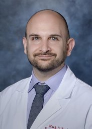 Mark Ewalt合作论文数Dept Pathol & Lab Med, Hematopathol Serv1
 Daniel Arber合作论文数Department of Pathology, The University of Chicago1Daniel A. Arber合作论文数Biological Sciences Division, The University of Chicago1
 Ronald M. Levy合作论文数Department of Chemistry, College of Science and Technology, Temple University1F Lee合作论文数1
 Sandra Horning合作论文数Department of Medicine, Stanford University1
 Roger Allen Warnke合作论文数Department of Pathology, School of Medicine, Stanford University1Hoyle C合作论文数1
 Sally Arai合作论文数Blood and Marrow Transplantation and Cellular Therapy Division, School of Medicine, Stanford University1
 Edmund Waller合作论文数Department of Hematology and Medical Oncology, School of Medicine, Emory University;Winship Cancer Institute, Emory University1Bodnar Andrea合作论文数Bermuda Institute of Ocean Sciences1Herron G S合作论文数1
 Gerald Berry合作论文数Department of Pathology Clinical, Stanford University1Scott Hall合作论文数1
 Manuel Lopez合作论文数Institut Paoli-Calmettes, Aix Marseille Université1Marilyn L. Slovak合作论文数1
 Tracy I. George合作论文数School of Medicine, University of Utah1Charles Lombard合作论文数1
 Bruno Medeiros合作论文数Department of Medicine, Stanford University1Ronald F. Dorfman合作论文数1
 Megan Troxell合作论文数Immunodiagnosis Laboratory, Stanford University;Department of Pathology, Stanford University1
 Gilbert Chu合作论文数Biochemistry Department, Stanford University/Medicine Department, Stanford University1Patty Jones合作论文数1
 Michael Khodadoust合作论文数Medicine Department, Stanford University;Dermatology Department, Stanford University1
 James L. Zehnder合作论文数Division of Hematology, School of Medicine, Stanford University;Department of Pathology, School of Medicine, Stanford University;Stanford Health Care1
 Christina Kong合作论文数Department of Pathology, School of Medicine, Stanford University;Stanford Medicine Children's Health;Stanford Health Care1Cary D. Austin合作论文数Developmental and Molecular Biology;Anatomy and Structural Biology;Einstein College of Medicine, Albert1
 Jason Gotlib合作论文数Department of Medicine, Stanford University1




