A new breed of swine, the Yucatan microswine, that was derived from repetitive inbreeding of selected, small Yucatan swine, was investigated as an animal model of advanced vascular atherosclerosis. Nineteen animals were fed an atherogenic diet for 9.9 +/- 1.5 (mean +/- SEM) weeks before and 19.9 +/- 1.8 weeks after balloon endothelial denudation of all four iliac arteries. In 18 (94.7%) of the 19 microswine, angiography performed at 33 to 87 weeks of age disclosed some degree of luminal diameter narrowing: six animals (33.3%) had one-vessel, six (33.3%) had two-vessel, four (22.2%) had three-vessel, and two (11.1%) had four-vessel disease. In 38 (50%) of 76 denuded arteries, angiographically apparent luminal diameter narrowing was observed as follows: three arteries (7.9%) were narrowed less than 50%; 10 arteries (26.3%) were narrowed 50% to 75%; seven arteries (18.4%) were narrowed 76% to 99%; and 18 arteries (47.3%) were occluded. Sixty-four arteries were harvested from 16 of the 18 microswine with angiographically apparent luminal narrowing, which yielded 748 histologic sections. Maximum cross-sectional area narrowing from atherosclerotic plaque exceeded 90% in 135 (18%) of the sections examined, while 65 sections (9%) were narrowed 76% to 90%, and 127 sections (17%) were narrowed 51% to 75%. Atherosclerotic plaque in these animals appeared histologically similar to the so-called "complex" lesion that is typical of human atherosclerosis, which consists predominantly of collagen with focal calcific deposits and a minor lipid component. The smaller size and lower weight of these animals, in comparison with full-size farm pigs and "minipigs," facilitated transportation, handling, and instrumentation. These findings establish the Yucatan microswine as a useful, representative, and economical atherosclerotic animal model for the evaluation of novel interventional techniques.
Percutaneous balloon mitral valvuloplasty has been shown to be an effective means of reducing mitral valve gradient and increasing mitral valve area in patients with mitral stenosis. Most techniques currently employed for performing this procedure involve delivery of one or two balloon valvuloplasty catheters through the interatrial septum en route to the mitral valve orifice. To determine the morphology of the resultant atrial septal defect (ASD), particularly as a function of the technique employed, we performed a series of in vitro experiments designed to simulate a variety of technical approaches. Ninety-eight experiments in total were performed in 19 normal adult hearts obtained in the fresh, nonpreserved state at necropsy. Transseptal delivery and withdrawal of two conventional, elliptical balloon catheters through two, individual septostomy sites was found to produce the largest ASD (combined area of two defects = 21.4 +/- 2.2 mm2). The defect resulting from transseptal delivery and tandem withdrawal of two elliptical balloon catheters through a single septostomy site measured 14.8 +/- 1.1 mm2, significantly (p = 0.0043) smaller than that produced by two septostomies. Transseptal delivery and withdrawal of a single, segmentally inflating (Inoue) balloon catheter produced a defect of intermediate size (17.5 +/- 1.2 mm2). ASD size was exacerbated by improper balloon withdrawal compared with tandem withdrawal of two completely deflated balloon catheters. Simultaneous withdrawal of two completely deflated balloon catheters through the same septostomy site increased ASD size from 14.8 +/- 1.1 mm2 to 23.6 +/- 2.3 mm2 (p = 0.0004). Simultaneous withdrawal of two incompletely deflated balloon catheters further increased ASD size to 45.8 +/- 2.6 mm2 (p less than 0.0001).(ABSTRACT TRUNCATED AT 250 WORDS)
Fields, Constance D. MD; Rosenfield, Kenneth MD; losordo, Douglas W. MD; Isner, Jeffrey M. MD Author Information
The development, in 1985, of phased array scanning in a linear format1 established the potential for acquiring high quality color flow Doppler (CFD) maps of the vasculature in the lower extremities. Subsequently, however, little information has been published2,3 describing examination of the peripheral arteries by CFD, whether in linear, sector or anular format. Accordingly, the present study was undertaken to evaluate the utility of CFD in combination with 2-dimensional ultrasound (2DU) for the assessment of peripheral vascular disease.
Balloon valvuloplasty has been shown to be an effective treatment for adults with aortic stenosis, typically providing a 50 to 80% increase in aortic valve area and marked improvement in exertional dyspnea, angina and syncope. However, the duration of this hemodynamic and clinical improvement is uncertain. Forty-two patients were followed for 10.2 +/- 0.5 months. Balloon valvuloplasty caused dramatic immediate reduction in the number of patients with moderate or severe dyspnea (80 to 14%), moderate or severe angina (39 to 2%) and syncope (30 to 2%). Furthermore, this improvement in symptoms continued for the duration of the follow-up period in most patients. Echocardiographic aortic valve mean gradient and area determined at 3-month intervals, however, showed a trend toward or return to prevalvuloplasty levels by 9 months' follow-up in 13 of 25 patients (52%), whereas 12 of 25 patients showed no deterioration in their hemodynamic parameters. This trend toward restenosis was accompanied by symptomatic deterioration in 5 of 13 patients (38%). This tendency toward restenosis in greater than 50% of patients by 9 months underscores the need for further technical improvements if balloon valvuloplasty is to be widely applied. Even with these limitations, however, balloon valvuloplasty seems to provide a significant improvement in actuarial survival compared with the natural history of elderly patients with severe aortic stenosis.
A dual balloon technique was studied in 16 patients with aortic stenosis in whom results with a single balloon (up to 20 mm, 5.5 cm or 25 mm, 3.0 cm in diameter and length, respectively) were judged to be suboptimal. Dual balloon valvulopasty was performed using 2 balloons advanced and inflated simultaneously across the stenotic aortic valve orifice. For the group as a whole, the average peak transvalvular gradient was reduced from 79 ± 8 to 57 ± 7 mm Hg (mean ± standard error) using a single balloon (p < 0.0005), and reduced further to 36 ± 4 mm Hg using dual balloons (p < 0.0005 compared with single balloon results). Similarly, calculated aortic valve orifice area was increased from 0.45 ± 0.04 to 0.57 ± 0.05 cm2 using a single balloon (p < 0.0005), and further increased to 0.77 ± 0.06 cm2 using dual balloons (p < 0.0005). Dual balloon dilation caused no complications directly attributable to the use of 2 balloons, including no exacerbation of aortic regurgitation. These results suggest that dual balloon valvuloplasty is safe and efficacious in selected patients with aortic stenosis.
Percutaneous laser angioplasty has now become a clinical reality, consisting chiefly of applications of thermal angioplasty in conjunction with balloon angioplasty for the recanalization of peripheral vascular obstructions. In conjunction with this development, various aspects of laser-tissue interactions, fibreoptic transmission and delivery catheter design pertinent to the cardiovascular system have come under closer scrutiny, resulting in the emergence of both noteworthy concepts and clinical achievements.
Balloon valvuloplasty has been shown to acutely reduce the hemodynamic and symptomatic severity of calcific aortic stenosis. The mechanism by which this improvement is accomplished is not known. At necropsy, three patients who died after hemodynamically successful aortic balloon valvuloplasty were found to have aortic valve calcific deposits fractured at one or more sites. These findings suggest that fracture of leaflet calcium represents the basis for successful aortic balloon valvuloplasty.
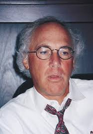 Jeffrey Michael Isner合作论文数St. Elizabeth's Medical Center11
 GA SLOVENKAI合作论文数7KR HALABURKA合作论文数5MR DESNOYERS合作论文数4Deeb N. Salem合作论文数4Tj Hougen合作论文数3
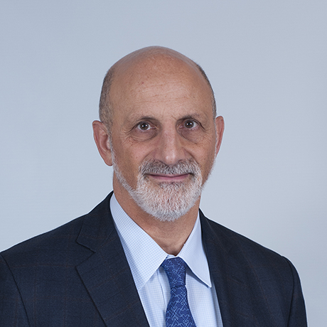 Kenneth Rosenfield合作论文数Massachusetts General Hospital;Mass General Research Institute3
 Alexandra R Lucas合作论文数2A DeFranco合作论文数1Ar Lucas合作论文数1ST DEJESUS合作论文数1Rf Donaldson合作论文数1Jo Pastore合作论文数1BD KOSOWSKY合作论文数1Re Langevin合作论文数1RH CLARKE合作论文数1N. Pandian合作论文数1AJ RONGIONE合作论文数1S Razvi合作论文数1D Gal合作论文数1
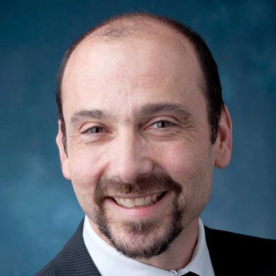 Eric J. Eichhorn合作论文数Dallas Cardiovascular Specialists1
 Douglas W. Losordo合作论文数Medicine at Northwestern University Feinberg School of Medicine1Paul Palefski合作论文数1Shan Shen Wang合作论文数1Robert Weinstein合作论文数1Susan M. Kelly合作论文数1




