Lymph nodes (LNs) containing only acellular mucin are considered negative for metastasis in treated colorectal cancers (CRCs). However, no data exist on how to stage these LNs in untreated CRCs. We collected 63 untreated CRC cases with LNs containing only acellular mucin from 27 US institutions and 23 additional cases from the Dutch Nationwide Pathology Databank. The practice patterns of pathologists in handling such cases, as well as the clinicopathological features of these cases, were analyzed. The survival of the study group was compared with 2 control groups: 102 pN0 and 76 pN1 untreated CRC cases. US and Dutch pathologists demonstrated similar practice patterns in assigning the pN stage and in the use of additional studies. Tumors in the study group were more likely to be located in the right colon, exhibit mucinous features, and be mismatch repair deficient compared with tumors in the pN0 and pN1 control groups. Compared with the pN1 control group, the study group showed significantly lower frequencies of lymphovascular invasion, local recurrence, and distant metastasis. No patient in the study group died of CRC, similar to the pN0 group. In contrast, the pN1 group had a significantly higher risk of CRC-related death. These findings suggest that LNs containing only acellular mucin in untreated CRCs should be interpreted as negative and staged as pN0, in line with current practice in neoadjuvant-treated CRCs.
Anastomotic doughnuts (ATD) are routinely submitted for pathological evaluation during colorectal surgery despite limited evidence supporting its clinical significance. This retrospective study aimed to analyze the pathological findings and cost-effectiveness of examining ATDs. A total of 870 pairs of ATDs from 870 patients who underwent colorectal surgery between 2012 and 2022 were included in the study. Microscopic examination was performed in all cases, and clinical charts and pathology reports were reviewed. The average cost of processing each case was conservatively estimated at US$ 59, with a total cost of US$ 51,185 during the study period. Of the 870 cases, 317 (36.4 %) were obtained from surgical procedures for benign conditions, whereas 553 (63.6 %) were from procedures related to malignant or neoplastic conditions. In cases of surgery for benign conditions (n = 317), no neoplastic or cancerous changes were observed in the ATDs. Among the malignant cases (n = 553), only 14 (1.6 % of the total 870 cases) showed neoplastic findings, including ovarian/endometrial carcinoma (n = 7), tubular adenoma (n = 4), colonic mucinous adenocarcinoma (n = 1), high-grade squamous intraepithelial lesion (n = 1), and low-grade B-cell lymphoma (n = 1). However, these findings did not interfere with the postsurgical or clinical management. The results suggest that routine pathological examination of ATDs provides limited clinical benefits and incurs significant costs. Pathology departments and surgeons should consider revising their protocols to limit examinations to a subset of high-risk cases where the results could potentially impact patient outcomes.
Background and AimsLocation-Based Resect and Discard (LBRD) and Polyp-Based Resect and Discard (PBRD) are two recently proposed strategies to minimize the cost of colonoscopy screening and surveillance. Our study applied these strategies to our colonoscopy database retrospectively to determine the applicability of these strategies in our screening and surveillance colonoscopy population.Methods6024 elective screening, surveillance, or diagnostic colonoscopies performed at the University of California, Irvine, were analyzed. We compared the LBRD and PBRD recommendations with long and short 2020 USMSTF surveillance interval recommendations. The primary outcome was the achievement of the 90% agreement threshold set by the American Society of Gastrointestinal Endoscopy (ASGE) Preservation and Incorporation of Valuable Endoscopic Innovations (PIVI).ResultsThe LBRD strategy achieved 88.0% and 71.6% concordance with the longer and shorter 2020 USMSTF recommendation guidelines, respectively. The PBRD strategy only applied to 65.4% of procedures, with the remaining third still requiring pathological evaluation. Among the applicable procedures, the PBRD strategy achieved 94.2% and 38.6% concordance with the longer and shorter USMSTF recommendation guidelines, respectively.ConclusionsThe PBRD strategy met the 90% PIVI threshold only when using the longer USMSTF recommendations, but concordance dropped to 38.6% when using the shorter surveillance intervals, which are commonly used in the United States. While resect and discard may decrease reliance on pathology, these two strategies do not achieve the level of concordance required to replace the use of pathology for diminutive polyps in our population.
Abstract Introduction/Objective Solitary fibrous tumor (SFT) is an uncommon fibroblastic tumor that originates from the mesenchymal tissue. It has been rarely reported within the stomach. None of these reported tumors have occurred in a background of autoimmune atrophic gastritis as was seen in our patient. Methods/Case Report Case report Results (if a Case Study enter NA) An 85-year-old female with history of anemia and autoimmune atrophic gastritis presented with two-month history of dysphagia and abdominal discomfort. Upper gastrointestinal endoscopy showed a 1 cm subepithelial lesion in the gastric body along the greater curvature. Upper endoscopic ultrasound showed mildly hypoechoic lesion in the submucosa of the stomach with the differential of leiomyoma, neuroendocrine tumor, gastrointestinal stromal tumor, and an atypical looking lipoma. The entire gastric lesion was lifted with submucosal saline injection and then resected using cap endoscopic mucosal resection (EMR) snare cautery. Gross examination showed a well-circumscribed lesion with firm, tan-white cut surface. On microscopy, a bland appearing spindle cell proliferation within a collagenous stroma was seen with alternating hypercellular and hypocellular areas and few dilated vessels. No significant mitosis, nuclear pleomorphism or necrosis was identified. Immunohistochemical stains were performed and the lesional cells were positive for STAT6. Markers for gastrointestinal stromal tumor (CD117, DOG1), smooth muscle tumor (desmin, smooth muscle actin) and schwannoma (S100, SOX10) were negative. Nuclear staining for STAT6 is a sensitive and specific marker for SFTs. This particular SFT was considered to be of low risk and non-malignant considering its small size, lack of mitotic activity and tumor necrosis. The etiology for SFT is not entirely clear but molecular studies have shown that the NAB2::STAT6 translocation in a spindle cell neoplasm is a diagnostic hallmark for SFTs. Conclusion Solitary fibrous tumors have been rarely reported within the stomach. Complete surgical resection with negative margins is the mainstay of treatment as they may recur after an incomplete resection.
Gallbladder is a common surgical pathology specimen. However, carcinomas of the gallbladder are relatively rare in most western countries. Hence, general surgical pathologists may not be that familiar and comfortable diagnosing these tumors. In this review, we discuss the morphological and immunohistochemical characteristics of gallbladder carcinomas, provide updates on tumor classification and staging of these tumors as per the most recent WHO classification, and focus on practical considerations that would be most relevant to diagnosis and clinical management of these tumors.
We present here a 66-year-old Caucasian male whose persistent abdominal pain thought to be due to appendicitis and associated acute splanchnic thrombosis. He was initially managed with antibiotics and anticoagulation. But further work up revealed a low-grade appendiceal mucinous neoplasm causing the splanchnic vein thrombosis. Additionally, diagnosis and management of this rare tumor and appropriate work up for splanchnic thrombosis will be briefly reviewed here.
Rising healthcare costs have been linked to overtreatment and overuse of available resources. Identifying and eliminating low-value services is vital for reducing such costs. At many institutions, including ours, all ileostomy and colostomy specimens are sent for pathological evaluation. It is estimated that approximately 120,000 ileostomy/colostomy procedures are done every year, and at least 1 million patients have stomas at any given time in North America. Hence, we decided to analyze the pathological findings and cost-benefit of undertaking the pathological evaluation of these colostomy and ileostomy specimens. The pathology database of our department was searched for all ileostomy and colostomy specimens received between 2000 and 2020, resulting in a total of 2762 cases (1944 ileostomy and 818 colostomy). We performed a cost-benefit analysis and pathologic review of these cases. The results of our study show that 99.38% of these specimens did not show any significant pathological abnormality, and non-neoplastic pathologic findings accounted for 99.63% of cases. Less than 1% of our cases showed any clinically significant pathological findings. All 10 cases that showed a neoplastic or malignant diagnosis showed some abnormal finding that was appreciated at the time of gross examination. We conclude that microscopic evaluation of ileostomy and colostomy specimens incurs significant costs and provides no clear value or relevant information for patient care. The results of our study provide support for ileostomy and colostomy specimens to be triaged by gross-only pathological examination in the first instance for the vast majority of cases.
Collagenous gastritis has been reported as a rare cause of nausea, diarrhea, weight changes, and early satiety in female patients. Here, we describe two women aged 43 and 71 years who presented with similar symptoms. Gastric biopsies from both individuals showed thickened, irregular subepithelial collagen bands (>10 μm). The pathogenesis of collagenous gastritis is poorly understood, but it may be the presenting symptom for many underlying autoimmune conditions. In particular, there is a well-established connection between collagenous disorders of the gastrointestinal tract and celiac sprue, Sjögren syndrome, and lymphocytic colitis; however, none of these conditions had been diagnosed in our patients. The older woman had incidentally discovered hypogammaglobinemia and IgA deficiency, whereas the younger woman suffered from fibromyalgia. Although a gluten-free diet and budesonide have been effective in some cases, there is no standardized therapy for collagenous gastritis. Our patients trialed diet modification and have required no additional medical interventions.
BACKGROUND:Percutaneous liver biopsy (P-bx) is the gold standard for diagnosing advanced fibrosis. Despite the proven technical feasibility of EUS-guided liver bx (EUS-bx) as a novel alternative way of liver biopsy, the clinical applicability remains to be determined. AIMS:The primary aim of this study is to evaluate if EUS-bx, compared to P-bx, can effectively and safely obtain adequate specimen and accurately predict hepatic fibrosis. METHODS:This is a single center, retrospective chart review among patients with liver diseases at a tertiary endoscopy center from February 2011 to March 2020. We assessed the EUS-bx versus P-bx outcomes by success rate, performance, and safety profile. The primary outcome was the association between EUS-bx clinical variables and the presence of histologic liver fibrosis stage ≥ 3. The secondary outcomes were the associations between EUS-bx and variables indicative of fibrosis. RESULTS:Fifty-nine patients underwent EUS-bx; and 59, P-bx. All EUS-bx procedures were successfully completed. All 56/56 (100%) of EUS-bx vs. 50/52 (96.2%) P-bx were considered adequate samples. Tissue lengths were significantly longer in the EUS-bx cohort (p < 0.0001) with a trend towards a greater number of portal tracts. 46/56 (82.1%) cases of EUS-bx vs. 32/52 (61.5%) of P-bx had > 10 portal tracts; 21/56 (37.5%) cases of EUS-bx vs. 14/52 (26.9%) of P-bx had > 15 portal tracts. There were 6 (10.2%) EUS-bx vs. 1 (1.7%) P-bx related complication leading to a phone call (p = 0.061). CONCLUSIONS:EUS-bx can safely performed and accurately predict liver fibrosis stage as the standard P-bx without being influenced by procedure-related factors.
University of California, Irvine, USA.
BACKGROUND & AIMS: Although transient bacteremia is common during dental and endoscopic procedures, infections developing during sterile diseases like acute pancreatitis (AP) can have grave consequences. We examined how impaired bacterial clearance may cause this transition. METHODS: Blood samples from patients with AP, normal controls, and rodents with pancreatitis or those administered different nonesterified fatty acids (NEFAs) were analyzed for albumin-unbound NEFAs, microbiome, and inflammatory cell injury. Macrophage uptake of unbound NEFAs using a novel coumarin tracer were done and the downstream effects-NEFA-membrane phospholipid (phosphatidylcholine) interactions-were studied on isothermal titration calorimetry. RESULTS: Patients with infected AP had higher circulating unsaturated NEFAs; unbound NEFAs, including linoleic acid (LA) and oleic acid (OA); higher bacterial 16S DNA; mitochondrial DNA; altered b-diversity; enrichment in Pseudomonadales; and increased annexin V-positive myeloid (CD14) and CD3-positive T cells on admission. These, and increased circulating dead inflammatory cells, were also noted in rodents with unbound, unsaturated NEFAs. Isothermal titration calorimetry showed progressively stronger unbound LA interactions with aqueous media, phosphatidylcholine, cardiolipin, and albumin. Unbound NEFAs were taken into protein-free membranes, cells, and mitochondria, inducing voltage-dependent anion channel oligomerization, reducing ATP, and impairing phagocytosis. These were reversed by albumin. In vivo, unbound LA and OA increased bacterial loads and impaired phagocytosis, causing infection. LA and OA were more potent for these amphipathic interactions than the hydrophobic palmitic acid. CONCLUSIONS: Release of stored LA and OA can increase their circulating unbound levels and cause amphipathic liponecrosis of immune cells via uptake by membrane phospholipids. This impairs bacterial clearance and causes infection during sterile inflammation.
 Tsung-Teh Wu (吴宗德)合作论文数Mayo Clinic Rochester Pathology28
 Shah Sejal合作论文数20
 Michael S. Torbenson合作论文数Mayo Clinic16Lee Whayoung合作论文数Thomas E. Starzl Transplantation Institute, University of Pittsburgh13Wenchang Guo合作论文数10
 Kenneth J. Chang合作论文数Division of Gastroenterology and Executive, School of Medicine, University of California8
 Nicolas Barry Delongchamps合作论文数7Gabriel P. Haas合作论文数7
 Gustavo de la Roza合作论文数7Jason B. Samarasena合作论文数7Mary Jumbelic合作论文数7Gregory Threatte合作论文数7Cary Johnson合作论文数6
 John G. Lee合作论文数6Sejal Shah合作论文数5Richard H. Jones合作论文数5Landas Steve K合作论文数5
 Lewis R. Roberts合作论文数Division of Gastroenterology and Hepatology, Mayo Clinic College of Medicine and Science5Lizhi Zhang合作论文数5Alyssa Y. Choi合作论文数4Hee Eun Lee合作论文数4
 Rondell P. Graham合作论文数Mayo Clinic4
 Joseph A. Murray合作论文数Mayo Clinic Comprehensive Cancer Center, Mayo Clinic;Division of Gastroenterology and Hepatology, Mayo Clinic4David C. Wilbur合作论文数Department of Pathology and Laboratory Medicine (D.C.W.), University of Rochester Medical Center4
 Ke-Qin Hu合作论文数Division of Gastroenterology & Hepatology, Department of Medicine, School of Medicine, University of California4
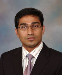 Sudhakar K. Venkatesh合作论文数Department of Radiology, Mayo Clinic4Sherehan Zada合作论文数3Kapitolina Semenova合作论文数3Joaquin Ponce-Zepeda合作论文数3Jana Tarabay合作论文数Univ Calif Irvine3Khurana Kamal K合作论文数3
 Roger Moreira合作论文数College of Physicians &, Columbia University;Surgeons, 630 W168th Street VC14-238B, New York, NY 10032, USA. rkm2124@columbia.edu;Department of Pathology, Columbia University College of Physicians & Surgeons3Mounajjed Taofic合作论文数3Xiaodong Li合作论文数3William C. Faquin合作论文数3Patricia J. Numann合作论文数2Liping Liu合作论文数2
 Sameer B. Shah合作论文数Department of Bioengineering, Jacobs School of Engineering, University of California, San Diego;Neuretix, Inc.;DelNova, Inc.2Ahmad Charifa合作论文数Genomic Testing Cooperative, Irvine, CA2Haijuan Gao合作论文数2Thomas Czeczok合作论文数2Richard Jones合作论文数2Lin Yuan合作论文数2Oana Madalina Mereuta合作论文数CURAM SFI Res Ctr Med Devices, Natl Univ Ireland Galway2
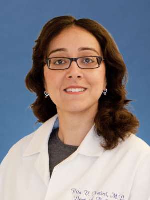 Bita V. Naini合作论文数Jonsson Comprehensive Cancer Center, University of California, Los Angeles2




