Percutaneous nephrostomy is widely used in kidney access surgeries. Despite its prevalence in urological interventions, it presents two operational challenges: 1) precise needle placement into the renal pelvis; and 2) avoiding hemorrhage from blood vessel rupture. In this study, we developed an endoscopic optical coherence tomography probe for needle navigation. We conducted experiments on thirty-one human kidneys for two aspects: 1) tissue recognition, and 2) blood vessel detection. Experimental results indicated that renal tissues including cortex, medulla, calyx, sinus fat, and pelvis could be effectively distinguished through structural optical coherence tomography imaging, and renal blood flow could be detected through the Doppler function. Deep learning methods were utilized to automate recognition procedures. For tissue classification, an Inception model was used, achieving a recognition accuracy of 99.6%. For blood vessel detection, an nnU-net model was applied, exhibiting an intersection over union value of 0.8917 for blood vessel and 0.9916 for background.
Human liver transplantation is constrained by a critical shortage of viable donor livers. In response to this shortage, marginal livers from extended criteria donors are increasingly used to expand the donor pool. Donor liver viability assessment remains limited by invasive, biopsy-based pathological sampling, and there is a need for more comprehensive and noninvasive evaluation techniques to meet the increasing demand for liver transplants. In this study, we propose the use of polarization-sensitive optical coherence tomography (PS-OCT) to perform a multiparameter viability evaluation across the entire surface of donor livers. PS-OCT imaging was conducted on multiple regions of human donor livers, and the findings were cross-validated with histopathological evaluations. Machine learning and texture analysis were used to evaluate hepatic steatosis, fibrosis, inflammation, and necrosis from PS-OCT measurements in comparison with traditional pathological assessments, finding a correlation (>80%) between PS-OCT quantifications and pathology. PS-OCT imaging findings demonstrated strong correlations with donor liver functional performance during normothermic machine perfusion and with clinical liver transplant outcomes. PS-OCT offers a noninvasive assessment of liver viability by quantifying hepatic parameters across the entire donor liver, effectively complementing current pathological analysis. These results suggest that PS-OCT provides a robust approach to assessing donor liver viability, which could potentially decrease the discard rate of high-risk livers, thereby expanding the donor pool and reducing the inadvertent use of unsuitable livers for transplantation.
End-stage liver disease (ESLD) is one of the leading causes of death worldwide. Currently, the only curative option for patients with ESLD is liver transplantation. However, the demand for donor livers far exceeds the available supply, partly because many potentially viable livers are discarded following biopsy evaluation. While biopsy is the gold standard for assessing liver histological features related to graft quality and transplant suitability, it often leads to high discard rates due to its susceptibility to sampling errors and limited spatial coverage. Besides, biopsy is invasive, time-consuming, and unavailable in clinical facilities with limited resources. Here, we present an AI-assisted photoacoustic/ultrasound (PA/US) imaging framework for quantitative assessment of human donor liver graft quality and transplant suitablity at the whole-organ scale. With multimodal volumetric PA/US images as the input, our deep-learning (DL) model accurately predicted the risk level of fibrosis and steatosis, which indicate the graft quality and transplant suitability, when comparing with true pathological scores. DL also identified the imaging modes (PAI wavelength and B-mode USI) that correlated the most with prediction accuracy, without relying on ill-posed spectral unmixing. Our method was evaluated in six discarded human donor livers comprising sixty spatially matched regions of interest. Our study will pave the way for a new standard of care in organ graft quality and transplant suitability that is fast, noninvasive, and spatially thorough to prevent unnecessary organ discards in liver transplantation.
Multi-contrast optical coherence tomography (MC-OCT), incorporating optical coherence tomography angiography (OCTA) and polarization-sensitive OCT (PS-OCT), offers a powerful non-invasive approach for interrogating ovarian microvascular and extracellular matrix (ECM) remodeling in vivo . However, its application to ovarian aging has remained largely unexplored. Here, we establish and validate MC-OCT as a robust imaging platform for visualizing and quantifying age-related changes in vascular architecture and collagen organization in mouse ovaries. Using in vivo OCTA, we demonstrate a progressive decline in ovarian microvascular density, complexity, and connectivity with advancing age. Complementary PS-OCT imaging reveals increased collagen-associated birefringence, indicative of fibrotic ECM remodeling. Quantitative OCT-derived metrics show strong agreement with histological validation using immunohistochemistry, second harmonic generation microscopy, and Sirius Red staining, confirming the accuracy and reproducibility of MC-OCT measurements. Importantly, MC-OCT enables longitudinal, real-time assessment of ovarian structure without tissue disruption, allowing detection of age-dependent remodeling that is not accessible with conventional ex vivo methods. Together, these findings establish MC-OCT as a sensitive and reliable tool for non-destructive evaluation of ovarian vascular and fibrotic changes, providing a valuable platform for studying ovarian aging and assessing interventions aimed at preserving reproductive health.
Multi-contrast polarization-sensitive optical coherence tomography (PS-OCT) provides complementary structural and polarization information that may improve epidural tissue classification. Here, we evaluated deep learning fusion of four PS-OCT channels, including intensity, phase retardation, degree of polarization uniformity (DOPU), and optic axis, using porcine (n = 6) and human (n = 5) spinal specimens. We benchmarked six multi-channel fusion strategies: Probability averaging, feature concatenation, trainable weighted output, shared-stage resnet, merged multi-channel input, and pooled data. Across subject-level nested cross-validation, multi-channel methods achieved modest but consistent accuracy improvements over the best single-channel baselines while reducing subject-to-subject variability. On porcine data, Probability Averaging increased mean validation accuracy by 3.46% (93.07% vs. 89.61% for the best single-channel baseline). On human data, fusion methods maintained the high single-channel baseline (approximately 97% to 98%) while modestly improving stability, with probability averaging achieving the highest mean validation accuracy (98.26%). In cross-testing, trainable weighted output achieved 92.32% versus 91.53% for the best porcine single-channel baseline, and probability averaging achieved 97.87% versus 97.75% for the best human single-channel baseline. On the human cohort, the gain in mean accuracy was small, but multi-channel fusion produced a statistically significant reduction in between-subject variance (Levene's p = 0.0108) and removed the need to know in advance which single channel would generalize best. Overall, multi-channel fusion improved classification performance and robustness, with probability averaging offering a favorable balance between accuracy and complexity because it requires no additional training beyond single-channel models.
BACKGROUND AND OBJECTIVES:The variability and biases in the real-world performance benchmarking of deep learning models for medical imaging compromise their trustworthiness for real-world deployment. The common approach of holding out a single fixed test set fails to quantify the variance in the estimation of test performance metrics. This study introduces NACHOS (Nested and Automated Cross-validation and Hyperparameter Optimization using Supercomputing) to reduce and quantify the variance of test performance metrics of deep learning models. METHODS:NACHOS integrates Nested Cross-Validation (NCV) and Automated Hyperparameter Optimization (AHPO) within a parallelized high-performance computing (HPC) framework. NACHOS was demonstrated on a chest X-ray repository and an Optical Coherence Tomography (OCT) dataset under multiple data partitioning schemes. Beyond performance estimation, DACHOS (Deployment with Automated Cross-validation and Hyperparameter Optimization using Supercomputing) is introduced to leverage AHPO and cross-validation to build the final model on the full dataset, improving expected deployment performance. RESULTS:The findings underscore the importance of NCV in quantifying and reducing estimation variance, AHPO in optimizing hyperparameters consistently across test folds, and HPC in ensuring computational feasibility. CONCLUSIONS:By integrating these methodologies, NACHOS and DACHOS provide a scalable, reproducible, and trustworthy framework for DL model evaluation and deployment in medical imaging. To maximize public availability, the full open-source codebase is provided at https://github.com/thepanlab/NACHOS.
Despite its widespread adoption, the success of radiofrequency (RF) ablation is often limited by challenges in achieving durable, transmural lesions while avoiding complications such as incomplete ablation or over-ablation. Optical coherence tomography (OCT) offers high-resolution visualization of cardiac tissue, enabling real-time assessment of lesion depth. Therefore, we investigated the impact of RF ablation on donated human left atrial tissue using an OCT imaging system to assess lesion formation in a controlled laboratory setting. RF ablation lesions were created at durations of 20, 40, 60, 80, and 100 seconds (s), and their effects were quantified by calculating the attenuation coefficient from OCT images. To identify tissue regions in the image, a U-Net model was employed for segmentation, achieving a mean intersection over union (IoU) of 0.9748 with an inference time of 82 milliseconds. Our results demonstrated clear differences in tissue response before and after ablation, with distinct lesion characteristics across the tested durations. One-way ANOVA analysis yielded a p-value of 1.09 × 10⁻⁸, confirming statistically significant differences among the ablation durations. These findings highlight the potential of SD-OCT for precise, real-time monitoring of ablation effects. Future work aims to integrate OCT with ablation catheters for clinical applications, enhancing the precision of catheter-based treatments for conditions like atrial fibrillation.
Human liver transplantation is severely constrained by a critical shortage of donor livers, with approximately one quarter of patients on the waiting list dying due to the scarcity of viable organs. Current liver viability assessments, which rely on invasive pathological methods, are hampered by limited sampling from biopsies, particularly in marginal livers from extended criteria donors (ECD) intended to expand the donor pool. Consequently, there is a pressing need for more comprehensive and non-invasive evaluation techniques to meet the escalating demand for liver transplants. In this study, we propose the use of polarization-sensitive optical coherence tomography (PS-OCT) to perform a thorough viability evaluation across the entire surface of donor livers. PS-OCT imaging was conducted on multiple regions, achieving near-complete coverage of the liver surface, and the findings were cross-validated with histopathological evaluations. The analysis of hepatic parameters derived from pathology highlighted tissue heterogeneity. Leveraging machine learning and texture analysis, we quantified hepatic steatosis, fibrosis, inflammation, and necrosis, and established strong correlations (≥ 80%) between PS-OCT quantifications and pathological assessments. PS-OCT offers a non-invasive assessment of liver viability by quantifying hepatic parenchymal parameters across the entire donor liver, significantly complementing current pathological analysis. These results suggest that PS-OCT provides a robust, non-invasive approach to assessing donor liver viability, which could potentially decrease the discard rate of higher risk livers, thereby expanding the donor pool and reducing the inadvertent use of those livers unsuitable for transplantation.
DNA barcode-based immunolabeling has revolutionized single-cell protein profiling. However, conventional multiplexed imaging methods are hindered by laborious probe exchange procedures involving buffer washing-based probe removal and prolonged hybridization cycles (requiring tens of minutes to 1.5 h per cycle), which limit throughput, specificity, and universality. Here, we introduce an electrophoresis-based in situ probe removal method that achieves high-specificity iterative probe imaging without washing steps, utilizing 2-min low-voltage electrophoresis for excess probes removal and 3-min high-voltage electrophoresis for hybridized probes dissociation. The robustness was validated through 19 rounds of cyclic electrophoresis, 10 rounds of repetitive imaging, and simultaneous processing of 14 probes (µM-level) across five rounds of multiprobe exchange, demonstrating exceptional specificity and efficiency. Applied to sequential color coding-based multiplexed imaging, this approach establishes voltage-programmed sequential fluorescence encoding (VPSFE), enabling multiplexed imaging of epithelial-mesenchymal transition (EMT)-related proteins. Furthermore, we developed a VPSFE-based Turing pattern coding strategy for multiplexed detection that requires only a single multicolor probe hybridization step. Using three voltage conditions and three fluorescence channels, this system generates 27 unique fluorescence Turing patterns to encode 27 distinct targets. This electric-field Turing pattern coding strategy represents a novel probe exchange-free approach for rapid, universal, and highly specific multiplexed in situ imaging.
Optical coherence tomography (OCT) imaging enables high resolution visualization of sub-surface tissue microstructures. However, OCT image analysis using deep learning is hampered by limited diverse training data to meet performance requirements and high inference latency for real-time applications. To address these challenges, we developed Octascope, a lightweight domain-specific convolutional neural network (CNN) - based model designed for OCT image analysis. Octascope was pre-trained using a curriculum learning approach, which involves sequential training, first on natural images (ImageNet), then on OCT images from retinal, abdominal, and renal tissues, to progressively acquire transferable knowledge. This multi-domain pre-training enables Octascope to generalize across varied tissue types. In two downstream tasks, Octascope demonstrated notable improvements in predictive accuracy compared to alternative approaches. In the epidural tissue detection task, our method surpassed single-task learning with fine-tuning by 9.13% and OCT-specific transfer learning by 5.95% in accuracy. Octascope outperformed VGG16 and ResNet50 by 5.36% and 6.66% in a retinal diagnosis task, respectively. In comparison to a Transformer-based OCT foundation model - RETFound, Octascope delivered 2 to 4.4 times faster inference speed with slightly better predictive accuracies in both downstream tasks. Octascope represented a significant advancement for OCT image analysis by providing an effective balance between computational efficiency and diagnostic accuracy for real-time clinical applications.
Ovarian cancer (OvCa) remains the leading cause of gynecological cancer mortality, with most patients developing chemoresistance. Drug repurposing offers promising alternatives, with mebendazole (MBZ) showing anticancer activity. This study evaluates MBZ efficacy using Spectral Domain Optical Coherence Tomography (SD-OCT). We conducted longitudinal imaging of 40 wild-type (WT) and cisplatin-resistant (CPR) OVCAR8 multicellular tumor spheroids over 11 days. Four analyses were performed: volume analysis, optical attenuation analysis, uniformity analysis, and texture feature analysis. Volume analysis showed MBZ reduced spheroid growth in both groups, with greater effects in CPR-MCTs. Optical attenuation analysis revealed increased necrotic tissue ratios in treated spheroids. Uniformity analysis demonstrated MBZ targets heterogeneous tissues effectively. Texture analysis identified significant structural changes, with 866 altered features in CPR spheroids versus 124 in WT spheroids. Cell viability assays confirmed MBZ's effectiveness against standard and chemo-resistant OVCAR8 tumors. This study demonstrates SD-OCT's utility for noninvasive therapy monitoring in 3D cancer models.
Optical Coherence Tomography Angiography (OCTA) is a non-invasive imaging technique with strong potential for studying ovarian physiology and pathology. Current in vivo methods are often invasive and lack sufficient resolution. Here, we validate OCT for visualizing and quantifying vascular features and collagen structure in mouse ovaries. Our approach provides high-resolution, real-time imaging of ovarian vasculature and collagen networks, with strong accuracy, reproducibility, and sensitivity to age-related changes. Comparison with histology confirms reliability of OCT enables longitudinal assessment and detection of intervention effects. These findings establish OCT as a powerful tool for advancing ovarian aging and reproductive health research.
Kidney transplantation remains the preferred treatment for patients with end-stage kidney disease. However, the ongoing shortage of donor organs continues to limit the availability of transplant treatments. Existing evaluation methods, such as the kidney donor profile index (KDPI) and pre-transplant donor biopsy (PTDB), have various limitations, including low discriminative power, invasiveness, and sampling errors, which reduce their effectiveness in organ quality assessment and contribute to the risk of unnecessary organ discard. In this study, we explored the dynamic optical coherence tomography (DOCT) as a label-free, non-invasive approach to monitor the viability of ex vivo mouse kidneys during static cold storage over 48 hours. The dynamic metrics logarithmic intensity variance (LIV), early OCT correlation decay speed (OCDS e ), and late OCT correlation decay speed (OCDS l ) were extracted from OCT signal fluctuations to quantify temporal and spatial tissue activity and deterioration. Our results demonstrate that DOCT provides complementary information relevant to tissue viability, in addition to the morphological assessment offered by conventional OCT imaging, showing potential to improve pre-transplant organ evaluation and clinic decision-making.
A three-dimensional convolutional neural network (3D-CNN) was developed for the analysis of volumetric optical coherence tomography (OCT) images to enhance endoscopic guidance during percutaneous nephrostomy. The model was performance-benchmarked using a 10-fold nested cross-validation procedure and achieved an average test accuracy of 90.57% across a dataset of 10 porcine kidneys. This performance significantly exceeded that of 2D-CNN models that attained average test accuracies ranging from 85.63% to 88.22% using 1, 10, or 100 radial sections extracted from the 3D OCT volumes. The 3D-CNN (~12 million parameters) was benchmarked against three state-of-the-art volumetric architectures: the 3D Vision Transformer (3D-ViT, ~45 million parameters), 3D-DenseNet121 (~12 million parameters), and the Multi-plane and Multi-slice Transformer (M3T, ~29 million parameters). While these models achieved comparable inferencing accuracy, the 3D-CNN exhibited lower inference latency (33 ms) than 3D-ViT (86 ms), 3D-DenseNet121 (58 ms), and M3T (93 ms), representing a critical advantage for real-time surgical guidance applications. These results demonstrate the 3D-CNN's capability as a powerful and practical tool for computer-aided diagnosis in OCT-guided surgical interventions.
Optical coherence tomography (OCT) is an ideal imaging technique for noninvasive and longitudinal monitoring of multicellular tumor spheroids (MCTS). However, the internal structure features within MCTS from OCT images are still not fully utilized. In this study, we developed cross-statistical, cross-screening, and composite-hyperparameter feature processing methods in conjunction with 12 machine learning models to assess changes within the MCTS internal structure. Our results indicated that the effective features combined with supervised learning models successfully classify OVCAR-8 MCTS culturing with 5,000 and 50,000 cell numbers, MCTS with pancreatic tumor cells (Panc02-H7) culturing with the ratio of 0%, 33%, 50%, and 67% of fibroblasts, and OVCAR-4 MCTS treated by 2-methoxyestradiol, AZD1208, and R-ketorolac with concentrations of 1, 10, and 25 µM. This approach holds promise for obtaining multi-dimensional physiological and functional evaluations for using OCT and MCTS in anticancer studies.
This article presents a comprehensive case report on an uncommon instance of metastatic clear cell sarcoma (CCS) originating from the pancreas. The high mortality rate of pancreatic carcinoma underscores the importance of precise diagnosis and early detection. The authors report a novel case of CCS with pancreatic metastasis, detailing successful surgical intervention through distal pancreatectomy and splenectomy, resulting in favourable outcomes. This study highlights the standard role of surgery in treating advanced CCS and emphasizes preoperative imaging and thorough patient history assessment. This article also underscores the necessity for long-term surveillance due to the potential for recurrence or metastasis. Despite the favourable recovery postsurgery, the absence of subsequent follow-up evaluation prompts consideration of the need for extended monitoring. This article raises questions about the nature of the pancreatic lesion and suggests the possibility of a primary lesion. Further evidence is crucial to establish the correlation between the features related to the development of the patient's primary and metastatic tumours. In conclusion, this study offers valuable insights into metastatic CCS of the pancreas, highlighting the importance of regular postoperative follow-up for improved outcomes through early detection and intervention.
Percutaneous renal biopsy (PRB) is commonly used for kidney cancer diagnosis. However, current PRB remains challenging in sampling accuracy. This study introduces a forward-viewing optical coherence tomography (OCT) probe for differentiating tumor and normal tissues, aiming at precise PRB guidance. Five human kidneys and renal carcinoma samples were used to evaluate the performance of our probe. Based on their distinct OCT imaging features, tumor and normal renal tissues can be accurately distinguished. We examined the attenuation coefficient for tissue classification and achieved 98.19% tumor recognition accuracy, but underperformed for distinguishing normal tissues. We further developed convolutional neural networks (CNN) and evaluated two CNN architectures: ResNet50 and InceptionV3, yielding 99.51% and 99.48% accuracies for tumor recognition, and over 98.90% for normal tissues recognition. In conclusion, combining OCT and CNN significantly enhanced the PRB guidance, offering a promising guidance technology for improved kidney cancer diagnosis.
 Qinggong Tang合作论文数44
 Feng Yan合作论文数Stephenson Sch Biomed Engn, Univ Oklahoma41
 Kar-Ming Fung合作论文数Department of Pathology, College of Medicine, The University of Oklahoma21
 Paul Calle合作论文数Pan Lab, University of Oklahoma14
 Yu Zhongxin合作论文数14
 Chongle Pan合作论文数Genome Science and Technology Graduate School, University of Tennessee Oak Ridge12Qinghao Zhang合作论文数8Trisha I. Valerio合作论文数7
 Justin C. Reynolds合作论文数Computer Science, University of Oklahoma Norman6Shashank S. Shettar合作论文数5Sam Ton合作论文数5Yuyang Yan合作论文数5Jayla A. Wilson合作论文数5
 Alberto de Armendi合作论文数5
 Wei R. Chen合作论文数Immunophotonics, Inc.;School of Biomedical Engineering, University of Oklahoma5Nu Bao Tran Ton合作论文数4Anthony M. Donaldson合作论文数4Zaid A. Alhajeri合作论文数4Noah Davidson合作论文数4Mourren Sibichan合作论文数4Bornface Mutembei合作论文数4
 Barry Y. Chen合作论文数International Computer Science Institute, University of California4Kari Chambers合作论文数3Michael O'Connell合作论文数3
 Avery D Ladymon合作论文数Stephenson Sch Biomed Engn, Univ Oklahoma3
 Gokhan Gunay合作论文数Stephenson Sch Biomed Engn, Univ Oklahoma3
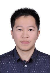 Zuyuan Zhang合作论文数Univ Oklahoma, Sch Compute Sci, Norman, OK 73070 USA3
 Sanjay Patel合作论文数Pathology and Laboratory Medicine, Weill Cornell Medicine, Cornell University3
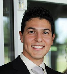 Stefano Tarantini合作论文数Reynolds Oklahoma Center on Aging, University of Oklahoma Health Sciences Center3
 Anna Csiszar合作论文数College of Medicine, The University of Oklahoma3
 Handan Acar合作论文数3Yunlong Liu合作论文数2Ahmed Ahmed合作论文数2
 Sharon Negri合作论文数Dept Biol & Biotechnol Lazzaro Spallanzani, Univ Pavia2
 Fan Zhang合作论文数Stephenson School of Biomedical Engineering, University of Oklahoma2Ha Ji Hee合作论文数2
 Chinthalapally V. Rao合作论文数Department of Pathology, College of Medicine, The University of Oklahoma;Department of Internal Medicine, College of Medicine, The University of Oklahoma2
 Madka Venkateshwar合作论文数Center for Chemoprevention and Cancer Drug Development, Department of Medicine, Oklahoma City, OK, USA.2
 Rajani Rai合作论文数2
 Jody Summers合作论文数Department of Cell Biology, University of Oklahoma Health Science Center2
 Qiang Zhou (周强)合作论文数Institute of Fundamental and Frontier Sciences, University of Electronic Science and Technology of China;School of Optoelectronic Science and Engineering, University of Electronic Science and Technology of China2
 Jun Su (苏君)合作论文数School of Optoelectronic Science and Engineering, University of Electronic Science and Technology of China2
 Shuangjin Shi (史双瑾)合作论文数School of Optoelectronic Science and Engineering, University of Electronic Science and Technology of China2Yunxiang Wang (王云祥)合作论文数2
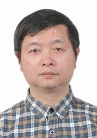 Zhiyong Wang (王智勇)合作论文数School of Optoelectronic Science and Engineering, University of Electronic Science and Technology of China2
 Qi Qiu (邱琪)合作论文数School of Optoelectronic Science and Engineering, University of Electronic Science and Technology of China2Pamela R. Roberts合作论文数2Reha S. Erzurumlu合作论文数Department of Psychobiology, University of California2Yu Chen合作论文数2




