Critical-sized bone defects, particularly segmental injuries such as radial defects, remain one of the most formidable challenges in orthopedic regeneration, as spontaneous healing is rarely achieved and existing grafts suffer from donor site morbidity, immune rejection, insufficient mechanical and biological performance. In this study, we present a biodegradable piezoelectric cryogel scaffold (PWH Gel) composed of a gelatin methacryloyl (GelMA) matrix embedded with piezoelectric whitlockite nanoparticles (PWH NP). Under physiological loading, PWH Gel generates localized electrical potentials and releases bioactive ions (Ca2+, Mg2+), creating a dynamic self-powered microenvironment that actively promotes bone regeneration. In vitro, the scaffold enhanced bone marrow mesenchymal stem cells (BMSCs) proliferation, migration, and osteogenic differentiation, facilitated endothelial tube formation, and triggered Piezo1-mediated Ca2+ influx and cytoskeletal remodeling. When implanted into a rat critical-size radial segmental defect, PWH Gel achieved complete bone bridging with markedly improved bone volume, trabecular organization, and vascularization. By coupling mechanical, electrical, and ionic stimulation within a single biodegradable system, this Mg2+-releasing piezoelectric cryogel establishes a new paradigm for functional reconstruction of segmental bone defects, representing a significant step toward clinically translatable electroactive biomaterials for large skeletal injuries.
PURPOSE:To investigate the association between patient characteristics (age, sex, and body mass index), the timing of posterior cruciate ligament reconstruction (PCLR), and the prevalence of concomitant cartilage and meniscal injuries. METHODS:Patients who underwent primary isolated PCLR between August 2016 and July 2024 were included. The presence of cartilage and meniscal injuries was confirmed via arthroscopic evaluation. The status of cartilage was, meanwhile, graded intraoperatively according to the International Cartilage Repair Society classification. Multivariable logistic regression analyses were performed to identify independent risk factors associated with both any-grade cartilage injuries and specifically high-grade (grades III-IV) cartilage injuries. RESULTS:A total of 1003 patients (787 males and 216 females) with a mean age of 33.18 years who underwent primary PCLR were retrospectively reviewed. Longer time from injury to surgery, particularly exceeding 24 months, was significantly associated with a higher risk of patellofemoral cartilage injury (odds ratio [OR], 1.794; P = .016), lateral compartment cartilage injury (OR, 2.141; P = .016), medial compartment cartilage injury (OR, 3.105; P < .001), and medial meniscal injury (OR, 1.990; P = .041) compared with PCLR performed within 3 months of injury. Additionally, older age significantly increased the prevalence of all cartilage and meniscal injuries (P < .001). Increased body mass index was independently associated with a higher incidence of patellofemoral cartilage injury (P = .006). Subgroup analysis revealed that patients with time from injury to surgery exceeding 24 months reported higher preoperative visual analog scale scores (P = .016). CONCLUSIONS:The incidence of concomitant injuries were 43.87% for patellofemoral cartilage, 10.87% for lateral compartment cartilage, 15.15% for medial compartment cartilage, 8.28% for lateral meniscus, and 10.47% for medial meniscus. Furthermore, delayed PCLR, especially beyond 24 months postinjury, coupled with older age and higher body mass index, were all crucial factors related to a higher prevalence of cartilage and meniscal injuries following grade III PCL injuries. LEVEL OF EVIDENCE:Level III, retrospective cohort study.
Septic arthritis is an orthopaedic emergency that rapidly destroys articular cartilage, and its incidence has increased in recent decades. Despite prompt joint drainage and antimicrobial therapy, progressive cartilage loss and joint dysfunction often persist, highlighting the need for strategies that preserve chondrocyte viability. Based on supportive evidence from other organ systems and non-infectious orthopaedic inflammation, chondrocyte fate in septic arthritis may reflect a multimodal landscape of regulated cell death (RCD) that could be therapeutically leveraged. This narrative review synthesizes mechanistic evidence from two complementary domains of septic arthritis biology: Staphylococcus aureus-driven RCD programs reported in infected joints and extra-articular tissues, and chondrocyte RCD mechanisms established in non-infectious arthritis, particularly osteoarthritis and rheumatoid arthritis. The evidence is organized around four canonical RCD modalities-apoptosis, necroptosis, pyroptosis, and ferroptosis-and their shared upstream stress signals linking infection, inflammation, and metabolic injury to cell-death execution. Direct cartilage-focused studies in septic arthritis suggest that apoptosis-associated processes may contribute to chondrocyte death, whereas evidence for necroptosis, pyroptosis, and ferroptosis in infected chondrocytes remains limited. By integrating direct and indirect evidence, we present a conceptual RCD framework in which crosstalk among multiple RCD pathways may sustain cartilage injury even after bacterial clearance. Short-term, locally delivered adjunctive therapies targeting selected RCD pathways after effective infection control may therefore help limit chondrocyte loss and cartilage collapse. Future priorities include direct pathway validation in infected cartilage, development of RCD-related biomarkers, and evaluation of appropriately timed host-directed interventions to improve long-term joint outcomes.
Objective:To investigate the impact of tibial tunnel position on postoperative knee function and stability in patients undergoing artificial posterior cruciate ligament (PCL) reconstruction. Methods:A retrospective analysis was conducted on patients who underwent single knee artificial PCL reconstruction between January 2018 and September 2024 and met the inclusion criteria. Based on postoperative three-dimensional (3D)-CT measurements of the tibial tunnel position, the patients were allocated into a low tunnel group ( n=35) and a high tunnel group ( n=30). Except for gender, there was no significant difference between groups ( P>0.05) in age, body mass index, injury side, time from injury to operation, preoperative posterior drawer test grade, knee range of motion (ROM), Tegner score, Lysholm score, International Knee Documentation Committee (IKDC) score, or the composition ratio of combined meniscus and cartilage injuries. The tibial tunnel positions were compared. Postoperative recovery of knee function was evaluated using the IKDC score, Lysholm score, Tegner score, and knee ROM. The differences between pre- and post-operative values (change values) for these indicators were calculated and compared between groups. Posterior knee stability was assessed using the posterior drawer test and the side-to-side difference (SSD) in tibial posterior translation measured on stress radiographs. Patient satisfaction was evaluated using the visual analogue scale (VAS) score. Postoperative complications such as graft failure were recorded. Results:The relative proximal-distal position of the tibial tunnel was significantly lower in the low tunnel group than in the high tunnel group ( P<0.05), while no significant difference was found in the relative medial-lateral position between groups ( P>0.05). All operations were successfully completed, and incisions healed by first intention. All patients were followed up 12-86 months, with a median follow-up of 23.0 months. The low tunnel group demonstrated superior posterior stability compared to the high tunnel group, showing a significantly lower SSD and lower posterior drawer test grade at last follow-up ( P<0.05). At last follow-up, the change value in the Tegner score and the VAS score for patient satisfaction were significantly higher in the low tunnel group than in the high tunnel group ( P<0.05). No significant difference was found between groups in the change values for IKDC score, Lysholm score, or knee ROM ( P>0.05). Two patients in the high tunnel group underwent revision surgery due to graft failure, whereas no such adverse events occurred in the low tunnel group. The difference in the incidence of complications between groups was not significant ( P>0.05). Conclusion:Compared with a high tibial tunnel, the low tibial tunnel technique can effectively reduce the graft's turning angle at the proximal tibia and lower the risk of graft failure, thereby significantly improving posterior knee stability and function recovery after artificial PCL reconstruction.
Abstract Purpose To track the 24‐month longitudinal changes in tibiofemoral alignment characterised by passive anterior tibial subluxation (PATS) following anterior cruciate ligament reconstruction (ACLR), and to investigate associated factors. Methods Fifty‐one patients who underwent primary ACLR using hamstring tendon autografts between March 2021 and February 2022 were enrolled. Clinical and magnetic resonance imaging (MRI) evaluations were performed at baseline (within 3 days preoperatively) and at 6‐, 12‐ and 24‐month follow‐ups to examine the graft integrity and tibiofemoral alignment. Lateral PATS (L‐PATS), medial PATS (M‐PATS), global PATS (G‐PATS) and rotational PATS (R‐PATS) were measured on serial MRI. The repeated‐measures one‐way analysis of variance was applied to test the longitudinal changes in PATS. Univariable and multivariable linear regression analyses were performed to identify associations between preoperative and postoperative PATS, adjusting for a priori‐defined covariates including time from injury to surgery, tibial slopes, meniscal injuries and anterolateral ligament (ALL) abnormality. Results The graft integrity was clinically and radiographically confirmed in all patients at the 24‐month follow‐up. However, serial MRI revealed significant increases in L‐PATS, M‐PATS and G‐PATS (all p < 0.001) following the primary ACLR. The increases in L‐PATS (1.5 mm, 95%CI [0.6,2.5], p < 0.001), M‐PATS (1.2 mm, 95%CI [0.5,1.9], p < 0.001) and G‐PATS (1.4 mm, 95%CI [0.6,2.1], p < 0.001) from baseline became the most prominent at 12 months and remained stable thereafter. Strong correlations were identified between preoperative and 24‐month postoperative values for L‐PATS (β = 0.60, p < 0.001), M‐PATS (β = 0.43, p < 0.001), G‐PATS (β = 0.48, p < 0.001) and R‐PATS (β = 0.70, p < 0.001). ALL abnormality was associated with increased L‐PATS (β = 1.85, p = 0.008) and G‐PATS (β = 1.31, p = 0.009), while medial meniscal injury was associated with increased M‐PATS (β = 1.12, p = 0.036) and G‐PATS (β = 0.96, p = 0.048) measured at 24 months postoperatively. Conclusions Residual tibiofemoral malalignment characterised by increased L‐PATS, M‐PATS and G‐PATS persists following ACLR using hamstring tendon autografts. Excessive preoperative PATS, ALL abnormality and medial meniscal injury are associated with increased postoperative PATS. Level of Evidence Level IV.
PURPOSE:To identify preoperative predictors for high-grade pivot shift under anesthesia in patients with primary anterior cruciate ligament (ACL) injury, with a particular focus on investigating the association between altered rotational tibiofemoral position measured on magnetic resonance imaging (MRI) and high-grade pivot shift. METHODS:Consecutive patients who underwent primary ACL reconstruction (ACLR) performed by the same senior surgeon between January 2022 and July 2024 were retrospectively reviewed. Patient characteristics, along with MRI measurements of anatomic features and tibiofemoral positions, were compared between the two groups. Multivariable logistic regression analysis was performed to identify predictors of high-grade pivot shift. Receiver operating characteristic (ROC) analyses were conducted to determine the optimal cutoff values for the identified predictors. RESULTS:A total of 52 patients with grade II or III pivot shift under anesthesia were included in this study, along with 156 age- and sex-matched controls exhibiting grade 0 or I pivot shift. ROC analysis indicated that the optimal cutoff value for internal rotational tibial subluxation (IRTS) to predict high-grade pivot shift was 5.5 mm (area under the curve [AUC] = 0.756). The multivariable prediction (AUC = 0.861) identified increased IRTS (odds ratio [OR] = 1.433), longer time from injury to surgery (OR = 1.737), Beighton score ≥ 4 (OR = 2.979), injury to the posterior horn of the lateral meniscus (OR = 3.587), and increased lateral femoral condyle ratio (LFCR; OR = 1.183) as significant predictors of high-grade pivot shift. CONCLUSIONS:Increased IRTS (threshold of 5.5 mm) showed notable diagnostic performance in predicting high-grade pivot shift. Additionally, longer time from injury to surgery, Beighton score ≥ 4, injury to the posterior horn of the lateral meniscus, and increased LFCR were also identified as significant predictors of high-grade pivot shift. LEVEL OF EVIDENCE:Level III, retrospective comparative case series.
Piezoelectric biomaterials can convert mechanical stimulation into electrical signals to regulate tissue regeneration. However, conventional piezoelectric materials often suffer from brittleness and poor biocompatibility, limiting their biomedical applications. Silk fibroin (SF) is a widely used biomaterial with excellent biocompatibility and mechanical robustness; while its electroactive potential has been successfully harnessed in various fields, the specific application of its intrinsic beta-sheet-derived piezoelectricity for bone repair remains largely unexplored. We propose a dual-crosslinking strategy to program piezoelectric functionality within the SF scaffold for bone regeneration. Chemical crosslinking constructs a stable porous microenvironment, while physical crosslinking activates the intrinsic piezoelectricity of the protein network. SF was fabricated into cryogel scaffolds through a chemical crosslinking step, forming a structurally stable porous architecture. Subsequent ethanol treatment induced beta-sheet crystallization, introducing physical crosslinking that increased structural order and encoded piezoelectricity into the scaffold, yielding a piezoelectric SF scaffold (Piezo-SF). To further enhance electrical signal transmission, a conductive poly(3,4-ethylenedioxythiophene) (PEDOT) network was introduced to construct a piezoelectric-conductive scaffold (PiezoC-SF). Under ultrasound (US) stimulation, the scaffold generated amplified electromechanical signals to significantly enhance bone regeneration in the rat calvarial defect model. This work establishes a bioinspired strategy for programming piezoelectric SF scaffolds through dual crosslinking, providing a promising platform for tissue engineering.
PURPOSE:To determine the maximal outcome improvement (MOI) thresholds for International Knee Documentation Committee (IKDC) score and Lysholm score anchored by patients' willingness to undergo posterior cruciate ligament (PCL) reconstruction again and identify predictors of failure to achieve these thresholds. METHODS:A retrospective review was conducted on patients who underwent primary PCL reconstruction. MOI was defined as the percentage of postoperative improvement relative to maximum possible improvement. Receiver-operating characteristic analyses were performed to determine the MOI thresholds based on patients' willingness to undergo PCL reconstruction as assessed through an anchor question at final follow-up. Multivariable logistic regression analyses were performed to identify predictors of failure to achieve these thresholds. RESULTS:A total of 217 patients were included, with a median follow-up of 63 months (range: 36-84 months). MOI thresholds were 34.5% for IKDC score (area under curve = 0.837) and 37.2% for Lysholm score (area under curve = 0.825), where the latter was calculated after excluding 11 patients with perfect preoperative scores. The proportions of patients achieving the MOI thresholds were 68.7% for IKDC score and 56.3% for Lysholm score. Independent predictors of failure to achieve these MOI thresholds included older age (odds ratio [OR] = 0.952), female sex (OR = 0.166), chronic injury (>12 months; OR = 0.377), and higher preoperative IKDC score (OR = 0.947). For Lysholm score, significant predictors included female sex (OR = 0.403), chronic injury (OR = 0.452), and higher preoperative Lysholm score (OR = 0.968). CONCLUSIONS:MOI thresholds for patients' willingness to undergo PCL reconstruction were 34.5% (IKDC score) and 37.2% (Lysholm score), achieved by 68.7% and 56.3% of patients, respectively. Older age, female sex, chronic injury (>12 months), and higher preoperative patient-reported outcome scores were negative predictors of achieving these MOI thresholds. LEVEL OF EVIDENCE:Level IV, retrospective case series.
With the establishment of key principles governing osteochondral structure, function, and reconstruction, researchers have gained an expanded toolkit for the precisein-vitroreconstruction of osteochondral tissues. As a convergence of tissue engineering and microphysiological modeling, the biomechanical heterogeneity of the osteochondral layers, which is critical to joint function, can be precisely engineered within osteochondral unit-on-a-chip (OC-OoCs), making them ideal tools for studying physiological activities. Specifically speaking, OC-OoCs are regarded as a promising platform for investigating the complex physiology of the osteochondral unit and its pathophysiology in disorders such as osteoarthritis (OA) and osteochondritis dissecans (OCDs). In OA, multiple forms of endochondral ossification, including chondrocalcinosis and osteophyte formation, disrupt the normal tissue relationship of cartilage, subchondral bone plate, and subchondral trabecular bone. Additionally, cellular and molecular communication networks between cartilage and subchondral bone are altered due to increased vascularization, porosity, microcracks, and fissures. Recapitulating these key physiological factors is therefore a critical objective in OC-OoC design. However, incorporation of increasing numbers of physiological parameters inevitably elevates system complexity, posing challenges to chip-to-chip reproducibility and batch-to-batch consistency. Robust quality control (QC) and standardization are thus essential to enhance the reliability and translational value of OC-OoC-derived data. This review summarizes the current advancements in OC-OoCs technology for osteochondral research and, from both diseases oriented as well as translational and clinical perspectives, highlights OC-OoCs' potential to advance our understanding of OA and facilitate the development of novel therapeutic strategies.
PURPOSE:To investigate whether primary anterior cruciate ligament reconstruction (ACLR) in the setting of adult irreducible tibial eminence fracture could achieve comparable clinical outcomes to suture fixation (SF) in reducible cases. METHODS:Patients who were diagnosed with displaced tibial eminence fracture and underwent either ACLR or SF were retrospectively analyzed. The treatment modalities were determined intra-operatively based on the intra-operative evaluation of the osseous and ligamentous status, according to which native ACL preservation by SF was applied in reducible cases and ACLR for irreducible cases. The primary outcome was patient-reported outcome measures at a minimum 2-year follow-up. Secondary outcomes include ipsilateral reinjury at the ACL-injured limb, contralateral injury, range of motion loss, arthrofibrosis, return to sport, and patient satisfaction at the most recent follow-up. Patient-acceptable symptomatic state was determined by the anchor question: "Taking into account your daily activities and functional performance, do you find your current condition satisfactory?". RESULTS:A total of 31 patients in the ACLR group and 43 patients in the SF group were included. Patients' characteristics were comparable between groups, except for time from injury to surgery (ACLR vs SF: mean [range], 11.8 [0.1, 84.0] months vs 1.2 [0.0, 12.0] months, P < .001) and lateral meniscal injury (ACLR vs SF: 45.2% vs 16.3%, P = .023). The mean follow-up was 77 months. Univariate comparison revealed no significant intergroup difference for the primary and secondary outcomes (all P > .05). Multivariable linear regressions revealed no significant association between treatment modalities and clinical outcomes in terms of the Tegner, Lysholm, and International Knee Documentation Committee scores (all P > .05). Subgroup analyses based on fracture types and sex also suggested similar results. Multivariable logistic regressions revealed no difference in ipsilateral reinjury, contralateral injury, range of motion loss, arthrofibrosis, return to sport, and patient satisfaction between groups (all P > .05). A total of 63 (85.1%) participants reported achieving patient-acceptable symptomatic state, with no significant difference between the ACLR and SF groups (26/31 vs 37/43, P > .999). CONCLUSIONS:Primary ACLR using hamstring autograft in the setting of adult irreducible tibial eminence fracture achieved clinical outcomes comparable to SF in reducible cases. The percentage of patients reported achieving patient-acceptable symptomatic state was similar between the 2 groups. LEVEL OF EVIDENCE:Level III, retrospective comparative case series.
Background: There is a lack of evidence and continuous debate on whether femoral tunnel displacement substantially influences the clinical efficacy of medial patellofemoral ligament reconstruction (MPFL-R) in addressing recurrent patellar dislocation. Purpose: To investigate possible associations between inaccurate femoral tunnel placement during MPFL-R and clinical outcomes, with a specific focus on proximal tunnel malpositioning. Study Design: Cohort study; Level of evidence, 3. Methods: Patients who were diagnosed with recurrent patellar dislocation and underwent MPFL-R were retrospectively analyzed. A true lateral view on 3-dimensional computed tomography was obtained, and the distance from the center of the femoral tunnel to the Schöttle point (DF-to-S) was measured. Accordingly, femoral tunnels were divided into the suboptimal group (DF-to-S > 10 mm) and the optimal group (DF-to-S≤ 10 mm). Patient data were collected and pathoanatomic risk factors were evaluated. Clinical assessments included patient-reported outcome measures, including the International Knee Documentation Committee (IKDC), Kujala, Lysholm, and Tegner scores; subjective satisfaction; and postoperative complications. A subgroup analysis was performed between proximally malpositioned tunnels and tunnels lying in other directions. Results: A total of 112 patients (112 knees) were included, with 34 in the suboptimal group and 78 in the optimal group. Patient characteristics and pathoanatomic abnormalities were comparable between the 2 arms, with mean follow-ups of 52.90 ± 23.80 months and 60.14 ± 27.88 months in the suboptimal and optimal groups, respectively ( P = .259). At the final follow-up, inferior IKDC (76.3 ± 12.8 vs 85.9 ± 6.3; P < .001), Kujala (87.7 ± 9.3 vs 93.8 ± 4.9; P = .001), and Lysholm (87.0 ± 11.8 vs 94.5 ± 5.3; P = .001) scores and subjective satisfaction (8.2 ± 1.4 vs 9.3 ± 0.9; P < .001) were observed in the suboptimal group compared with the optimal group. Lower proportions of patients met the minimal clinically important difference for the IKDC (82.4% vs 100.0%; P = .001) and Lysholm (79.4% vs 98.7%; P = .001) scores in the suboptimal group compared with the optimal group. Subgroup analysis demonstrated that the final IKDC ( P = .002), Kujala ( P = .001), and Lysholm ( P = .001) scores and subjective satisfaction ( P = .031) were statistically worse in the proximally located group, with insignificant improvements compared with preoperative levels. The suboptimal group showed a higher rate of overall complications, and anterior knee pain was more often reported in the proximally located subgroup. Conclusion: Inaccurate femoral tunnel positioning was associated with inferior midterm subjective functional scores and a higher rate of postoperative complications after MPFL-R. Proximal displacement in particular was associated with adverse clinical outcomes.
Background Few reports have documented left apical ventricular aneurysm attributable solely to aortic valve stenosis, rapidly exacerbated by prosthesis-patient mismatch (PPM), and necessitating unconventional redo surgical strategies. Case Summary A 42-year-old woman with a 17-year cardiac disease history was admitted to our institution. In 2018, she received a 17-mm mechanical aortic valve. In 2024, the unconventional redo procedures comprised left-ventricular aneurysmoplasty, direct-visualization endocardial radiofrequency ablation, Y-incision aortic root enlargement, and aortic and mitral valve replacement. Discussion During the initial operation, implantation of a 17-mm prosthesis created moderate PPM, deteriorating over time. Achieving a balance between avoiding PPM and the risk of iatrogenic mitral compromise is often challenging but clinically essential. Take-Home Messages Isolated aortic valve stenosis gives rise to an apical ventricular aneurysm, and PPM accelerates its expansion. The complex redo surgery demands meticulous preoperative planning and tactical intraoperative decision-making.
Total hip replacement (THR) surgery has achieved significant success in alleviating pain from hip joint diseases and improving patients' quality of life. However, the challenge of precisely placing the acetabular implant during surgery can lead to complications, such as early loosening and wear of the implant. Traditionally, this critical step relies on the surgeon's experience and subjective judgment, lacking objective tools to monitor the pressure field within the acetabulum in real-time. This study presents the development of a highly sensitive micropyramid flexible sensor array, fabricated using microfine photolithography precision molding technology, for assisting pressure field monitoring in the acetabulum during THR. The compression of the pyramid microstructure results in large changes in contact area, allowing our single sensor to exhibit an ultrahigh sensitivity of 11 711.95 kPa(-1) within the 0-90-kPa pressure range, with a response/recovery time of 120/81 ms. Six independent sensors were integrated into a customized acetabular implant model, enabling the collection of pressure field data from the acetabulum. To simulate the surgical process, we modified the test platform, developed corresponding signal acquisition circuits, and a software system for real-time visualization of pressure distribution in the acetabulum, demonstrating the potential application of this system in assisting surgeons in precisely placing implants during THR surgery.
With the increasing incidence of joint diseases and the growing demand for surgical precision, robotic-assisted surgery has emerged as a promising advancement in orthopedic training. The Mako surgical robot, known for its precision in preoperative planning and intraoperative guidance, may offer significant educational benefits to joint surgery fellows. However, its potential impact on enhancing traditional surgical skills and research capabilities remains underexplored. To evaluate whether systematic training in Mako robotic-assisted hip replacement improves surgical performance and research skills among joint surgery fellows compared to traditional training. From October 2021 to May 2023, 40 joint surgery fellows were randomly divided into two groups, the experimental group received a comprehensive and systematic mako robot-assisted hip replacement training course. The control group received traditional training in hip replacement surgery within one month. The results of operation simulation and questionnaire were used to evaluate the training effect. The test results showed that in the simulation test of acetabular prosthesis placement, the overall pass rate of the experimental group was 90
Repairing chondral defects remains challenging due to the avascular and acellular nature of cartilage. Advances in tissue engineering based on hydrogels offer significant potential for high-quality chondrogenesis, with injectable hydrogels emerging as a prominent area of research, because they meet the requirements for minimally invasive administration. This review provides an overview of recent progress in injectable hydrogels for cartilage repair and regeneration and summarizes the synthesis strategies. We specifically focused on key challenges including gelation techniques, robust bioadhesion, and bioactive functionalization, which are primary obstacles to achieving superior chondral repair in recent studies. By exploring current strategies to address these challenges and their underlying mechanism, we aim to inspire continuous advancements in promoting the application of injectable hydrogels for cartilage regeneration.
Background:There is a risk of convergence between the anterior cruciate ligament reconstruction (ACLR) femoral tunnel and the lateral extra-articular tenodesis (LET) tunnel, which may cause damage to the graft and fixation device. Purpose:To develop a simple and safe method for drilling the LET tunnel to avoid convergence between the ACL femoral tunnel and the LET tunnel. Study Design:Descriptive laboratory study. Methods:Three-dimensional computed tomography reconstructed images of 60 knees after ACLR and 10 knees after ACLR and LET between August 2023 and July 2024 were analyzed. The possibility and utility of using the anterior cartilage edge (ACE) of the lateral femoral condyle medial wall as an intra-articular landmark of the LET tunnel exit were analyzed in 60 knees grouped by ACL tunnel diameter: the 7-mm group (n = 20); the 8-mm group (n = 20); and the 9-mm group (n = 20). The safe zone is defined as the area of the LET bone tunnel exit at the medial wall of the lateral condyle of the femur where the 2 bone tunnels do not intersect. In addition, the bone bridge thickness was measured in 10 knees after ACLR and LET. Results:The x-coordinate of the highest point in the cartilage edge was significantly smaller than that of the intersection point between the safe line and the cartilage edge in each group (7-mm group: 57.27 ± 7.32 vs 88.27 ± 6.17; P = .000; 8-mm group: 56.37 ± 6.90 vs 81.10 ± 8.23; P = .000; 9-mm group: 56.05 ± 3.98 vs 70.99 ± 14.85; P = .000). The ACE was beyond the safe zone. The safe zone increased as the ACL bone tunnel diameter decreased. There was no tunnel conflict in all 10 patients who underwent ACLR and LET. The bone bridge thickness ranged from 3.15 to 8.92 mm. Conclusion/Clinical Relevance:To avoid tunnel convergence in ACLR combined with LET, the ACE of the lateral femoral condyle medial wall is a useful landmark to drill the LET bone tunnel.
The washing prescription for lower limb sprains (WPFLLS) has acceptable effects on chronic ankle sprains (CAS). To provide additional evidence supporting the efficacy of the WPFLLS prescription, we designed a multicenter, double-blind, randomized controlled trial with professional ballet dancers who have a high incidence of chronic ankle sprain as the study subjects. This is a multicenter, double-blind, randomized controlled trial. According to the strict inclusion and exclusion criteria, 120 ballet dancers with CAS will be selected and randomly assigned to the traditional Chinese medicine (TCM) group or to the placebo group. Patients will be treated for 14 consecutive days and followed up for 28 days. The primary outcome is the ankle-hindfoot score of the American Orthopaedic Foot & Ankle Society (AOFAS). The secondary outcomes encompass the Visual Analog Scale (VAS), the Symptom Severity and Dance Performance Quality Index, and the frequency and dosage of externally applied rescue medication. This trial will provide high-quality clinical evidence on the efficacy and safety of WPFLLS in the treatment of CAS and provide clinical recommendations for subsequent studies on WPFLLS.
Bone marrow mesenchymal stem cells (MSCs) serve a pivotal role in the hematopoietic niche. The present study collected bone marrow samples from individuals across various age groups to investigate the biological characteristics of MSCs. By modifying the bone marrow microenvironment through co-culture techniques, changes in the stemness of MSCs were examined. An in vitro hematopoietic co-culture system was established to simulate the impact of MSCs on hematopoietic stem cells. The results demonstrated that the mode of cell-to-cell contact among stem cells is more influential in shaping bone marrow function compared with the effects of aging on these stem cells. Transcriptomic analysis revealed that MSCs serve as essential mediators, with their growth variations being both a consequence and a cause of changes in the bone marrow microenvironment. Furthermore, the decline in hematopoietic function observed in the elderly is a manifestation of this phenomenon. Data from the present study suggest that targeting MSCs is essential for enhancing bone marrow function and improving the outcomes of bone marrow transplantation.
 Jianquan Wang (王健全)合作论文数Peking University Third Hospital34
 Xi Gong (龚熹)合作论文数25
 Hua Tian (田华)合作论文数Peking University Third Hospital23
 Jiang Peng (彭江)合作论文数Chinese PLA General Hospital19史尉利合作论文数北京大学第三医院17
 Feng Li (李锋)合作论文数Department of Orthopaedics, Peking University Third Hospital15
 Yingfang Ao (敖英芳)合作论文数Peking University Third Hospital15Qingyang Meng合作论文数13Haoye MENG (孟昊业)合作论文数13Aiyuan Wang (汪爱媛)合作论文数Chinese PLA General Hospital13
 Shibi Lu (卢世璧)合作论文数Chinese PLA General Hospital12
 Yuping Yang (杨渝平)合作论文数Department of Sports Medicine, Peking University Third Hospital12
 Yong Ma (马勇)合作论文数Department of Sports Medicine, Peking University Third Hospital12
 Minwei Zhao (赵旻暐)合作论文数Department of Orthopedics, Peking University Third Hospital11
 Quanyi Guo (郭全义)合作论文数Chinese Pla General Hospital11Zhang Zhiyu (张智昱)合作论文数10
 Haijun Wang (王海军)合作论文数Department of Sports Medicine, Peking University Third Hospital10Yu Wang (王玉)合作论文数Chinese PLA General Hospital9
 Junyang Li合作论文数9Pan Xiaoyu合作论文数8Xueling Yuan (袁雪凌)合作论文数8
 Jian Wang (王健)合作论文数Institute of Sports Medicine, Peking University Third Hospital8
 Weishi Li (李危石)合作论文数Peking University Third Hospital8Wen-bin Bai合作论文数7Xinguang Wang (王鑫光)合作论文数7Hong-Jie Huang合作论文数7
 Yuelin Hu (胡跃林)合作论文数Peking University Third Hospital7Yitian Gao合作论文数6Xin Wang合作论文数6
 Xin Zhang (张辛)合作论文数Department of Sports Medicine, Peking University Third Hospital6
 Yang Li (李杨)合作论文数Department of Orthopedics, Peking University Third Hospital6Yanfang Jiang合作论文数5Nayun Chen (陈拿云)合作论文数5
 Qinwei Guo (郭秦炜)合作论文数Department of Sports Medicine, Peking University Third Hospital;Institute of Sports Medicine of Peking University5
 Ping Liu (刘平)合作论文数Reproductive Medicine Center, Peking University Third Hospital;State Key Laboratory of Female Fertility Promotion, Peking University Third Hospital5Pakezhati Maimaitijiang合作论文数4Ke-Ying Zhang合作论文数4Fengyi Hu合作论文数4
 Xiao Geng合作论文数4Wenli Dai合作论文数4Sun Weijia合作论文数4Wenlong Gou (苟文隆)合作论文数4
 Xiaoqing Hu (胡晓青)合作论文数Institute of Sports Medicine, Peking University Third Hospital4
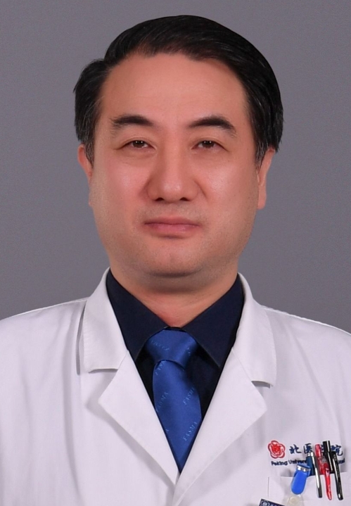 Chen Jiao (焦晨)合作论文数Institute of Sports Medicine, Peking University Third Hospital4Dong Sun (孫東)合作论文数4Wang Peng (王鹏)合作论文数3Li Yang合作论文数3
 Guanying Gao合作论文数3




