We present a case of a port malposition into the azygos vein resulting in both a broncho-esophageal and veno-bronchial fistula. While complications of central venous catheter malposition into the azygos vein are well documented in literature, these unique complications have not yet been described. This case underscores how utilizing state of the art technology like intra-cavity electrocardiography rather that reliance on fluoroscopy can help eliminate catheter malposition and its potential catastrophic consequences.
Purpose To demonstrate that magnetic resonance‐guided focused ultrasound (MRgFUS) facilitates blood‐spinal cord barrier (BSCB) permeability and develop observer‐independent MRI quantification of BSCB permeability after MRgFUS for spinal cord injury (SCI). Methods Noninjured Sprague‐Dawley rats (n = 3) underwent MRgFUS and were administered Evans blue post‐MRgFUS to confirm BSCB opening. Absorbance was measured by spectrophotometry and correlated with its corresponding image intensity. Rats (n = 21) underwent T8–T10 laminectomy and extradural compression of the spinal cord (23g weighted aneurysm‐type clip, 1 min). The intervention group (n = 11) was placed on a preclinical MRgFUS system, administered microbubbles (Optison, 0.2 mL/kg), and received 3 MRgFUS sonications (25 ms bursts, 1 Hz pulses for 3 min, 3 acoustic W, approximately 1.0–2.1 MPa peak pressure as measured via hydrophone). The sham group (n = 10) received equivalent procedures with no sonications. T1w MRI was obtained both pre‐ and post‐MRgFUS BSCB opening. Spinal cords were segmented manually or semiautomatically and a Pearson correlation with P ≤ 0.001 was used to correlate the two segmentation methods. MRgFUS sonication and control regions intensity values were evaluated with a paired t ‐test with a P ≤ 0.01. Results Semiautomatic segmentation reduced computational time by 95% and was correlated with manual segmentation (Pearson = 0.92, P < 0.001, n = 71 regions). In the noninjured rat group, Evans blue absorbance correlated with image intensity in the MRgFUS and control regions (Pearson = 0.82, P = 0.02, n = 6). In rats that underwent the SCI procedure, an increase in signal intensity in the MRgFUS targeted region relative to control was seen in all SCI rats (10.65 ± 12.4%, range: 0.96–43.9%, n = 11, P = 0.002). SCI sham MRgFUS revealed no change (0.63 ± 0.52%, 95% CI 0.320.95, n = 10). This result was significant between both groups ( P = 0.003). Conclusion The implemented semiautomatic segmentation procedure improved data analysis efficiency. Quantitative methods using contrast‐enhanced MRI with histological validation are sensitive for detection of blood‐spinal cord barrier opening induced by magnetic resonance‐guided focused ultrasound.
BACKGROUND:Microtubule stabilizing drugs, commonly used as anti-cancer therapeutics, have been proposed for treatment of Alzheimer's disease (AD); however, many do not cross the blood-brain barrier.OBJECTIVE:This research investigated if paclitaxel (PTX) delivered via the intranasal (IN) route could alter the phenotypic progression of AD in 3xTg-AD mice.METHODS:We administered intranasal PTX in 3XTg-AD mice (3xTg-AD n = 15, 10 weeks and n = 10, 44 weeks, PTX: 0.6 mg/kg or 0.9%saline (SAL)) at 2-week intervals. After treatment, 3XTg-AD mice underwent manganese-enhanced magnetic resonance imaging to measure in vivo axonal transport. In a separate 3XTg-AD cohort, PTX-treated mice were tested in a radial water tread maze at 52 weeks of age after four treatments, and at 72 weeks of age, anxiety was assessed by an elevated-plus maze after 14 total treatments.RESULTS:PTX increased axonal transport rates in treated 3XTg-AD compared to controls (p≤0.003). Further investigation using an in vitro neuron model of Aβ-induced axonal transport disruption confirmed PTX prevented axonal transport deficits. Confocal microscopy after treatment found fewer phospho-tau containing neurons (5.25±3.8 versus 8.33±2.5, p < 0.04) in the CA1, altered microglia, and reduced reactive astrocytes. PTX improved performance of 3xTg-AD on the water tread maze compared to controls and not significantly different from WT (Day 5, 143.8±43 versus 91.5±77s and Day 12, 138.3±52 versus 107.7±75s for SAL versus PTX). Elevated plus maze revealed that PTX-treated 3xTg-AD mice spent more time exploring open arms (Open arm 129.1±80 versus 20.9±31s for PTX versus SAL, p≤0.05).CONCLUSION:Taken collectively, these findings indicate that intranasal-administered microtubule-stabilizing drugs may offer a potential therapeutic option for treating AD.
Repetitive mild traumatic brain injury (rmTBI) is known to disturb axonal integrity and may play an important role in the pathogenic cascades leading to neurodegeneration. One critical approach to reduce the future onset of neurodegeneration is to intervene in this process at an early stage following a brain injury. Previously we showed that direct application of the microtubule-stabilizing drug, paclitaxel, on the brain following controlled cortical impact improved motor function and reduced lesion size. Herein, we extended these findings to a model of mild brain injury induced by repeated closed-skull impacts. Paclitaxel was administered intranasally to circumvent its poor transport across the blood-brain barrier. Mice received five mild closed-skull impacts (one per day for five days). Intranasal paclitaxel was administered once only, immediately after the first impact. We found that paclitaxel prevented injury-induced deficits in a spatial memory task in a water tread maze. In vivo magnetic resonance imaging (MRI) and positron emission tomography with 18F-flurodeoxyglucose (FDG-PET) revealed that paclitaxel prevented structural injury and hypometabolism. On MRI, apparent, injury-induced microbleeds were observed in 100% of vehicle-treated rmTBI mice, but not in paclitaxel-treated subjects. FDG-PET revealed a 42% increase in whole brain glucose metabolism in paclitaxel-treated mice as compared to vehicle-treated rmTBI. Immunohistochemistry found reduced evidence of axonal injury and synaptic loss. Our results indicate that intranasal paclitaxel administration imparts neuroprotection against brain injury and cognitive impairment in mice. The results from this study support the idea that microtubule-stabilization strategies hold therapeutic promise in mitigating traumatic brain injury.
OBJECTIVE:Autosomal-dominant familial Alzheimer disease (AD) is caused by by variants in presenilin 1 (PSEN1), presenilin 2 (PSEN2), and amyloid precursor protein (APP). Previously, we reported a rare PSEN2 frameshift variant in an early-onset AD case (PSEN2 p.K115Efs*11). In this study, we characterize a second family with the same variant and analyze cellular transcripts from both patient fibroblasts and brain lysates. METHODS:We combined genomic, neuropathological, clinical, and molecular techniques to characterize the PSEN2 K115Efs*11 variant in two families. RESULTS:Neuropathological and clinical evaluation confirmed the AD diagnosis in two individuals carrying the PSEN2 K115Efs*11 variant. A truncated transcript from the variant allele is detectable in patient fibroblasts while levels of wild-type PSEN2 transcript and protein are reduced compared to controls. Functional studies to assess biological consequences of the variant demonstrated that PSEN2 K115Efs*11 fibroblasts secrete less Aβ 1-40 compared to controls, indicating abnormal γ-secretase activity. Analysis of PSEN2 transcript levels in brain tissue revealed alternatively spliced PSEN2 products in patient brain as well as in sporadic AD and age-matched control brain. INTERPRETATION:These data suggest that PSEN2 K115Efs*11 is a likely pathogenic variant associated with AD. We uncovered novel PSEN2 alternative transcripts in addition to previously reported PSEN2 splice isoforms associated with sporadic AD. In the context of a frameshift, these alternative transcripts return to the canonical reading frame with potential to generate deleterious protein products. Our findings suggest novel potential mechanisms by which PSEN variants may influence AD pathogenesis, highlighting the complexity underlying genetic contribution to disease risk.
Despite the recent increase in use of mouse models in scientific research, researchers continue to use cognitive tasks that were originally designed and validated for rat use. The Radial Water Tread (RWT) maze test of spatial memory (designed specifically for mice and requiring no swimming) has been shown previously to successfully distinguish between controlled cortical impact-induced TBI mice and sham controls. Here, a detailed protocol for this task is presented. The RWT maze capitalizes on the natural tendency of mice to avoid open areas in favor of hugging the sides of an apparatus (thigmotaxis). The walls of the maze are lined with nine escape holes placed above the floor of the apparatus, and mice are trained to use visual cues to locate the escape hole that leads out of the maze. The maze is filled with an inch of cold water, sufficient to motivate escape but not deep enough to require that the mouse swim. The acquisition period takes only four training days, with a test of memory retention on day five and a long-term memory test on day 12. The results reported here suggest that the RWT maze is a feasible alternative to rat-validated, swimming-based cognitive tests in the assessment of spatial memory deficits in mouse models of TBI.
416 Objectives Alzheimer’s disease (AD) therapeutic investigations require preclinical assessment in transgenic mice. Imaging provides complementary evidence to support cognitive tests and histopathology that can be translated to clinical trials. This research investigated the therapeutic efficacy of a non-amyloid targeting microtubule-stabilizing drug paclitaxel with in vivo imaging and cognitive tests. Methods Mice (3xTg-AD n=25) treated by intranasal lavage with paclitaxel (0.6mg/kg) (TgTAX) or saline (TgSAL) at 14 d intervals from 10-22wks (n=17) OR 47-58wks (n=8) and compared to WT (n=20). Therapeutic outcome was assessed by FDG-PET: mice (n=13) had 250µCi FDG ip with microPET after 60min uptake and 14T MRI: MEMRI of axonal transport: T1-weighted TR/TE: 5000ms/1.9ms, 0.140x0.140x0.25mm3 at 100min. and 280-350min after intranasal administration of MnCl2, and DTI of white matter: 0.195x0.195x0.5mm3 FA/TR/TE: 90deg/8750ms/17.8ms, 30 directions. Water tread maze had 3 trials/day for 4 days, short-term memory test at day 5 and long-term at day 12. MEMRI of VOIs in the olfactory tract estimated drug effect on axonal transport. FDG-PET normalized to tracer dose and weight assessed global brain metabolism. Results MEMRI indicated increased axonal transport in TgTAX vs TgSAL (p≤0.05). DTI fractional anisotropy was 16% decreased in 3xTg-AD vs WT at 6 mos (mn±sd 0.32±0.03 vs 0.38±0.02 p≤0.01) with no significant effect of drug. FDG-PET at 12mos found decreased brain metabolism in 3xTg-AD vs WT mice (18%, 114.2±15 vs 140.6±8, p≤0.05), but also did not distinguish drug vs saline. TgSAL mice exhibited short and long-term memory deficits compared to both WT and TgTAX at 12 mos (5 day 143.8±43s vs 64.2±59s and 91.5±77.4: day 12 138.3±52s vs 33.4±49s and 107.7±74 p≤0.05). Conclusions The results indicate that microtubule stabilization may be a therapeutic option for AD. Preclinical testing in AD is critical to evaluate drugs for clinical trials. Cognitive and imaging tests must be able to distinguish disease from controls and detect outcome improvements from drug. Research Support Support for this research was provided by the Alzheimer9s Association.
 Donna J. Cross合作论文数Dept Radiology, Univ of Washington5
 Satoshi Minoshima合作论文数5
 Marcella Melinda Cline合作论文数4David G Cook合作论文数2Carole L Smith合作论文数2Leah Osnis合作论文数2Tsuang Debby W合作论文数Dept Psychiat & Behav Sci, Univ Washington2
 Caitlin S. Latimer合作论文数Department of Laboratory Medicine and Pathology, University of Washington2Dorschner Michael O合作论文数2Young Jessica E合作论文数Department of Cellular and Molecular Medicine, University of California2Scherpelz Kathryn P合作论文数Department of Biochemistry and Molecular Biophysics, The University of Chicago2
 Thomas D. Bird合作论文数Division of Medical Genetics, University of Washington;Medicine Memory and Brain Wellness Center, University of Washington2
 Gwenn Garden合作论文数Department of Neurology and Center for Neurogenetics and Neurotherapeutics, University of Washington2Bryce L. Sopher合作论文数2
 Gregory G Garwin合作论文数2Suman Jayadev合作论文数2
 C. Dirk Keene合作论文数Department of Laboratory Medicine & Pathology, Division of Neuropathology, School of Medicine, University of Washington;Memory and Brain Wellness Center, School of Medicine, University of Washington2
 Luis F. Gonzalez-Cuyar合作论文数Department of Pathology, University of Maryland2Stephanie A. Bucks合作论文数1Marie-Christin Metz合作论文数1Trevor B Gill合作论文数1Kiel Shuey合作论文数1Uday Kumar Marri合作论文数1Sebastian Roehrich合作论文数1Christopher J R Gallo合作论文数1Mattie Salim合作论文数1Josh Yumul合作论文数1Megan A Ostlie合作论文数1Rahul Cheeniyil合作论文数1Stephanie Bucks合作论文数1Federica Vernuccio合作论文数1Kimiko Domoto-Reilly合作论文数1
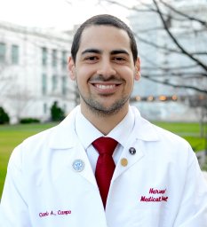 Camilo A Campo合作论文数University of Wisconsin1
 Amanda J Stump合作论文数1
 Evan Masutani合作论文数Sch Med, Univ Calif San Diego1Dalia Murra合作论文数Michigan Neuroscience Institute, University of Michigan1
 Asser Abou Elkassem合作论文数Department of Radiology, University of Alabama at Birmingham1Sowmya Varada合作论文数1
 Simon S. Martin合作论文数1Christina Caso合作论文数1
 Meredith M Course合作论文数Department of Molecular Biology, Colorado College1Jacquelyn E. Braggin合作论文数1Ishan Garg合作论文数Division of Internal Medicine, University of New Mexico Health Sciences Center1
 Bertrand Huber合作论文数VA Bedford Healthcare Syst1Yang Zhang合作论文数1
 Dooman Arefan合作论文数Univ Pittsburgh, Dept Radiol, 3362 Fifth Ave, Pittsburgh, PA 15213 USA1Matthew T MacLean合作论文数Perelman School of Medicine, University of Pennsylvania;Perelman School of Medicine, University of Pennsylvania;University of Pennsylvania, University of Pennsylvania1
 Yi Yan (闫艺)合作论文数Wuhan Children's Hospital, Tongji Medical College, Huazhong University of Science and Technology1
 Shanshan Gao合作论文数1




