Physical activity induces systemic benefits for brain and muscle function, but how the healthy spinal cord adapts to exercise remains largely unknown. Here, we combine bulk proteomics, single-nucleus RNA sequencing, and cellular communication inference to map exercise- induced molecular adaptations in the mouse lumbar spinal cord. Endurance training elicited robust baseline remodeling, dominated by glial transcriptional changes. Acute exhaustive exercise triggered biphasic responses: widespread metabolic and synaptic gene upregulation at 6 h followed by balanced suppression at 24 h, with trained animals exhibiting enhanced amplitude and faster resolution. Cell–cell communication analysis revealed that exercise reshaped signaling networks in both magnitude and composition. While glia emerged as primary transcriptional responders, cholinergic neurons—despite minimal transcriptional changes—were central signaling hubs, engaging various pathways with astrocytes, oligodendrocyte precursor cells, and oligodendrocytes in a training-dependent and temporally restricted manner. Glial-derived communication further diversified these responses, with astrocytes, oligodendrocytes, and microglia shifting toward pathways supporting synaptic remodeling, axon guidance, and growth factor signaling while dampening inflammatory cues. Together, these findings identify neuron–glia communication as potential driver of spinal cord adaptation to exercise, suggesting pathways through which glial plasticity may serve as a key mediator linking motor activity to spinal cord resilience.
Exercise stimulates the release of bioactive factors, termed exerkines, that contribute to local and systemic adaptation. Circulating exerkines are often interpreted as direct readouts of muscle secretion, overlooking regulatory processes within tissues that shape their production, transformation, and release. Using skeletal muscle and endurance exercise as a model system, we define a local-systemic secretome axis shaped by spatial organization, stimulus-specific programs, temporal dynamics, extracellular processing, and paracrine circuitry. By integrating evidence from recent transcriptomics, proteomics, interstitial fluid, and extracellular vesicle studies, we outline how these processes govern signal propagation from muscle to circulation and inform the interpretation of circulating exerkines as biomarkers and therapeutic targets.
Dysferlin is a transmembrane protein that plays a prominent role in membrane repair of damaged muscle fibers. Accordingly, mutations in the dysferlin gene cause progressive muscular dystrophies, collectively referred to as dysferlinopathies for which no effective treatment exists. Unexpectedly, experimental approaches that successfully restore membrane repair fail to prevent a dystrophic phenotype, suggesting that additional, hitherto unknown dysferlin-dependent functions contribute to the development of the pathology. Our experiments revealed an altered metabolic phenotype in dysferlin-deficient muscles, characterized by (1) mitochondrial abnormalities and elevated death signaling and (2) increased glucose uptake, reduced glycolytic protein levels, and pronounced glycogen accumulation. Strikingly, elevating mitochondrial volume density and muscle glycogen accelerates disease progression; whereas, improvement of mitochondrial function and recruitment of muscle glycogen with exercise ameliorated functional parameters in a mouse model of dysferlinopathy. Collectively, our results not only shed light on a metabolic function of dysferlin but also imply new therapeutic avenues aimed at promoting mitochondrial function and normalizing muscle glycogen to ameliorate dysferlinopathies, complementing efforts that target membrane repair.
OBJECTIVES:Endurance training is known to elicit numerous changes in skeletal muscle to enhance performance and function. Many of these adaptations are controlled by the modulation of transcriptional programs in myonuclei. While previous studies have explored alterations in DNA methylation and histone modifications in response to exercise, the specific changes in chromatin restructuring and accessibility, a prerequisite for transcription, are still poorly understood. METHODS:A multi-omics analysis was performed: ATAC-sequencing was used to map chromatin accessibility in myonuclei isolated from endurance-trained and untrained mice at multiple time points (0 h, 6 h, and 72 h) post-exercise. Gene expression was assessed via RNA-sequencing, and motif activity analysis identified regulatory factors involved in exercise-induced chromatin remodeling and transcriptomic response. RESULTS:Endurance training amplified rapid chromatin closing immediately after exercise, with trained muscle exhibiting a more pronounced loss of chromatin accessibility at 0 h and 6 h post-exercise compared to untrained muscle. These chromatin accessibility changes persisted longer in trained muscle, with significant retention until 72 h post-exercise. Immediate early transcription factors, such as Fos and Jun, showed a training state-dependent shift in activation dynamics. Similarly, specific modulation of genes involved in metabolism, insulin response and angiogenesis was observed. CONCLUSIONS:Endurance training triggers rapid and persistent chromatin remodeling in muscle, contributing to the transcriptional response to exercise. Our findings suggest that training induces long-lasting epigenetic changes, potentially underpinning muscle memory and improved physiological resilience. These new insights into the molecular mechanisms of muscle adaptation help to understand the training response, and might become relevant in disease prevention.
Muscle spindles are stretch receptors lying deep within the muscle belly involved in detecting changes in muscle length and playing a fundamental role in motor control, posture and synchronized gait. They are made up of an external capsule surrounding three to five intrafusal muscle fibres and a nuclear bag complex. Dysfunction of muscle spindles leads to abnormal proprioceptor function, which has been linked to aberrant bone and cartilage development, scoliosis, kyphosis and joint contractures. RYR1, the gene encoding the calcium release channel of the sarcoplasmic reticulum, is the most common target of mutations linked to human congenital myopathies, a condition often accompanied by skeleton alterations and joint contractures. So far, the link between RYR1 mutations, altered muscle spindles and skeletal defects has not been investigated. To this end, we investigated heterozygous mice carrying recessive Ryr1 mutations isogenic to those present in a severely affected child. Here, we show that: (i) the RyR1 protein localizes to the polar regions of intrafusal fibres and exhibits a doubled row distribution pattern, typical for junctional sarcoplasmic reticulum proteins; (ii) muscle spindles of compound heterozygous mice show structural defects; and (iii) in intrafusal muscle fibres from dHT mice, RyR1-mediated Ca2+ release is significantly impaired and RyR1 protein content is reduced by 54%. These results support the hypothesis that Ryr1 mutations not only affect the function of extrafusal muscles, but also might affect that of intrafusal muscles. The latter may be one of the underlying causes of skeletal abnormalities seen in patients affected by recessive RYR1 mutations. KEY POINTS: Dysfunction of muscle spindles leads to abnormal proprioceptor function, which can cause abnormal bone and cartilage development, scoliosis, kyphosis and joint contractures. Muscle spindles contain intrafusal muscle fibres, which are made up of nuclear bag and nuclear chain fibres; the polar regions of intrafusal muscle fibres contain contractile filaments and sarcotubular membranes. Patients with RYR1 mutations often present skeleton alterations and joint contractures from birth. Using the dHT mouse model carrying recessive Ryr1 mutations, we investigated whether muscle spindles from dHT mice show structural defects and altered RyR1 content and function. Our results support the hypothesis that RYR1 mutations also affect the function of intrafusal muscles and this may be one of the underlying causes of the skeletal abnormalities seen in patients.
Skeletal muscle possesses extraordinary plasticity of structure, metabolism, and function in response to repeated contractile activity. As a syncytium embedded within a complex microenvironment, muscle relies on the coordination of distinct myonuclear populations and diverse mononucleated cell types. Here, we present a high-resolution single-nucleus RNA-sequencing atlas of 550000 skeletal muscle nuclei, capturing the longitudinal transcriptional responses of 17 distinct myonuclear and 21 mononuclear cell populations at multiple time points after one bout of exhaustive exercise in trained and sedentary mice. The transcriptional programs of these populations are further shaped by training status into divergent adaptive trajectories. A subset of oxidative myonuclei enters a delayed regenerative state post-exercise, reflecting a bifurcated response to a disproportionate metabolic load on fibers during endurance exercise. Prior training accelerates homeostatic recovery and shields oxidative nuclei from exacerbated damage signatures. In parallel, mononucleated cells emerge as the primary mediators of intercellular communication during recovery. Together, this dataset establishes that training adaptation emerges through a coordinated interplay of intrinsic adaptive programs of multicellular remodeling, and provides a foundational resource for mechanistic insights into muscle plasticity.
Many countries face an unprecedented challenge in aging demographics. This has led to an exponential growth in research on aging, which, coupled to a massive financial influx of funding in the private and public sectors, has resulted in seminal insights into the underpinnings of this biological process. However, critical validation in humans has been hampered by the limited translatability of results obtained in model organisms, additionally confined by the need for extremely time-consuming clinical studies in the ostensible absence of robust biomarkers that would allow monitoring in shorter time frames. In the future, molecular parameters might hold great promise in this regard. In contrast, biomarkers centered on function, resilience, and frailty are available at the present time, with proven predictive value for morbidity and mortality. In this review, the current knowledge of molecular and physiological aspects of human aging, potential antiaging strategies, and the basis, evidence, and potential application of physiological biomarkers in human aging are discussed.
Regular physical activity induces a variety of health benefits, preventing and counteracting diseases caused by a sedentary lifestyle. However, the molecular underpinnings of skeletal muscle plasticity in exercise remain poorly understood. We identified a role of the Krüppel-Like Factor 5 (Klf5) in this process, in particular in the regulation of lipid homeostasis. Surprisingly, gain- and loss-of-function studies in muscle in vivo revealed seemingly opposite functions of Klf5 in the response to an acute exercise bout and chronic training, modulating lipid oxidation and synthesis, respectively. Thus, even though only transiently induced, the function of Klf5 is complex and fundamental for a normal long-term training response. These findings highlight the importance of this mediator of external stress response to adaptive remodeling of skeletal muscle tissue.
Skeletal muscle is one of the tissues with the highest range of variability in metabolic rate, which, to a large extent, is critically dependent on tightly controlled and fine-tuned mitochondrial activity. Besides energy production, other mitochondrial processes, including calcium buffering, generation of heat, redox and reactive oxygen species homeostasis, intermediate metabolism, substrate biosynthesis, and anaplerosis, are essential for proper muscle contractility and performance. It is thus not surprising that adequate mitochondrial function is ensured by a plethora of mechanisms, aimed at balancing mitochondrial biogenesis, proteostasis, dynamics, and degradation. The fine-tuning of such maintenance mechanisms ranges from proper folding or degradation of individual proteins to the elimination of whole organelles, and in extremis, apoptosis of cells. In this review, the present knowledge on these processes in the context of skeletal muscle biology is summarized. Moreover, existing gaps in knowledge are highlighted, alluding to potential future studies and therapeutic implications.
Biomarkers of aging are indispensable for testing interventions. While promising, the recent focus on molecular aspects should not detract from the functional parameters for which excellent correlation with mortality, and ample clinical human data exist.
In recent years, a plethora of different data-independent acquisition methods have been developed for proteomics to cover a wide range of requirements. Current deep proteome profiling methods rely on fractionations, elaborate chromatography, and mass spectrometry setups or display suboptimal quantitative precision. We set out to develop an easy-to-use one shot DIA method that achieves high quantitative precision and high proteome coverage. We achieve this by focusing on a small mass range of 430–670 m/z using small isolation windows without overlap. With this new method, we were able to quantify >9200 protein groups in HEK lysates with an average coefficient of variance of 3.2%. To demonstrate the power of our newly developed narrow mass range method, we applied it to investigate the effect of PGC-1α knockout on the skeletal muscle proteome in mice. Compared to a standard data-dependent acquisition method, we could double proteome coverage and, most importantly, achieve a significantly higher quantitative precision, as compared to a previously proposed DIA method. We believe that our method will be especially helpful in quantifying low abundant proteins in samples with a high dynamic range. All raw and result files are available at massive.ucsd.edu (MSV000092186).
Skeletal muscle plasticity enables an enormous potential to adapt to various internal and external stimuli and perturbations. Most notably, changes in contractile activity evoke a massive remodeling of biochemical, metabolic and force-generating properties. In recent years, a large number of signals, sensors, regulators and effectors have been implicated in these adaptive processes. Nevertheless, our understanding of the molecular underpinnings of training adaptation remains rudimentary. Specifically, the mechanisms that underlie signal integration, output coordination, functional redundancy and other complex traits of muscle adaptation are unknown. In fact, it is even unclear how stimulus-dependent specification is brought about in endurance or resistance exercise. In this review, we will provide an overview on the events that describe the acute perturbations in single endurance and resistance exercise bouts. Furthermore, we will provide insights into the molecular principles of long-term training adaptation. Finally, current gaps in knowledge will be identified, and strategies for a multi-omic and -cellular analyses of the molecular mechanisms of skeletal muscle plasticity that are engaged in individual, acute exercise bouts and chronic training adaptation discussed.
Circadian rhythms, governed by the dominant central clock, in addition to various peripheral clocks, regulate almost all biological processes, including sleep-wake cycles, hormone secretion and metabolism. In certain contexts, the regulation and function of the peripheral oscillations can be decoupled from the central clock. However, the specific mechanisms underlying muscle-intrinsic clock-dependent modulation of muscle function and metabolism remain unclear. We investigated the outcome of perturbations of the primary and secondary feedback loops of the molecular clock in skeletal muscle by specific gene ablation of Period circadian regulator 2 (Per2) and RAR-related orphan receptor alpha (Rorα), respectively. In both models, a dampening of core clock gene oscillation was observed, while the phase was preserved. Moreover, both loops seem to be involved in the homeostasis of amine groups. Highly divergent outcomes were seen for overall muscle gene expression, primarily affecting circadian rhythmicity in the PER2 knockouts and non-oscillating genes in the RORα knockouts, leading to distinct outcomes in terms of metabolome and phenotype. These results highlight the entanglement of the molecular clock and muscle plasticity and allude to specific functions of different clock components, i.e. the primary and secondary feedback loops, in this context. The reciprocal interaction between muscle contractility and circadian clocks might therefore be instrumental to determining a finely tuned adaptation of muscle tissue to perturbations in health and disease. KEY POINTS: Specific perturbations of the primary and secondary feedback loop of the molecular clock result in specific outcomes on muscle metabolism and function. Ablation of Per2 (primary loop) or Rorα (secondary loop) blunts the amplitude of core clock genes, in absence of a shift in phase. Perturbation of the primary feedback loop by deletion of PER2 primarily affects muscle gene oscillation. Knockout of RORα and the ensuing modulation of the secondary loop results in the aberrant expression of a large number of non-clock genes and proteins. The deletion of PER2 and RORα affects muscle metabolism and contractile function in a circadian manner, highlighting the central role of the molecular clock in modulating muscle plasticity.
Satellite cells, the stem cells of skeletal muscle tissue, hold a remarkable regeneration capacity and therapeutic potential in regenerative medicine. However, low satellite cell yield from autologous or donor -derived muscles hinders the adoption of satellite cell transplantation for the treatment of muscle diseases, including Duchenne muscular dystrophy (DMD). To address this limitation, here we investigated whether satellite cells can be derived in allogeneic or xenogeneic animal hosts. First, injection of CRISPR/Cas9-corrected Dmd mdx mouse induced pluripotent stem cells (iPSCs) into mouse blastocysts carrying an ablation system of host satellite cells gave rise to intraspecies chimeras exclusively carrying iPSC-derived satellite cells. Furthermore, injection of genetically corrected DMD iPSCs into rat blastocysts resulted in the formation of interspecies rat -mouse chimeras harboring mouse satellite cells. Notably, iPSC-derived satellite cells or derivative myoblasts produced in intraspecies or interspecies chimeras restored dystrophin expression in DMD mice following intramuscular transplantation and contributed to the satellite cell pool. Collectively, this study demonstrates the feasibility of producing therapeutically competent stem cells across divergent animal species, raising the possibility of generating human muscle stem cells in large animals for regenerative medicine purposes.
Human skeletal muscle demonstrates remarkable plasticity, adapting to numerous external stimuli including the habitual level of contractile loading. Accordingly, muscle function and exercise capacity encompass a broad spectrum, from inactive individuals with low levels of endurance and strength to elite athletes who produce prodigious performances underpinned by pleiotropic training-induced muscular adaptations. Our current understanding of the signal integration, interpretation, and output coordination of the cellular and molecular mechanisms that govern muscle plasticity across this continuum is incomplete. As such, training methods and their application to elite athletes largely rely on a "trial-and-error" approach, with the experience and practices of successful coaches and athletes often providing the bases for "post hoc" scientific enquiry and research. This review provides a synopsis of the morphological and functional changes along with the molecular mechanisms underlying exercise adaptation to endurance- and resistance-based training. These traits are placed in the context of innate genetic and interindividual differences in exercise capacity and performance, with special consideration given to aging athletes. Collectively, we provide a comprehensive overview of skeletal muscle plasticity in response to different modes of exercise and how such adaptations translate from "molecules to medals."
Abstract Exercise effectively promotes and preserves cardiorespiratory, neuromuscular, metabolic, and cognitive functions throughout life. The molecular mechanisms underlying the beneficial adaptations to exercise training are, however, still poorly understood. To improve the mechanistic study of specific exercise training adaptations, standardized, physiological, and well‐characterized training interventions are required. Therefore, we performed a comprehensive interrogation of systemic changes and muscle‐specific cellular and molecular adaptations to voluntary low‐resistance wheel running (Run) and progressive high‐resistance wheel running (RR) in young male mice. Following 10 weeks of training, both groups showed similar improvements in body composition and peak oxygen uptake (V̇O2peak), as well as elevated mitochondrial proteins and capillarization markers in the M. plantaris. Run mice clearly outperformed RR mice in a forced treadmill running capacity test, while RR mice displayed increased grip strength as well as superior mass gains in the M. soleus, associated with distinct proteomic changes specifying the two paradigms. Thus, even though both training modalities induce overlapping adaptations, Run interventions preferably improve submaximal running performance, while progressive RR is a valid model to study training‐induced gains in grip strength and plantar flexor hypertrophy.
Der Autor nimmt Bezug auf einen Artikel im "Kurz und bündig" und findet: Körperliche Aktivität sollte nicht weniger («no sports»), sondern ganz im Gegenteil mehr ausgeübt und gefördert werden – auch von den Ärztinnen und Ärzten.
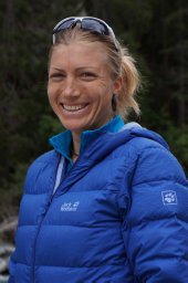 Furrer Regula合作论文数MOVE Research Institute Amsterdam, VU University Amsterdam27
 Bruce M. Spiegelman合作论文数Department of Cancer Biology, Dana-Farber Cancer Institute;Blavatnik Institute, Harvard Medical School24
 Gesa Santos合作论文数23
 Urs A. Meyer合作论文数17
 Joaquin Perez-Schindler合作论文数Biozentrum, Univ Basel16
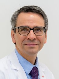 Eberli Daniel合作论文数16
 Tullio Sulser合作论文数Faculty of Medicine, University of Zurich;Zentrum für Urologie Zürich – Klinik Hirslanden15Serge Summermatter合作论文数14Delezie Julien合作论文数Institute of Cellular and Integrative Neurosciences, University of Strasbourg13Danilo Ritz合作论文数12
 Jiandie Lin合作论文数Department of Cell & Developmental Biology, University of Michigan Medical School;Jiandie Lin Lab, Life Sciences Institute, University of Michigan12
 Svensson Kristoffer合作论文数12
 Svenia Schnyder合作论文数12
 Michael Podvinec合作论文数12
 Jonathan Gill合作论文数9
 Souzan Salemi合作论文数Zürich9Julie St-Pierre合作论文数8
 Bettina Karrer-Cardel合作论文数8
 Salatino Silvia合作论文数8Cardel Bettina合作论文数8Francesco Zorzato合作论文数University of Ferrara8
 Vamsi Mootha合作论文数Department of Systems Biology, Harvard Medical School;Department of Medicine, Massachusetts General Hospital;Broad Institute of MIT and Harvard;Howard Hughes Medical Institute8
 Anne-Sophie Arnold合作论文数8Ivana Dinulovic合作论文数7
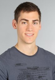 Aurel B. Leuchtmann合作论文数Biozentrum, Univ Basel7Deana Haralampieva合作论文数6Elyzabeth Vargas-Fernández合作论文数6Stefan Steurer合作论文数6Petra S Eisele合作论文数6Markus Beer合作论文数6
 Barbara Heim-Kupr合作论文数6Wenli Yang合作论文数6
 Renate Looser合作论文数6Sherry Chin合作论文数6I. Dinulovic合作论文数5
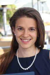 Geraldine Maier合作论文数Biozentrum, Univ Basel5
 Markus Rüegg合作论文数The Center for Molecular Life Sciences, University of Basel;SEAL Therapeutics Ltd.5
 Verena Albert合作论文数Biozentrum, University of Basel5
 Pere Puigserver合作论文数Department of Cancer Biology, Dana-Faber Cancer Institute, Harvard Medical School5Betzel Thomas合作论文数5
 M. Alexander Schmidt合作论文数Department of Infectiology, University of Munster;Institute of Infectiology, University of Munster5Carmela Gnerre合作论文数5
 James Rhee合作论文数Dana-Farber Cancer Institute and Department of Cell Biology, Harvard Medical School5Simon M. Ametamey合作论文数4D. Haralampieva合作论文数4Martine Christe合作论文数4
 Sedat Dilbaz合作论文数4
 Oliver Baum合作论文数4
 Susan Treves合作论文数Department of Biomedicine, University of Basel4




