BACKGROUND CONTEXT Cervical-to-sacrum (CTS) fusion is an extensive surgical option often used in patients with prior fusion or multiregional deformity. Whether outcomes differ compared with shorter constructs such as upper thoracic-to-sacrum (UTS) fusion remains unclear. PURPOSE To assess whether CTS fusion yields inferior outcomes compared with UTS constructs after adjusting for patient complexity. STUDY DESIGN/SETTING Retrospective study of prospectively collected multicenter data. PATIENT SAMPLE Adults undergoing ASD surgery with constructs spanning either cervical-to-sacrum/pelvis (CTS) or upper thoracic (T2–T5) to sacrum/pelvis (UTS). Total N=399, including 54 CTS patients. OUTCOME MEASURES Patient-reported outcomes (NRS Back, NRS Leg, ODI, PCS, SRS domains), radiographic parameters (Cobb angle, sagittal vertical axis [SVA], L1 pelvic angle [L1PA]), complications, and revision rates. METHODS Patients were grouped by construct (CTS vs UTS). Outcomes were assessed at baseline, 6 weeks, 1 year, and limited 2-year follow-up using mixed-effects models adjusted for demographics, ASA class, estimated blood loss, alignment parameters, and baseline PROs. Subanalyses evaluated staged vs single-stage CTS procedures and upper instrumented vertebra (C1–C3 vs C4+) subgroups. RESULTS Among 399 patients, CTS patients were more complex, with higher rates of osteoporosis, higher ASA scores, and more prior fusions (all p<0.05). Unadjusted early outcomes favored CTS, with improved 6-week NRS Back, NRS Leg, and ODI scores (all p<0.05). However, by 1 year, outcomes favored UTS patients across NRS Leg, ODI, PCS, and SRS domains. Mixed-effects modeling confirmed that CTS patients demonstrated greater early improvement but worse longitudinal trajectories for NRS Leg, ODI, SRS Total, PCS, and sagittal alignment (all p<0.05). At 1 year, CTS patients had worse adjusted NRS Leg scores (p=0.023). At 2 years, CTS patients had worse ODI (p=0.013), PCS (p<0.001), and SVA (p=0.041). Coronal alignment (Cobb angle) was similar, while sagittal alignment (SVA, L1PA) was worse in CTS patients. Wound complications were higher in CTS (13% vs 6%; p=0.049). In subanalysis, single-stage CTS procedures were associated with higher implant-related complications, radiographic adverse events, and revision rates (p<0.006). UIV subgroup analysis showed no clear advantages, although C1–C3 constructs demonstrated worse sagittal alignment trajectories. CONCLUSIONS Although CTS fusion results in meaningful early improvement, it is associated with worse long-term patient-reported and radiographic outcomes compared with shorter UTS constructs, as well as higher complication rates. These findings can inform surgical planning and patient counseling in complex ASD cases. FDA Device/Drug Status This abstract does not discuss or include any applicable devices or drugs.
OBJECTIVE:Adult spinal deformity (ASD) surgery patients maintain upright posture by using numerous compensatory mechanisms. The distribution of this compensation throughout the skeleton has not been fully investigated. METHODS:Patients with lumbar deformity curves undergoing fusion from T10 to the pelvis were included. Groups were stratified by Scoliosis Research Society (SRS)-Schwab sagittal deformity severity (mild, moderate, and severe). Compensation was determined based on the published values of asymptomatic individuals by Bao et al. (2018), with patients outside 1 standard deviation of the mean values deemed to be compensating. Adequate deformity correction was determined based on matching published sagittal age-adjusted score (SAAS) criteria. Means comparisons tests assessed differences between cohorts at each time point. RESULTS:In total, 379 ASD patients were included (mean age 66.7 ± 10.2 years, body mass index 28.4 ± 5.4 kg/m2, Charlson Comorbidity Index 1.20 ± 1.73). In total, 23.8% of patients had mild deformity, 19.2% moderate, and 57% severe. The severe deformity cohort generally demonstrated the highest rates of compensation across all regions at different time points. At baseline, the severe and moderate cohorts demonstrated predominantly lower limb-dominant compensation, with the highest frequencies of compensation seen at the knee and pelvis. In the mild cohort, knee compensation was relieved by 1 year when adequate correction was achieved. For the moderate cohort, hip and pelvic compensation were relieved first, with knee compensatory relief occurring by 2 years. For the severe cohort, pelvic compensation was relieved first, with global lower limb and thoracic compensation subsequently occurring. CONCLUSIONS:There is notable variation in how ASD patients compensate as the severity of their deformity progresses. There is also variation in how these patterns are altered postoperatively.
BACKGROUND CONTEXT Failure to achieve nominal distal lumbar lordosis (DLL) of ≥35° and lumbar distribution index (LDI) of 50-80% after short-segment fusions is associated with high rates of iatrogenic sagittal deformity, adjacent segment disease, and revision surgery. PURPOSE This study evaluates the ability of personalized interbody devices (PIDs) with patient-specific alignment plans to correct or preserve DLL and LDI to nominal postoperative values and to minimize reoperations for mechanical complications or adjacent segment disease (ASD). STUDY DESIGN/SETTING Retrospective review of consecutive patients. PATIENT SAMPLE Consecutive patients treated for degenerative disorders with 1- to 3-level fusions and UIV L3, L4, or L5 using personalized interbody spacers and supplemental fixation from an IRB-approved multicenter registry were included. The study cohort had 182 subjects from 16 centers, with median age of 65 years (range: 29-91), 95 females (62%), and mean 26-month follow-up (range: 15-47, 110 patients with >24-month follow-up). OUTCOME MEASURES Preoperative surgeon-approved planned DLL and LDI for each patient was recorded from the personalized device digital design platform and compared to postoperative radiographic DLL and LDI measures. Complications and revisions were extracted from medical records. METHODS Preoperative and planned DLL and LDI were compared to postoperative radiographs measured by an independent core laboratory and adjudicated by 3 experienced spine surgeons. This study was retrospective and noninterventional; ie, planning for nominal DLL or LDI was not uniformly asserted nor considered during surgical planning or execution. Frequency of planned and achieved nominal DLL and LDI were calculated. Mechanical or ASD complications that required reoperation were enumerated. RESULTS Among 72 cases with preoperative nominal DLL ≥35°, 71 were planned to preserve DLL and 60 of these (85%) achieved nominal postoperative DLL. Among 110 cases with preoperative suboptimal DLL <35°, 41 were planned to correct DLL and 27 of these (66%) were restored to nominal DLL ≥35°. Among 99 cases with preoperative nominal LDI, 92 were planned to preserve LDI and 75 of these (82%) achieved nominal postoperative LDI. Among 78 cases with preoperative suboptimal (ie, hyper or hypo) LDI, 18 were planned to correct LDI and 15 of these (83%) were restored to nominal LDI. Three reoperations were reported (ie, 3/182 = 1.6% of cases) for ASD. Two of the 3 revisions were in patients with preoperative low DLL, for whom a correction to nominal DLL was not planned. The third revision was in a patient having both low DLL and LDI, who was planned and corrected to nominal LDI and corrected to nominal DLL. CONCLUSIONS In this study, cases with preoperative suboptimal DLL or LDI and PID-planned corrections were restored to nominal in 66% and 83% of patients, respectively, compared to published rates of 10% to 37% for patients treated with stock devices. The reoperation rate for ASD or mechanical complications in the current study (1.6%) was significantly lower than the referenced studies (17-22%, each p<0.001). Personalized interbody devices can predictably achieve surgeon-planned DLL and LDI in short-segment lumbar fusions. Favorably low reoperation rates in this study may be attributable to high rates of postoperative nominal DLL and LDI that protect against ASD. Results reflect both the value of surgical planning to preserve or restore DLL and LDI and the utility of personalized implants for achieving planned alignment. FDA Device/Drug Status aprevo (Approved for this indication).
OBJECTIVE:The aim of this study was to determine predictors of the minimum clinically important difference (MCID) in the Neck Disability Index (NDI) following cervical spinal deformity surgery. METHODS:A retrospective review was performed of a prospective, multicenter adult cervical spinal deformity database. All patients had baseline and 1-year NDI scores. Patients met MCID with an improvement of NDI by 7 points between baseline and 1 year, as previously established. Baseline demographics, comorbidities, and both baseline and 1-year spinopelvic parameters were evaluated for statistical significance in a univariate logistic regression analysis. Significant variables, in addition to baseline NDI, were analyzed in a multivariable logistic regression model by backward selection with Akaike information criterion minimization. RESULTS:A total of 122 patients were included with a median age of 62 (IQR 56, 69) years; 62% of patients were female. Of the 122 patients, 72 (59%) achieved NDI MCID at 1 year. Predictors of achieving MCID on univariate analysis included a lower Charlson Comorbidity Index (CCI) total score (OR 0.70, p = 0.03), depression as a comorbidity (OR 2.9, p = 0.02), lower C2 tilt at the 1-year follow-up (OR 0.92, p = 0.02), and a greater difference between 1-year postoperative C2-7 sagittal vertical axis (SVA) and preoperative C2-7 SVA (OR 0.98, p = 0.0495). On multivariable logistic regression analysis, predictors of achieving MCID included a lower CCI (OR 0.62, p = 0.03), depression as a comorbidity (OR 3.1, p = 0.059), a greater change in C2-7 SVA at the 1-year follow-up compared with baseline (OR 0.97, p = 0.055), and baseline NDI (OR 1.02, p = 0.24) with an area under the curve of 0.74. CONCLUSIONS:The best-fit multivariable model included higher baseline NDI, a greater change in C2-7 SVA, patient-reported baseline depression, and lower CCI as important factors in predicting NDI MCID.
BACKGROUND AND OBJECTIVES:The purpose of this study was to analyze the impact of patient-specific rods on mechanical complications after adult thoracolumbar spinal deformity surgery. METHODS:A consecutive series was analyzed of 200 adult thoracolumbar spinal deformity patients. Half of the patients (n = 100) had surgery just before implementing patient-specific rods, and the other 100 patients had surgery immediately after adoption of patient-specific rods. All patients had instrumented fusions from the thoracic spine to the pelvis. Mechanical complications were defined as rod fracture, proximal junctional kyphosis, proximal junctional failure (revision surgery requiring proximal fusion extension), and/or revision surgery for rod fracture or pseudoarthrosis. Minimum follow-up was 2 years. RESULTS:A total of 200 patients were included with a median (IQR) age of 68 (61, 73), body mass index of 28 (25, 32), and 73% were female. The patients with patient-specific rods compared with those without had less preoperative kyphosis (39° vs 44°, P = .01), a higher median number of rods (4 vs 2, P = .04), more often a combined anterior-posterior approach (61% vs 46%, P = .047), more commonly a upper instrumented vertebra vertebroplasty (67% vs 45% P = .003), and postoperatively had less thoracic kyphosis (45% vs 49%, P = .02). Patient-specific rod patients had a lower rate of mechanical complications (29% vs 47%, P = .01), a lower rate of rod fracture (3% vs 16%, P = .003), and shorter operative times (215 minutes vs 250 minutes, P = .04). On multivariable analysis, the only 2 independent predictors of reducing the rate of mechanical complications were the utilization of patient-specific rods (odds ratio 0.47, P = .013, CI 0.26-0.85) and lower postoperative thoracic kyphosis (odds ratio 1.02, P = .04, CI 1.00-1.05). CONCLUSION:In a consecutive series of adult thoracolumbar spinal deformity patients, patient-specific rods reduced the odds of mechanical complications by over 50%. Adoption of patient-specific rods should be considered to optimize patient outcomes after adult spinal deformity surgery.
Adult spinal deformity patients undergoing total hip arthroplasty experience higher hip dislocation rates than those with normal spinal alignment. The traditional Lewinnek safe zone does not account for spinopelvic variation such as pelvic retroversion. To address this, three patient-specific normative zones for acetabular anteversion were defined. A multicenter retrospective analysis of 146 adult spinal deformity patients and 47 asymptomatic controls was performed using three-dimensional biplanar radiograph reconstructions to measure spinopelvic alignment and acetabular orientation. Normative Zone 1, for patients not undergoing spinal realignment, was delineated by the 95% confidence interval limits: minimum anteversion = 0.3182 × pelvic tilt +2.947 and maximum anteversion = 0.3317 × pelvic tilt +25.823. Normative Zone 2, for patients following spinal realignment, was based on pelvic incidence: minimum anteversion = 0.0682 × pelvic incidence +9.7749 and maximum anteversion = 0.0698 × pelvic incidence +21.5218. Normative Zone 3, intended for cases with uncertain spinal correction plans, was defined as the intersection of Zones 1 and 2, yielding a narrower target anteversion range. These zones enable patient-specific cup placement that accounts for existing or planned spinal alignment, with the potential to reduce dislocation risk. Clinical Significance: This study provides acetabular cup orientation tailored to each patient's spinopelvic alignment and surgical plan, potentially reducing dislocation rates in spinal malalignment patients.
Lower limbs can play a major compensating role for sagittal malalignment; however, little is known about the different types of compensation. This study aimed to identify different patterns of lower limb compensation and to determine which parameters may affect the recruitment of knee flexion versus hip extension. This study included adult spinal deformity (ASD) patients with full-body X-rays in erect position from a multicentric prospective database. All parameters were measured at baseline: demographics, clinical scores and radiographic parameters: pelvic parameters, pelvic incidence-lumbar lordosis (PI-LL) mismatch, T1 pelvic angle (TPA), sacro-femoral angle (SFA), knee flexion angle (KA), ankle dorsi-flexion angle (AA), pelvic shift (PSh), hip and knee osteoarthritis (OA) grade. A K-means cluster analysis was conducted to identify patterns of lower limb compensation based on SFA and KA. The optimal number of clusters was determined using the silhouette score. The different parameters were then compared across clusters. 871 ASD patients were included, of whom 66.9
BACKGROUND CONTEXT BMP reduces pseudarthrosis and revision surgeries in adult spinal deformity (ASD) surgery. The long-term cost effectiveness for BMP use in ASD surgery remains uncertain. PURPOSE To investigate BMP cost savings in a cohort with an average 4.5-year follow-up. BMP is used more often and in higher doses in more complex surgeries. By reducing revision surgery, BMP may reduce the aggregate cost of a patient’s care over time. STUDY DESIGN/SETTING Retrospective analysis of a multicenter prospective database. PATIENT SAMPLE Patients with adult spinal deformity who received BMP during surgery. OUTCOME MEASURES Cost, mechanical complications. METHODS A multicenter ASD surgical database of 1355 patients with 4 or more levels of fusion and a minimum 2-year follow-up was analyzed. The total cost of surgery was calculated as index surgery cost plus all revisions; if no revisions occurred, then revision cost was 0. Logistic regression was performed on 10 variables for medical and surgical complexity—including number of levels fused, number of osteotomies, and age—which were compiled into a propensity score. Researchers performed 1:1 nearest neighbor propensity score matching (PSM) to create BMP and noBMP cohorts. Average total cost and time to mechanical complications were calculated. Regression of revision cost on BMP usage and surgical complexity, with interaction terms, was used to determine a cost saving threshold. RESULTS A total of 1029 ASD patients (60.6±14.2 yo; 74.8% F) had cost data, 623 received BMP (mean follow-up 4.56 years ± 1.33). Prior to PSM, the BMP group was more surgically complex (p < 0.001), following 1:1 matching BMP and noBMP cohorts (n=288) had similar surgical complexity. Revisions were significantly reduced in the BMP group (16.4% BMP, 26.7% noBMP, OR 0.55, p=0.002) as well as pseudoarthrosis (3.4% BMP, 7.8% noBMP, OR 0.41, p=0.03), with no increase in other complications. The mean time-to-revision was 1.7 years in the BMP group and 1.2 years in the noBMP group (HR 0.59, p=0.002). In 2025 $USD, BMP index surgeries were similar in cost ($100,917 noBMP vs $92,757 BMP; p=0.08) but revisions were significantly cheaper ($12,697 noBMP vs $7,114 BMP; p=0.005) with total cost saving of $13,743 (p=0.015) overall. Univariate regression showed as surgical complexity increased, the cost savings from BMP increased (r=0.38, p = 0.004), directly mediated by a relative reduction in revision rate. BMP dosages scaled with both complexity and cost reduction. A point estimate of break-even revision cost suggests that BMP reduces revision cost after 6 levels fused (6.18 ± 2.4), any SPO (0.91 ± 1.5), any 3CO (0.27 ± 0.5), or pelvic fixation (0.05 ± 0.21), with an optimal dosage of 2.4mg per level. CONCLUSIONS BMP is cost-effective over a long follow-up period, by preventing revision surgeries in more complex patients. The BMP surgical population is becoming more complex over time, likely due to appropriate patient selection by surgeons; thus, cost savings may be increasingly obscured over time. More than 6 levels of fusion, pelvic fixation, or any osteotomies result in cost-saving with BMP, with an optimal dosage of 2.4mg per level. FDA Device/Drug Status This abstract does not discuss or include any applicable devices or drugs.
BACKGROUND CONTEXT Advanced cervical deformity (CD) includes distinct sagittal alignment patterns based on subaxial cervical alignment, including global cervical kyphosis (GK) and preserved lordosis (PL) morphotypes. Comparative evidence describing how these phenotypes influence surgical strategy, radiographic correction, patient-reported outcomes (PROs), and complications remains limited. PURPOSE To compare demographics, surgical characteristics, radiographic parameters, PROs, and complication profiles between GK and PL morphotypes following surgical correction of advanced CD. STUDY DESIGN/SETTING Retrospective analysis of a prospective multicenter cervical deformity cohort. PATIENT SAMPLE Adults undergoing CD surgery with available T1 slope (T1S) and C2 slope (C2S) measurements. Patients were categorized as PL (T1S > C2S and T1S > 35°) or GK (T1S < C2S). Patients with focal or compensated deformity (C2–7 SVA < 4 lt; 4 cm) were excluded. OUTCOME MEASURES Radiographic parameters (preoperative, postoperative, and 1-year), PROs, complications, and revision surgery. METHODS Group comparisons were performed using appropriate statistical tests. Multivariable logistic regression evaluated associations with surgical complications and revision surgery, adjusting for demographic, surgical, and alignment variables. RESULTS The cohort included 110 patients (PL n=55; GK n=55) with similar demographics. PL patients had more prior thoracolumbar fusions (51% vs 25%, p<0.01). At baseline, PL demonstrated preserved cervical lordosis (18° vs −22°, p<0.01), greater thoracic kyphosis (54° vs 38°, p<0.01), higher T1 slope (56° vs 23°), and greater global sagittal malalignment compared to GK. GK more frequently had upper cervical UIVs, while PL more often had mid/lower thoracic LIVs (both p<0.01). PL patients underwent more three-column osteotomies (55% vs 25%, p<0.01) and had higher estimated blood loss (1000 vs 400 mL, p=0.01). At 6 weeks, PL had worse Neck Disability Index scores (46 vs 38, p=0.03), with no differences in PROs at later time points. Overall complication and revision rates were similar between groups. Distal junctional kyphosis (DJK) was more common in GK (38% vs 15%, p<0.01). On multivariable analysis, deformity type was not independently associated with complications or revision surgery. CONCLUSIONS GK and PL represent distinct morphotypes of advanced cervical deformity with differing baseline alignment and surgical strategies. While overall outcomes were similar, GK demonstrated higher rates of distal junctional kyphosis. These differences should be considered in preoperative planning and patient counseling. FDA Device/Drug Status This abstract does not discuss or include any applicable devices or drugs.
STUDY DESIGN:Retrospective review of multicenter, prospective cervical deformity database. OBJECTIVE:To compare outcomes of extension of fusion to the cervical spine versus the upper thoracic (UT) spine. SUMMARY OF BACKGROUND DATA:Proximal junctional kyphosis (PJK) management after thoracolumbar fusion requires extension of fusion to the proximal spinal segments. Unlike extensions to the less mobile thoracic segments, crossing the cervicothoracic junction (CTJ) involves more mobile cervical segments and creates different biomechanical influences and clinical outcomes. No study has compared the outcomes of extending fusion to the cervical versus the UT spine. METHODS:Patients with thoracic PJK who underwent revision with extension of fusion to either the cervical or UT (T1 or T2) spine were identified in a multicenter, prospective cervical deformity database. Patients with cervical upper instrumented vertebra (UIV) were subdivided into lower cervical (LC; C4-7) and upper cervical (UC; and occiput-C3) groups. Baseline demographics, surgical variables, radiographic outcomes, 2-year health-related quality-of-life scores, complications, and revision rates were analyzed. RESULTS:Fifty-one patients (mean age: 60.4±12.9 y; 91% female) with at least 2 years of follow-up were included. Twelve had extension to the UT, 20 to the LC, and 19 to the UC spine. Demographic data, Charlson Comorbidity Index, follow-up duration, surgical parameters, radiographic measurements, recurrent PJK and reoperation rates, and 2-year patient-reported outcome scores were similar across groups. The instrumentation failure rate was higher in the LC (25%) than in the UT (0%) and UC (8%) groups (P=0.03). CONCLUSIONS:Stopping fusion at T1 or T2 did not result in greater complication or reoperation rates than extending to the cervical spine. The instrumentation-related complication rate was higher for extension to the LC than to the UC or UT spine. Crossing the CTJ should be individualized, but may not prevent additional proximal junctional-level problems in the management of thoracolumbar fusion PJK. LEVEL OF EVIDENCE:Level IV.
OBJECTIVE:There is no gold standard for assessing sarcopenia. The authors aimed to quantify agreement and reliability among common sarcopenia measures based on muscle function, body composition, and imaging characteristics in a cohort of adult spinal deformity (ASD) patients. METHODS:This was a cross-sectional study. Preoperative ASD patients at a single tertiary-care center underwent the following sarcopenia assessments: 2 functional muscle assessments (grip strength and gait speed), 3 bioelectrical impedance (BIA) measures (skeletal muscle index [SMI], phase angle, and extracellular water [ECW]/total body water [TBW] ratio), and 2 imaging-based assessments (psoas muscle index [PMI] and total psoas area [TPA]/vertebral body area [VBA] ratio). Spearman's correlation analysis was used to test for associations. Measurements were standardized into sex-specific z-scores. Bland-Altman analysis was used to quantify agreement and Cronbach's alpha analysis was used to quantify reliability between sarcopenia measures. RESULTS:Between 2017 and 2025, 272 ASD patients were enrolled in the study. Of these, 88 ASD patients (59.1% female, median age 67.7 years) completed all sarcopenia assessments and thus were included in the analysis. Most sarcopenia measures were weakly correlated, except for PMI and TPA/VBA, which were moderately correlated (ρ = 0.84, p < 0.001), and phase angle and ECW/TBW (ρ = -0.82, p < 0.001). Bland-Altman analysis demonstrated poor agreement between all pairs of sarcopenia measures. Cronbach's alpha analysis showed poor reliability (α < 0.7) between all pairs of sarcopenia measures except for PMI and TPA/VBA (α = 0.89). These results did not significantly change when all 272 patients with imputed missing data were included. CONCLUSIONS:The authors found low agreement and reliability between 7 commonly used sarcopenia measures, except for good reliability between PMI and TPA/VBA. These sarcopenia measures are not interchangeable and may not be measuring the same underlying clinical entity. This is the first study to quantify agreement and reliability between sarcopenia measures. Future studies are needed to determine which sarcopenia measures best predict clinical outcomes in ASD patients.
BACKGROUND CONTEXT T1 slope (T1S) is a key determinant of cervical deformity (CD). Despite correction of cervical lordosis (CL) and thoracic kyphosis (TK), T1S may remain unchanged or increase postoperatively. PURPOSE To determine whether the effect of cervicothoracic correction on T1S is modified by preoperative pelvic tilt (PT), a surrogate of pelvic compensation reserve. STUDY DESIGN/SETTING Retrospective multicenter analysis of a cervical deformity surgical cohort. PATIENT SAMPLE Adults undergoing CD surgery with preoperative and early postoperative (3-month) imaging. OUTCOME MEASURES Change in T1S at early postoperative imaging. METHODS A total of 272 adult CD patients with complete pre- and early postoperative imaging were analyzed. T1S outcomes were categorized as reduced (>5° decrease), stable (±5°), or increased (>5° increase). Variables evaluated included CL restoration, TK correction, preoperative PT, C2 tilt, and global alignment parameters. Hierarchical regression assessed main effects and interactions. Postoperative PT change (ΔPT) was analyzed to validate compensation behavior. RESULTS Mean T1S change was −0.3° ± 21.4°, with 32% achieving T1S reduction (>5°). A three-tier gradient was observed: T1S reduced (n=87, −19° ± 12°), T1S stable (n=79, −1° ± 3°), and T1S increased (n=106, +16° ± 9°; p<0.001). The T1S-increased group underwent greater CL correction than the T1S-reduced group (+26° vs +3°, p<0.001), yet achieved similar postoperative CL (+8° vs +6°, p>0.1). TK correction differed significantly across groups (T1S reduced +6° ± 15°, stable −2° ± 6°, increased −6° ± 11°; p<0.001), with both CL and TK showing moderate correlations with ΔT1S (r=0.5–0.6). Upper thoracic lower instrumented vertebrae (LIV) predominated in the T1S-increased group (74%), whereas lower thoracic LIV was more common in the T1S-reduced group (47%). Upper thoracic three-column osteotomy was more frequent in the T1S-reduced group (21% vs 3%, p<0.001). Preoperative PT demonstrated a gradient, highest in the T1S-reduced group (24.0° ± 10.9°) and lowest in the T1S-increased group (19.0° ± 9.3°, p=0.003). Postoperative PT changes were minimal and not significantly different between groups. Baseline PT and C2 tilt remained independently associated with ΔT1S after adjusting for CL and TK correction. The full model explained 62% of variance (p<0.001). CONCLUSIONS T1S can be reduced with greater TK correction, upper thoracic osteotomy, and more caudal LIV selection, but is also significantly influenced by preoperative pelvic compensation reserve. Higher preoperative PT facilitates T1S reduction, whereas limited reserve predisposes to paradoxical T1S increase despite greater cervicothoracic correction. Preoperative pelvic parameters should be incorporated into surgical planning and risk stratification. FDA Device/Drug Status This abstract does not discuss or include any applicable devices or drugs.
Epigenetic changes, such as DNA methylation (DNAm), offer a measure of biological age distinct from chronological age. DNAm PhenoAge is one such biomarker that is more strongly related to morbidity, mortality, and physical function than chronological age. More accurate risk stratification methods are needed for ASD surgeries, where complications remain difficult to predict with an increasingly aged population. A multicenter ASD registry was queried. DNAm PhenoAge was calculated as per Levine et al. (6). Multivariable logistic regression examined the associations of DNAm PhenoAge and chronological age with perioperative adverse events (AE). The relative improvements in model discrimination, fit, and classification performance were compared. Adjusted odds ratios compared the risk of 55 versus 75 years for each age metric. Laboratory data were available for 200 patients. Mean DNAm PhenoAge was lower than chronological (DNAm PhenoAge, 53.7 ± 18.1; chronological, 61.1 ± 15.4; p < 0.001; 95
STUDY DESIGN:Prospective multicenter study. OBJECTIVE:To determine the incidence of all-cause mortality after adult spinal deformity surgery. SUMMARY OF BACKGROUND DATA:Patients undergoing adult spinal deformity surgery are often frail and the procedures are invasive. The incidence of all-cause mortality among patients undergoing cervical or thoracolumbar deformity surgery is unclear. MATERIALS AND METHODS:Using two prospective, multicenter databases, we identified patients who underwent surgery for cervical deformity surgery from 2013 to 2020 (n=169) or thoracolumbar deformity from 2008 to 2020 (n=1507). Mortality incidence density was calculated as follows: 100×(number of deaths)/(sum of total years of follow-up for all patients). RESULTS:Of 169 participants in the cervical group (mean±SD age, 61±10 yr), death occurred in 19 (11%). The mean time to death was 25±19 months. Mortality incidence density was 4.4 deaths per 100 person-years. The 30-day mortality rate was 0.6% (1/169) and the 90-day mortality rate was 1.2% (2/169). The three most common causes of death were arrhythmia/cardiac arrest (16%), congestive heart failure (11%), and pneumonia (11%). There were no intraoperative deaths. Of 1507 participants in the thoracolumbar group (mean±SD age, 61±14 yr), death occurred in 53 (3.5%). The mean time to death was 32.5±21.5 months. Mean duration of follow-up was 1.8±1.5 years. The mortality incidence density was 0.8 deaths per 100 person-years. The 30-day mortality rate was 0.1% (1/1507) and 90-day mortality rate was 0.3% (4/1507). The three most common causes of death were nonspine malignancy (13%), pneumonia (9%), and arrhythmia/cardiac arrest (6%). CONCLUSIONS:The number of deaths per year was higher among cervical deformity patients (4.4 per 100 person-years) than among thoracolumbar deformity patients (0.8 per 100 person-years). Pneumonia and arrhythmia/cardiac arrest were among the most common causes of death in both groups. LEVEL OF EVIDENCE:Level III.
BACKGROUND:Kinematic alignment in total knee arthroplasty has been increasingly investigated for its potential improved functional outcomes. The coronal plane alignment of the knee (CPAK) classification was developed to better define native coronal alignment (to act as a target) using joint line obliquity (joint line apex [distal, neutral, or proximal]) and the arithmetic hip-knee-ankle angle (varus, neutral, or valgus). Since the literature is limited in addressing how spinal deformity influences CPAKs, this study examined the distribution of CPAK types in patients who had adult spinal deformity and evaluated whether surgical correction of the spinal deformity alters this distribution. METHODS:A total of 264 patients (528 knees) from a prospectively maintained multicenter database were included based on the availability of full-body biplanar radiographs both before spinal realignment surgery and at 1-year follow-up. The CPAK classification was assigned at each time point and compared to the distribution reported in a healthy population. Statistical analyses included Chi-square goodness-of-fit testing, univariate correlation analyses, and multivariate regressions. RESULTS:Preoperative and postoperative CPAK distributions differed significantly from the healthy population (P < 0.001), but the overall distribution of the cohort did not change significantly following surgery. However, on an individual level, 36% of knees showed a change in CPAK classification. These patients were older and had higher body mass indexes and greater changes in global spinal alignment, pelvic shift, and knee flexion. Multivariate analyses identified changes in lumbar lordosis, pelvic shift, and sagittal knee angle as independent predictors of change in arithmetic hip-knee-ankle angle. CONCLUSIONS:Patients who had adult spinal deformity demonstrate a distinct CPAK profile. Though global distribution remains stable following spinal realignment, substantial individual variability exists, with some patients experiencing major changes to coronal knee alignment postoperatively. Clinicians should interpret CPAK classification cautiously in this population, especially in those at risk for or undergoing spinal realignment surgery.
BACKGROUND CONTEXT Adult cervical deformity (CD) surgery aims to restore sagittal alignment and biomechanical stability across the cervicothoracic junction. Prior work has demonstrated that upper cervical extension reserve is associated with postoperative functional outcomes; however, the contribution of dynamic cervical alignment to distal junctional kyphosis (DJK) remains incompletely understood. PURPOSE To determine whether dynamic cervical sagittal vertical axis (cSVA) is associated with distal junctional kyphosis following cervical deformity surgery. STUDY DESIGN/SETTING Retrospective analysis of a multicenter cervical deformity surgical cohort. PATIENT SAMPLE Adults undergoing cervical deformity surgery with 2-year follow-up and available preoperative dynamic imaging. OUTCOME MEASURES Distal junctional kyphosis (DJK). METHODS A total of 360 patients undergoing cervical deformity surgery were included (mean age 61.3 ± 10.8 years; 64% female). DJK at one year was defined as a distal junctional angle ≥ 10° with a change of ≥ 10° from baseline. Dynamic cervical parameters included C0–2, C2–7, and T1 slope range of motion (ROM) on flexion-extension radiographs, as well as dynamic cSVA range (difference between flexion and extension C2–7 cSVA). Multivariable logistic regression models adjusted for age and baseline alignment were constructed, with missing data addressed using multiple imputation. RESULTS Dynamic imaging was available in 82% of patients, with DJK occurring in 39 patients (10.8%). On univariable analysis, greater C0–2 ROM was associated with lower odds of DJK (OR 0.96, p = 0.02), while increased dynamic cSVA range was associated with higher DJK risk (OR 1.2 per 10 mm, p = 0.01). In multivariable analysis, dynamic cSVA range remained the strongest independent predictor of DJK (OR 1.6 per 10 mm, 95% CI 1.2–2.3), with C0–2 ROM demonstrating a protective trend. A parsimonious model confirmed both variables as independently significant. Quartile analysis demonstrated a dose-response relationship, with DJK rates increasing from 8.3% in the lowest quartile to 15.1% in the highest quartile of dynamic cSVA range. CONCLUSIONS Increased dynamic cervical sagittal vertical axis range is independently associated with distal junctional kyphosis following adult cervical deformity surgery. Incorporating dynamic cervical alignment and upper cervical mobility into preoperative evaluation may improve risk stratification and surgical planning to reduce distal junctional complications. FDA Device/Drug Status This abstract does not discuss or include any applicable devices or drugs.
OBJECTIVE:Rates of revision surgery following operative treatment for adult symptomatic lumbar scoliosis (ASLS) are high, mostly due to mechanical complications, including proximal junctional failure (PJF) and pseudarthrosis/rod fracture (RF). How these revisions impact long-term clinical outcomes remains unclear. The aim of this study was to assess revision rates for the two most common mechanical complications (PJF and RF) and the potential impact of these revisions on patient-reported outcome measures over 8 years of follow-up for operatively treated patients with ASLS. METHODS:This retrospective review used data from a multicenter prospective ASLS study to assess operative versus nonoperative ASLS treatment. Patients were 40-80 years of age with ASLS (Cobb ≥ 30° and Oswestry Disability Index [ODI] score ≥ 20 or revised Scoliosis Research Society 22-item questionnaire [SRS-22r] score ≤ 4.0 in pain, function/activity, and/or self-image domains). Patients who underwent long-segment posterior fusion (thoracic spine to sacrum) were assessed for the impact of revision due to mechanical complications on outcomes (SRS-22r subscore and ODI score). RESULTS:Overall, 160 patients (141 female, median age 61.31 years) met inclusion criteria. Of these, 53 (33.1%) required revision (71 revisions, 25 for PJF and 46 for RF) for mechanical complications, with 1, 2, and 3 revisions in 39, 10, and 4 patients, respectively. By 8 years of follow-up, patients had a 38% estimated risk of revision for mechanical complications. The mean time to the first and second revisions was 3.0 years (SD 2.1) and 4.8 years (SD 2.3), respectively. In unadjusted analyses, patients with ≥ 1 revision had a significant negative impact on their 8-year ODI score (mean difference 9.40, 95% CI 3.68-15.13; p = 0.0013) and SRS-22r subscore (mean difference -0.27, 95% CI -0.49 to -0.05; p = 0.0141). Patients with ≥ 2 revisions experienced the greatest impact on both their ODI score (mean difference 14.48, 95% CI 4.89-24.07; p = 0.0031) and SRS-22r subscore (mean difference -0.38, 95% CI -0.74 to -0.02; p = 0.0361), with the impact exceeding the minimum detectable measurement difference for the ODI score (7) but not the SRS-22r subscore (0.4). In adjusted analyses, these differences were attenuated for patients with ≥ 2 revisions: ODI score (mean difference 14.14, 95% CI 4.52-23.75; p = 0.0040) and SRS-22r subscore (mean difference -0.30, 95% CI -0.67 to 0.06; p = 0.1001). CONCLUSIONS:By the 8-year follow-up, revision surgery for mechanical complications was required in an estimated 38% of operative ASLS patients. Patients who underwent ≥ 1 revision had a significant negative impact on ODI score and SRS-22r subscore, and this impact was greatest with ≥ 2 revisions. These findings emphasize the need for better techniques to reduce mechanical complications in ASLS surgery.
 Justin S Smith合作论文数742
 Virginie Lafage合作论文数Hosp Special Surg, Dept Orthoped, 535 E 70th St, New York, NY 10021 USA705
 Shay Bess合作论文数648
 Christopher Ignatius Shaffrey合作论文数Department of Neurosurgery, School of Medicine, Duke University636
 Eric O. Klineberg合作论文数University of California Davis Medical Center, University of California564Douglas C. Burton合作论文数465Schwab Frank合作论文数420
 Munish C. Gupta合作论文数Department of Orthopedics, Washington University in St. Louis, School of Medicine393
 Han Jo Kim合作论文数Hospital for Special Surgery;Orthopaedic Surgery Department, Weill Cornell Medical College391Lafage Renaud合作论文数Department of Orthopaedic Surgery, New York University Hospital for Joint Diseases382
 Gregory Mundis合作论文数Scripps Health378Peter G Passias合作论文数375Richard Hostin合作论文数338
 Robert Hart合作论文数Swedish Medical Center311
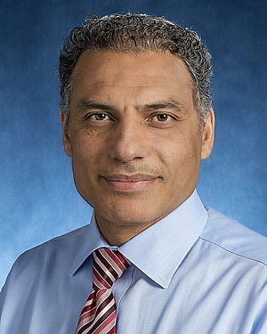 Khaled M Kebaish合作论文数Department of Otolaryngology-Head and Neck Surgery, School of Medicine, Johns Hopkins University273Themistocles S Protopsaltis合作论文数253Scheer Justin K合作论文数University of California246
 Vedat Deviren合作论文数University of California237Frank J Schwab合作论文数229
 Alan Daniels合作论文数Department of Molecular Pharmacology, Physiology, and Biotechnology, Brown University208Breton Line合作论文数207
 Michael Kelly合作论文数Division of Orthopedics & Scoliosis, Rady Children's Hospital-San Diego205Gum Jeffrey L合作论文数Northwestern University, Duke University Medical Center195D. Kojo Hamilton合作论文数194Robert K. Eastlack合作论文数188
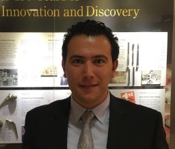 Bassel G. Diebo合作论文数New York University Langone Medical Center145
 Lawrence G Lenke合作论文数Department of Orthopedic Surgery, Columbia University Irving Medical Center;NewYork-Presbyterian Och Spine Hospital;NewYork-Presbyterian Morgan Stanley Children's Hospital111
 Themistocles Protopsaltis合作论文数Department of Neurosurgery, Grossman School of Medicine, New York University106
 Daniel M. Sciubba合作论文数Johns Hopkins University86Praveen V. Mummaneni合作论文数Dept. of Neurosurgery, Emory University85
 Oheneba Boachie-Adjei合作论文数FOCOS81Soroceanu Alex合作论文数80Dean Chou合作论文数79
 Sigurd Berven合作论文数Department of Orthopaedic Surgery, University of California, San Francisco76
 Brian J Neuman合作论文数Dept Orthopaed Surg, Johns Hopkins Univ75Soroceanu Alexandra合作论文数74Darryl Lau合作论文数Department of Neurosurgery, University of California at San Francisco68
 Stephen Lewis合作论文数Department of Surgery, Temerty Faculty of Medicine, University of Toronto65International Spine Study Group合作论文数60Frank L. Acosta合作论文数51
 Aaron Clark合作论文数50
 Michael Fehlings合作论文数Department of Surgery, Temerty Faculty of Medicine, University of Toronto;University Health Network;McEwen Centre for Regenerative Medicine;Toronto Western Hospital49Cole Bortz合作论文数46
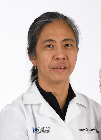 Leah Y. Carreon合作论文数Norton Leatherman Spine Center46
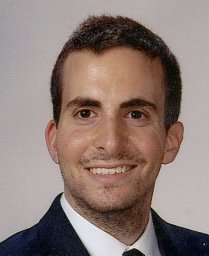 Safaee Michael合作论文数Departments of Neurological Surgery and.45Burch Shane合作论文数42Amit Jain (Jain A)合作论文数41Buell Thomas J合作论文数Department of Neurosurgery, University of Virginia40




