BACKGROUND AND OBJECTIVES:The purpose of this study was to analyze the impact of patient-specific rods on mechanical complications after adult thoracolumbar spinal deformity surgery. METHODS:A consecutive series was analyzed of 200 adult thoracolumbar spinal deformity patients. Half of the patients (n = 100) had surgery just before implementing patient-specific rods, and the other 100 patients had surgery immediately after adoption of patient-specific rods. All patients had instrumented fusions from the thoracic spine to the pelvis. Mechanical complications were defined as rod fracture, proximal junctional kyphosis, proximal junctional failure (revision surgery requiring proximal fusion extension), and/or revision surgery for rod fracture or pseudoarthrosis. Minimum follow-up was 2 years. RESULTS:A total of 200 patients were included with a median (IQR) age of 68 (61, 73), body mass index of 28 (25, 32), and 73% were female. The patients with patient-specific rods compared with those without had less preoperative kyphosis (39° vs 44°, P = .01), a higher median number of rods (4 vs 2, P = .04), more often a combined anterior-posterior approach (61% vs 46%, P = .047), more commonly a upper instrumented vertebra vertebroplasty (67% vs 45% P = .003), and postoperatively had less thoracic kyphosis (45% vs 49%, P = .02). Patient-specific rod patients had a lower rate of mechanical complications (29% vs 47%, P = .01), a lower rate of rod fracture (3% vs 16%, P = .003), and shorter operative times (215 minutes vs 250 minutes, P = .04). On multivariable analysis, the only 2 independent predictors of reducing the rate of mechanical complications were the utilization of patient-specific rods (odds ratio 0.47, P = .013, CI 0.26-0.85) and lower postoperative thoracic kyphosis (odds ratio 1.02, P = .04, CI 1.00-1.05). CONCLUSION:In a consecutive series of adult thoracolumbar spinal deformity patients, patient-specific rods reduced the odds of mechanical complications by over 50%. Adoption of patient-specific rods should be considered to optimize patient outcomes after adult spinal deformity surgery.
OBJECTIVE:There is no gold standard for assessing sarcopenia. The authors aimed to quantify agreement and reliability among common sarcopenia measures based on muscle function, body composition, and imaging characteristics in a cohort of adult spinal deformity (ASD) patients. METHODS:This was a cross-sectional study. Preoperative ASD patients at a single tertiary-care center underwent the following sarcopenia assessments: 2 functional muscle assessments (grip strength and gait speed), 3 bioelectrical impedance (BIA) measures (skeletal muscle index [SMI], phase angle, and extracellular water [ECW]/total body water [TBW] ratio), and 2 imaging-based assessments (psoas muscle index [PMI] and total psoas area [TPA]/vertebral body area [VBA] ratio). Spearman's correlation analysis was used to test for associations. Measurements were standardized into sex-specific z-scores. Bland-Altman analysis was used to quantify agreement and Cronbach's alpha analysis was used to quantify reliability between sarcopenia measures. RESULTS:Between 2017 and 2025, 272 ASD patients were enrolled in the study. Of these, 88 ASD patients (59.1% female, median age 67.7 years) completed all sarcopenia assessments and thus were included in the analysis. Most sarcopenia measures were weakly correlated, except for PMI and TPA/VBA, which were moderately correlated (ρ = 0.84, p < 0.001), and phase angle and ECW/TBW (ρ = -0.82, p < 0.001). Bland-Altman analysis demonstrated poor agreement between all pairs of sarcopenia measures. Cronbach's alpha analysis showed poor reliability (α < 0.7) between all pairs of sarcopenia measures except for PMI and TPA/VBA (α = 0.89). These results did not significantly change when all 272 patients with imputed missing data were included. CONCLUSIONS:The authors found low agreement and reliability between 7 commonly used sarcopenia measures, except for good reliability between PMI and TPA/VBA. These sarcopenia measures are not interchangeable and may not be measuring the same underlying clinical entity. This is the first study to quantify agreement and reliability between sarcopenia measures. Future studies are needed to determine which sarcopenia measures best predict clinical outcomes in ASD patients.
BACKGROUND CONTEXT:A common concern is that the stress induced by adult spinal deformity (ASD) surgery may cause a postoperative decrease in cognitive function, especially in the elderly patients with some component of cognitive impairment. On the other hand, it is possible that ASD surgery could stabilize cognitive function by increasing activity and decreasing pain. PURPOSE:Here, we evaluate the effect of ASD surgery on cognitive outcome in a prospective study. STUDY DESIGN/SETTING:This is a prospective study of patients undergoing ASD surgery at a single institution over a 5-year period. PATIENT SAMPLE:ASD patients treated with posterior spinal fusion of greater or equal to 7 vertebral segments for adult deformity were included. Only patients with 12 month follow up are included in this study. OUTCOME MEASURES:The primary outcome variable was performance on the Montreal Cognitive Assessment (MoCA) test of dementia and cognitive impairment, collected prospectively preoperatively and at 12-month follow-up. We also collected outcome metrics including the Oswestry Disability Index (ODI), Scoliosis Research Society questionnaire (SRS-22) with mental health (MH), activity (ACT), pain (P), and self-image (SI) subcomponents. Preoperative and postoperative morphine equivalent dose (MED) of narcotic medication was collected using patient surveys and verified using prescription data. METHODS:The primary outcome was assessed using a paired t-test. Further analyses included performing univariate and multivariable analyses comparing patients with improved versus nonimproved MoCA scores across demographic, radiographic, surgical, outcome data, and opioid usage. RESULTS:We enrolled 55 patients who met inclusion criteria. There was a significant increase in MoCA scores at 12-month follow-up compared to preoperative MoCA scores (p<.001). Overall, 60% of patients exhibited an increase in MoCA scores, and 47.2% met minimally clinically important difference (MCID). More severely cognitively impaired patients tended to improve to a greater degree than less severely impaired patients (p=.003). While there was no clear association between reduction in postoperative opioid use and cognitive improvement, we observed a possible association between postoperative delirium and cognitive decline among patients with baseline cognitive impairment (p=.01). CONCLUSIONS:Our prospective data suggests that ASD surgery is associated with an improvement in cognitive function at one-year follow-up. Further work is required to understand the drivers associated with cognitive improvement and worsening after ASD surgery.
OBJECTIVE:The purpose of this study was to evaluate whether level selection for a three-column osteotomy (3CO) impacts cervical deformity correction outcomes, including neurological, radiographic, and patient-reported outcomes. METHODS:A retrospective review was performed of patients who underwent a cervical or upper thoracic 3CO for cervical deformity correction by the senior author from 2008 to 2024. Collected outcome measures included neurological outcomes, mechanical complication rates, spinopelvic alignment, and patient-reported outcomes. The minimum follow-up was 1 year. RESULTS:One hundred fifteen patients were identified who underwent a cervical or upper thoracic 3CO for cervical deformity correction, of whom 77 met inclusion criteria for this study with a minimum follow-up of 1 year. The median age was 66 years, the median BMI was 27, and 43% of patients were male. Sixteen patients underwent a 3CO at the C7-T1 levels and 61 patients at T2-6 levels. Patients who underwent C7-T1 3CO were more likely to experience a new postoperative neurological deficit compared with those who underwent T2-6 3CO (56% vs 18%, p = 0.004), had less correction in their T1 slope (6° vs 18°, p = 0.027), had less correction in C2-T4 sagittal vertical axis (2.8 cm vs 4.9 cm, p = 0.043), and had a worse Neck Disability Index (NDI) score at 1 year compared with baseline (an increase of 7 vs a decrease of 12, p = 0.033). CONCLUSIONS:Cervical deformity patients who underwent a 3CO at C7-T1 had a higher rate of postoperative neurological deficits, less radiographic correction, and worse NDI scores at 1 year compared with patients who underwent a 3CO from T2 to T6. Although 3CO level selection is multifactorial and patient specific, surgeons should consider a 3CO level caudal to T1 when feasible.
This narrative review focuses on advances in clinical outcomes following adult cervical spinal deformity (CSD) surgery over the past decade. Progress is highlighted in 4 domains: radiographic alignment targets, preoperative risk stratification, surgical techniques, and long-term operative outcomes. The predominant trend across these advances has been towards a patient-specific approach for addressing cervical deformity. We anticipate that future progress will continue this patient-specific approach with refined preoperative optimization, patient-specific biomechanical modeling including digital twins with associated tailored instrumentation, and widespread adoption of artificial intelligence (AI) to predict postoperative outcomes.
OBJECTIVE:The purpose of this study was to determine risk factors for distal junctional failure (DJF) following three-column osteotomy (3CO) for the correction of cervical deformity. METHODS:A retrospective review was performed of patients who underwent a cervical or upper thoracic 3CO for cervical deformity correction by the senior author from 2008 to 2023. The main outcome of interest was DJF, defined as revision surgery with extension of the distal end of the fusion construct. Patients were excluded if the lowest instrumented vertebra (LIV) was the sacrum/pelvis, and if patients had prior autofusion throughout the distal part of the spine to the sacrum (e.g., ankylosing spondylitis). The minimum follow-up duration was 1 year. RESULTS:One hundred fourteen patients were identified who underwent a cervical or upper thoracic 3CO for cervical deformity correction, 41 of whom met inclusion criteria for this study. The median patient age was 66 years, median BMI was 27, and 61% were male. Ten patients (24%) experienced DJF, requiring reoperation and distal extension of the construct to the pelvis in 9 cases and to L1 in 1 case. On univariable analysis, patients who experienced DJF compared with those who did not were more likely to be female (80% vs 35%, p = 0.007), had lower LIV Hounsfield units (HUs; 127 vs 167, p = 0.041), were less likely to have an LIV in a small autofused segment (10% vs 45%, p = 0.02), had an LIV that was closer to the first lordotic level (one level above vs three, p = 0.043), and had a longer length of fusion (17 vs 13 levels, p = 0.033). A stepwise multivariable regression model showed that having an LIV closer to the first lordotic vertebra was the only statistically significant predictor of DJF (OR 0.49, p = 0.013) and low LIV HUs did not reach statistical significance (OR 0.97, p = 0.09). CONCLUSIONS:Patients with cervical deformity undergoing a 3CO are at higher risk for DJF with constructs terminating near the first lordotic vertebra. While LIV selection is complex and patient specific, choosing an LIV at least two levels above the first lordotic vertebra may help prevent DJF.
To explore the impact of different lumbar pedicle subtraction osteotomy (L-PSO) levels on Global Alignment and Proportion (GAP) scores. Adults at a single center who underwent lumbar PSOs with revision instrumentation [thoracolumbar junction (T9-L1) to pelvis] and a minimum 2-year follow-up were reviewed. The patients were divided by level of PSO (L2, L3, L4, and L5) and compared with respect to demographic and surgical data, sagittal parameters, GAP scores, and mechanical complications requiring revision operations. 152 patients (average age 64.4 ± 10.6 years, average follow-up 9.0 ± 4.1 years) were included for analysis. L3 (40.8
INTRODUCTION: Three-column osteotomy (3CO) is a powerful corrective technique in adult spinal deformity. Unfortunately, they carry high complication rates including mechanical complications of rod fracture and pseudoarthrosis. Due to the rapidly aging population, this intervention will only increase in use. METHODS: Single high-volume academic institution retrospective review of an experienced surgeon’s database. Patients with 3CO of the lumbar spine performed by the senior author were identified. Collected data included demographics, BMI, diagnosis, past surgeries, neurological assessments, smoking status, comorbidities, operative time, EBL, LOS, complications, and radiographic measurements. RESULTS: A total of 196 patients were identified with 78 (39.8%) 4-rod, 24 (12.2%) 5-rod, 54 (27.6%) 6-rod, and 40 (20.4%) 7-rod constructs. There was statistical significance in mechanical complications with 4-rod constructs having greater complications (p=0.010). This was primarily due to rod fractures (p<0.001) and, secondarily, due to pseudoarthrosis. There was also statistical significance in the change of lumbar lordosis from immediate postoperative radiographs to follow-up radiographs 4- and 7-rod constructs with 4-rod constructs losing 6.2 degrees compared to 1.8 degrees in 7-rod constructs (p=0.009). CONCLUSIONS: We found that the number of rods across a lumbar 3CO site can affect the number of mechanical complications. We also found that the number of rods prevents the loss of lordosis over time following correction. We recommend using a 7-rod construct for lumbar 3CO to minimize mechanical complications and loss of lordosis over time.
BACKGROUND:Hip osteoarthritis (OA) is common in patients with adult spinal deformity (ASD). Limited data exist on the prevalence of hip OA in patients with ASD, or on its impact on baseline and postoperative alignment and patient-reported outcome measures (PROMs). Therefore, this paper will assess the prevalence and impact of hip OA on alignment and PROMs. METHODS:Patients with ASD who underwent L1-pelvis or longer fusions were included. Two independent reviewers graded hip OA with the Kellgren-Lawrence (KL) classification and stratified it by severity into non-severe (KL grade 1 or 2) and severe (KL grade 3 or 4). Radiographic parameters and PROMs were compared among 3 patient groups: Hip-Spine (hip KL grade 3 or 4 bilaterally), Unilateral (UL)-Hip (hip KL grade 3 or 4 unilaterally), or Spine (hip KL grade 1 or 2 bilaterally). RESULTS:Of 520 patients with ASD who met inclusion criteria for an OA prevalence analysis, 34% (177 of 520) had severe bilateral hip OA and unilateral or bilateral hip arthroplasty had been performed in 8.7% (45 of 520). A subset of 165 patients had all data components and were examined: 68 Hip-Spine, 32 UL-Hip, and 65 Spine. Hip-Spine patients were older (67.9 ± 9.5 years, versus 59.6 ± 10.1 years for Spine and 65.8 ± 7.5 years for UL-Hip; p < 0.001) and had a higher frailty index (4.3 ± 2.6, versus 2.7 ± 2.0 for UL-Hip and 2.9 ± 2.0 for Spine; p < 0.001). At 1 year, the groups had similar lumbar lordosis, yet the Hip-Spine patients had a worse sagittal vertebral axis (SVA) measurement (45.9 ± 45.5 mm, versus 25.1 ± 37.1 mm for UL-Hip and 19.0 ± 39.3 mm for Spine; p = 0.001). Hip-Spine patients also had worse Veterans RAND-12 Physical Component Summary scores at baseline (25.7 ± 9.3, versus 28.7 ± 9.8 for UL-Hip and 31.3 ± 10.5 for Spine; p = 0.005) and 1 year postoperatively (34.5 ± 11.4, versus 40.3 ± 10.4 for UL-Hip and 40.1 ± 10.9 for Spine; p = 0.006). CONCLUSIONS:This study of operatively treated ASD revealed that 1 in 3 patients had severe hip OA bilaterally. Such patients with severe bilateral hip OA had worse baseline SVA and PROMs that persisted 1 year following ASD surgery, despite correction of lordosis. LEVEL OF EVIDENCE:Prognostic Level III. See Instructions for Authors for a complete description of levels of evidence.
BACKGROUND CONTEXT Adult spinal deformity necessitates surgical intervention to restore spinal alignment and prevent associated complications. The Global Alignment and Proportion (GAP) scoring system was created to offer insight into predicting mechanical complications. Lumbar pedicle subtraction osteotomies (PSOs) have been extensively used to correct sagittal-plane deformities and restore proper alignment. PURPOSE To explore the impact of different PSO levels on the GAP score in adult patients with spinal deformities. STUDY DESIGN/SETTING Retrospective cohort analysis at a single academic medical center. PATIENT SAMPLE Adults at a single center who underwent posterior instrumented fusions from the thoracolumbar junction (T9-L1) and included a lumbar PSO between 2008 and 2019. OUTCOME MEASURES Demographics, operative details, and the following preop and postop radiographic parameters: pelvic incidence (PI), sacral slope, L4-S1 lordosis, L1-S1 lordosis, and global tilt. The total GAP score and its subdomains were derived, including relative pelvic version (RPV), lordosis distribution index (LDI), relative lumbar lordosis (RLL), and relative spinopelvic alignment (RSA). METHODS A retrospective analysis of consecutive adults at a single center who underwent lumbar PSOs between 2008 and 2019 were reviewed. Inclusion criteria included revision operations with instrumentation from the pelvis to the thoracolumbar (TL) junction (T9-T1) with minimum 2-year follow-up. Patients were divided by level of PSO (L2, L3, L4, L5) and compared with respect to demographic, surgical, and the following preop and postop radiographic parameters: pelvic incidence (PI), sacral slope, L4-S1 lordosis, L1-S1 lordosis, and global tilt. From these measurements, the total GAP score and its subdomains were derived, including relative pelvic version (RPV), lordosis distribution index (LDI), relative lumbar lordosis (RLL), and relative spinopelvic alignment (RSA). Student's T-test and Chi-square tests were used for statistical comparisons. RESULTS A total of 152 patients (average age 64.4 ± 10.6 years, 43% female; mean BMI 29.7 ± 5.8) were included for analysis. The majority of PSOs were at the L3 (n=62; 40.8%) and L4 level (n=69; 45.4%), with the minority being at L2 (n=7; 4.6%) and L5 (n=14; 9.2%). Average follow-up was 9.0 ± 4.1 years with L2 and L3 PSOs having significantly longer follow-up. Total preop and postop GAP scores were similar between all groups. However, postop LDI scores were significantly greater (i.e. higher prevalence of moderate and severe hypolordotic maldistributions) in patients who underwent higher PSOs (L2 and L3). This was a result of lower PSOs (L4 and L5) improving L4-S1 lordosis to a greater extent than higher PSOs (L2 and L3). Rates of revision operations for proximal junctional failure (PJF) and rod fractures were found to be higher in patients who underwent L2 and L3 PSOs, with only PJF rates being significantly different. CONCLUSIONS In adults undergoing revision multi-level thoracolumbar instrumented fusions from the TL junction to the pelvis, lower PSOs (L4 and L5) improved L4-S1 lordosis to a greater extent than higher PSOs (L2 and L3), which in turn resulted in better distribution of lordosis and lower rates of revision operations for mechanical complications, particularly for PJF. FDA Device/Drug Status This abstract does not discuss or include any applicable devices or drugs.
To assess the effect of various pelvic fixation techniques and number of rods on biomechanics of the proximal junction of long thoracolumbar posterior instrumented fusions. A validated spinopelvic finite-element (FE) model was instrumented with L5–S1 ALIF and one of the following 9 posterior instrumentation configurations: (A) one traditional iliac screw bilaterally (“2 Iliac/2 Rods”); (B) T10 to S1 (“Sacral Only”); (C) unilateral traditional iliac screw (“1 Iliac/2 Rods”); (D) one traditional iliac screw bilaterally with one midline accessory rod (“2 Iliac/3 rods”); (E) S2AI screws connected directly to the midline rods (“2 S2AI/2 Rods”); and two traditional iliac screws bilaterally with two lateral accessory rods connected to the main rods at varying locations (F1: T10–11, F2: T11–12, F3: T12–L1, F4: L1–2) (“4 Iliac/4 Rods”). Range of motions (ROM) at T10–S1 and T9–T10 were recorded and compared between models. The T9–T10 intradiscal pressures and stresses of the T9–10 disc’s annulus in addition to the von Mises stresses of the T9 and T10 vertebral bodies were recorded and compared. For T10–S1 ROM, 4 iliac/4 rods had lowest ROM in flexion and extension, while 2 S2AI/2 rods showed lowest ROM in rotation. Constructs with 3 or 4 rods had lower stresses on the primary rods compared to 2-rod constructs. At the proximal adjacent disc (T9–10), 4 iliac/4 rods showed lowest ROM, lowest intradiscal pressures, and lowest annular stress in all directions (most pronounced in flexion–extension). Under flexion and extension, 4 iliac/4 rods also showed the lowest von Mises stresses on the T10 vertebral body but the highest stresses on the T9 vertebral body. Dual iliac screws with 4 rods across the lumbosacral junction and extending to the thoracolumbar junction demonstrated the lowest T10–S1 ROM, the lowest adjacent segment disc (T9–T10) ROM, intradiscal pressures, and annular stresses, and the lowest UIV stresses, albeit with the highest UIV + 1 stresses. Additional studies are needed to confirm whether these biomechanical findings dictate clinical outcomes and effect rates of proximal junctional kyphosis and failure.
OBJECTIVE This study aimed to assess whether elderly patients (aged ≥ 70 years) face an elevated risk of complications following pedicle subtraction osteotomy (PSO) for adult spinal deformity (ASD) compared with younger patients (< 70 years) and to evaluate if clinical and radiological outcomes differ between these age groups. METHODS A retrospective analysis of 513 patients undergoing PSO for ASD by a single surgical team between January 2006 and January 2023 was conducted. Patients were categorized by age (≥ 70 years and < 70 years). Data on clinical, demographic, comorbidity, and radiographic details were collected and compared between the groups. For health-related quality of life assessment, the authors recorded the Oswestry Disability Index (ODI), numeric rating scale (NRS), and Scoliosis Research Society–22 revised (SRS-22r) scores preoperatively and at 6 weeks and 1 year postoperatively. Perioperative complications included major (neurological deficit, death, acute myocardial infarction, stroke), minor (ileus, arrhythmia, delirium), and intraoperative (durotomy, vascular injury). RESULTS Of 513 patients, 412 were included in the study. Clinical outcomes, as measured by NRS, ODI, and SRS-22r scores, were comparable between groups, with both groups showing significant improvements postoperatively. Radiographic outcomes also showed significant and comparable improvements in sagittal balance and spinopelvic harmony in both groups. Deformity corrections were also well maintained at 1 year postoperatively. The elderly group (mean age 75.48 years) had a higher rate of perioperative complications (44.64%) than the younger group (mean age 59.60 years; 30.33%) (p = 0.0030), primarily minor complications such as delirium and arrhythmia (16.07% vs 8.61%, p = 0.0279). There was no significant difference between groups regarding the major complication rate (elderly group: 20.83% vs younger group: 14.34%, p = 0.1087), intraoperative complication rate (2.98% vs 3.69%, p = 0.6949), short-term complication rate (10.12% vs 8.20%, p = 0.5024), mechanical complication rate (30.95% vs 32.79%, p = 0.6949), and reoperation rate due to mechanical complications (38.46% vs 43.75% p = 0.5470). CONCLUSIONS Elderly patients undergoing PSO for ASD experience a higher rate of minor complications but can achieve clinical and radiological outcomes that are comparable to those of younger patients. The authors found no significant increase in major, intraoperative, short-term, or mechanical complication rates and their subsequent reoperation rates among the elderly. These findings underscore the effectiveness of PSO in improving the quality of life for patients with ASD across age groups, emphasizing the critical role of personalized perioperative management in enhancing outcomes and minimizing risks for all patients.
Background: The objective of this study was to evaluate if imbalance influences complication rates, radiological outcomes, and patient-reported outcomes (PROMs) following adult spinal deformity (ASD) surgery. Methods: ASD patients with baseline and 2-year radiographic and PROMs were included. Patients were grouped according to whether they answered yes or no to a recent history of pre-operative loss of balance. The groups were propensity-matched by age, pelvic incidence–lumbar lordosis (PI-LL), and surgical invasiveness score. Results: In total, 212 patients were examined (106 in each group). Patients with gait imbalance had worse baseline PROM measures, including Oswestry disability index (45.2 vs. 36.6), SF-36 mental component score (44 vs. 51.8), and SF-36 physical component score (p < 0.001 for all). After 2 years, patients with gait imbalance had less pelvic tilt correction (−1.2 vs. −3.6°, p = 0.039) for a comparable PI-LL correction (−11.9 vs. −15.1°, p = 0.144). Gait imbalance patients had higher rates of radiographic proximal junctional kyphosis (PJK) (26.4% vs. 14.2%) and implant-related complications (47.2% vs. 34.0%). After controlling for age, baseline sagittal parameters, PI-LL correction, and comorbidities, patients with imbalance had 2.2-times-increased odds of PJK after 2 years. Conclusions: Patients with a self-reported loss of balance/unsteady gait have significantly worse PROMs and higher risk of PJK.
Study Design. Cross-sectional survey. Objective. To assess the reliability of a proposed novel classification system for thoracic disc herniations (TDHs). Summary of Background Data. TDHs are complex entities varying substantially in many factors, including size, location, and calcification. To date, no comprehensive system exists to categorize these lesions. Methods. Our proposed system classifies 5 types of TDHs using anatomical and clinical characteristics, with subtypes for calcification. Type 0 herniations are small (≤40% of spinal canal) TDHs without significant spinal cord or nerve root effacement; type 1 are small and paracentral; type 2 are small and central; type 3 are giant (>40% of spinal canal) and paracentral; and type 4 are giant and central. Patients with types 1-4 TDHs have correlative clinical and radiographic evidence of spinal cord compression. Twenty-one US spine surgeons with substantial TDH experience rated 10 illustrative cases to determine the system’s reliability. Interobserver and intraobserver reliability were determined using the Fleiss kappa coefficient. Surgeons were also surveyed to obtain consensus on surgical approaches for the various TDH types. Results. High agreement was found for the classification system, with 80% (range 62-95%) overall agreement and high interrater and intrarater reliability (kappa 0.604 [moderate to substantial agreement] and kappa 0.630 [substantial agreement], respectively). All surgeons reported nonoperative management of type 0 TDHs. For type 1 TDHs, most respondents (71%) preferred posterior approaches. For type 2 TDHs, responses were roughly equivalent for anterolateral and posterior options. For types 3 and 4 TDHs, most respondents (72% and 68%, respectively) preferred anterolateral approaches. Conclusions. This novel classification system can be used to reliably categorize TDHs, standardize description, and potentially guide the selection of surgical approach. Validation of this system with regard to treatment and clinical outcomes represent lines of future study.
Background Assess correlation between preoperative cervical sagittal alignment (T1 slope [T1S] and C2-C7 cervical sagittal vertical axis [cSVA]) and postoperative cervical sagittal balance after posterior cervical laminoplasty. Methods Consecutive patients who underwent laminoplasty at a single institution with >6 weeks postoperative follow-up were divided into 4 groups based on preoperative cSVA and T1S (Group 1: cSVA <4 cm/T1S <20°; Group 2: cSVA ≥4 cm/T1S ≥20°; Group 3: cSVA <4 cm/T1S ≥20°; Group 4: cSVA <4 cm/T1S <20°). Radiographic analyses were conducted at 3 timepoints, and changes in cSVA, C2-C7 cervical lordosis (CL), and T1S -CL were compared. Results A total of 214 patients met inclusion criteria (28 patients had cSVA <4 cm/T1S <20° [Group 1]; 47 patients had cSVA ≥4 cm/T1S ≥20° [Group 2]; 139 patients had cSVA <4 cm/T1S ≥20° [Group 3]). No patients had cSVA ≥4 cm/T1S <20° (Group 4). Patients either had a C4-C6 (60.7%) or C3-C6 (39.3%) laminoplasty. Mean follow-up was 1.6 ± 1.32 years. For all patients, mean cSVA increased 6 mm postoperatively. cSVA significantly increased postoperatively for both groups with a preoperative cSVA <4 cm (ie, Groups 1 and 3 [P < 0.01]). For all patients, mean CL decreased 2° postoperatively. Groups 1 and 2 had significant differences in preoperative CL but nonsignificant differences at 6 weeks (P = 0.41) and last follow-up (P = 0.06). Conclusion Cervical laminoplasty resulted in a mean decrease in CL. Patients with high preoperative T1S, irrespective of cSVA, were at risk of loss of CL postoperatively. While patients with low preoperative T1S and cSVA <4 cm experienced a decrease in global sagittal cervical alignment, CL was not jeopardized. Clinical Relevance The results of this study may facilitate preoperative planning for patients undergoing posterior cervical laminoplasty. Level of Evidence 3.
BACKGROUND: For transforaminal lumbar interbody fusion (TLIF), there are equally good open and minimally invasive surgery (MIS) options. OBJECTIVE: To determine if frailty has a differential effect on outcome for open vs MIS TLIF. METHODS: We performed a retrospective review of 115 TLIF surgeries (1-3 levels) for lumbar degenerative disease performed at a single center; 44 MIS transforaminal interbody fusions and 71 open TLIFs were included. All patients had at least a 2-year follow up, and any revision surgery during that time was recorded. The Adult Spinal Deformity Frailty Index (ASD-FI) was used to separate patients into nonfrail (ASD-FI < 0.3) and frail (ASD-FI > 0.3) cohorts. The primary outcome variables were revision surgery and discharge disposition. Univariate analyses were performed to reveal associations in demographic, radiographic, and surgical data with the outcome variables. Multivariate logistic regression was used to assess independent predictors of outcome. RESULTS: Frailty uniquely predicted both reoperation (odds ratio 8.1, 95% CI 2.5-26.1, P = .0005) and discharge to a location other than home (odds ratio 3.9, 95% CI 1.2-12.7, P = .0239). Post hoc analysis indicated that frail patients undergoing open TLIF had a higher revision surgery rate (51.72%) compared with frail patients undergoing MIS-TLIF (16.7%). Nonfrail patients undergoing open and MIS TLIF had a revision surgery rate of 7.5% and 7.7%, respectively. CONCLUSION: Frailty was associated with increased revision rate and increased probability to discharge to a location other than home after open transforaminal interbody fusions, but not MIS transforaminal interbody fusions. These data suggest that patients with high frailty scores may benefit from MIS-TLIF procedures.
Purpose To define the prevalence, characteristics, and treatment approach for proximal junction failure secondary to odontoid fractures in patients with prior C2-pelvis posterior instrumented fusions (PSF). Methods A single institution’s database was queried for multi-level fusions (6+ levels), including a cervical component. Posterior instrumentation from C2-pelvis and minimum 6-month follow-up was inclusion criteria. Patients who sustained dens fractures were identified; each fracture was subdivided based on Anderson & D’Alonzo and Grauer’s classifications. Comparisons between the groups were performed using Chi-square and T tests. Results 80 patients (71.3% female; average age 68.1 ± 8.1 years; 45.0% osteoporosis) were included. Average follow-up was 59.8 ± 42.7 months. Six patients (7.5%) suffered an odontoid fracture post-operatively. Cause of fracture in all patients was a mechanical fall. Average time to fracture was 23 ± 23.1 months. Average follow-up after initiation of fracture management was 5.84 ± 4 years (minimum 1 year). Three patients sustained type IIA fractures one of which had a concomitant unilateral C2 pars fracture. Three patients sustained comminuted type III fractures with concomitant unilateral C2 pars fractures. Initial treatment included operative care in 2 patients, and an attempt at non-operative care in 4. Non-operative care failed in 75% of patients who ultimately required revision with proximal extension. All patients with a concomitant pars fracture had failure of non-operative care. Patients with an intact pars were more stable, but 50% required revision for pain. Conclusions In this 11-year experience at a single institution, the prevalence of odontoid fractures above a C2-pelvis PSF was 7.5%. Fracture morphology varied, but 50% were complex, comminuted C2 body fractures with concomitant pars fractures. While nonoperative management may be suitable for type II fractures with simple patterns, more complex and unstable fractures likely benefit from upfront surgical intervention to prevent fracture displacement and neural compression. As all fractures occurred secondary to a mechanical fall, inpatient and community measures aimed to minimize risk and prevent mechanical falls would be beneficial in this high-risk group.
OBJECTIVE Vertebral osteomyelitis is a rare complication of coccidioidomycosis infection. Surgical intervention is indicated when there is failure of medical management or presence of neurological deficit, epidural abscess, or spinal instability. The relationship between timing of surgical intervention and recovery of neurological function has not been previously described. The purpose of this study was to investigate if the duration of neurological deficits at presentation affects neurological recovery after surgical intervention. METHODS This was a retrospective study of all patients diagnosed with coccidioidomycosis involving the spine at a single tertiary care center between 2012 and 2021. Data collected included patient demographics, clinical presentation, radiographic information, and surgical intervention. The primary outcome was change in neurological examination after surgical intervention, quantified according to the American Spinal Injury Association Impairment Scale. The secondary outcome was the complication rate. Logistic regression was used to test if the duration of neurological deficits was associated with improvement in the neurological examination after surgery. RESULTS Twenty-seven patients presented with spinal coccidioidomycosis between 2012 and 2021; 20 of these patients had vertebral involvement on spinal imaging with a median follow- up of 8.7 months (IQR 1.7-71.2 months). Of the 20 patients with vertebral involvement, 12 (60.0%) presented with a neurological deficit with a median duration of 20 days (range 1-61 days). Most patients presenting with neurological deficit (11/12, 91.7%) underwent surgical intervention. Nine (81.2%) of these 11 patients had an improved neurological examination after surgery and the other 2 had stable deficits. Seven patients had improved recovery sufficient to improve by 1 grade according to the AIS. The duration of neurological deficits on presentation was not significantly associated with neurological improvement after surgery (p = 0.49, Fisher's exact test). CONCLUSIONS The duration of neurological deficits on presentation should not deter surgeons from operative intervention in cases of spinal coccidioidomycosis.
STUDY DESIGN:Retrospective comparative study.OBJECTIVE:To assess the relationship of fusion mass bone density on computed tomography (CT) and the development of rod fractures (RFs) and proximal junctional kyphosis (PJK).SUMMARY OF BACKGROUND DATA:Few studies have evaluated the relationship of fusion mass bone density to mechanical complications.MATERIALS AND METHODS:A retrospective review of adult spinal deformity patients who underwent thoracolumbar three-column osteotomy from 2007 to 2017 was performed. All patients underwent routine 1-year CT imaging and had at least 24 months follow-up. Posterior fusion mass bone density was evaluated by measuring hounsfield unit (HU) on CT in three different regions [upper instrumented vertebra (UIV), lower instrumented vertebra, and osteotomy site], and were compared between patients with and without mechanical complications.RESULTS:A total of 165 patients (63.2 years, 33.5% male) were included. Overall PJK rate was 18.8%, and 35.5% of these underwent PJK revision. There was significantly lower density of posterior fusion mass at the UIV in patients who experienced PJK compared with patients without PJK (431.5HU vs. 537.4HU, P =0.026). Overall RF rate was 34.5% and 61.4% of these underwent revision for RFs. Among 57 patients with RFs, 71.9% had pseudarthrosis. Fusion mass density did not differ between patients with or without RFs. However, in RF patients with pseudarthrosis, there was significantly higher bone mass density near the osteotomy compared with those without pseudarthrosis (515.7HU vs. 354.2HU, P =0.012). There were no differences in radiographic sagittal measures between the patients with and without RF or PJK.CONCLUSIONS:Patients with PJK tend to have less dense posterior fusion mass at the UIV. Fusion mass density does not correlate with RF, but greater bone density near the osteotomy was correlated with accompanying pseudarthrosis in patients with RFs. Assessing density of posterior fusion mass on CT may be helpful in assessing risk for PJK and provide insight as to the causes of RFs.
 Christopher P. Ames合作论文数School of Medicine, University of California, San Francisco;Helen Diller Family Comprehensive Cancer Center, University of California, San Francisco;Weill Institute for Neurosciences, University of California, San Francisco;UCSF Medical Center237
 Christopher Ignatius Shaffrey合作论文数Department of Neurosurgery, School of Medicine, Duke University125
 Virginie Lafage合作论文数Hosp Special Surg, Dept Orthoped, 535 E 70th St, New York, NY 10021 USA118
 Justin S Smith合作论文数115
 Gregory Mundis合作论文数Scripps Health107
 Shay Bess合作论文数106
 Sigurd Berven合作论文数Department of Orthopaedic Surgery, University of California, San Francisco101
 Eric O. Klineberg合作论文数University of California Davis Medical Center, University of California92Richard Hostin合作论文数87Scheer Justin K合作论文数University of California78
 Munish C. Gupta合作论文数Department of Orthopedics, Washington University in St. Louis, School of Medicine71Douglas C. Burton合作论文数70Schwab Frank合作论文数64Praveen V. Mummaneni合作论文数Dept. of Neurosurgery, Emory University61Burch Shane合作论文数60
 Robert Hart合作论文数Swedish Medical Center60Dean Chou合作论文数57
 Serena Hu合作论文数Neurosurgery Department, Stanford University;Department of Orthopaedic Surgery, Stanford University54Bobby Tay合作论文数50
 Han Jo Kim合作论文数Hospital for Special Surgery;Orthopaedic Surgery Department, Weill Cornell Medical College48
 Khaled M Kebaish合作论文数Department of Otolaryngology-Head and Neck Surgery, School of Medicine, Johns Hopkins University47Robert K. Eastlack合作论文数45Frank J Schwab合作论文数42Juan S. Uribe合作论文数42Themistocles S Protopsaltis合作论文数38
 Behrooz A Akbarnia合作论文数Department of Orthopaedic Surgery, University of California, San Diego;Department of Orthopaedic Surgery, Rady Children's Hospital37Darryl Lau合作论文数Department of Neurosurgery, University of California at San Francisco33Lafage Renaud合作论文数Department of Orthopaedic Surgery, New York University Hospital for Joint Diseases32
 Michael Kelly合作论文数Division of Orthopedics & Scoliosis, Rady Children's Hospital-San Diego32D. Kojo Hamilton合作论文数32Peter G Passias合作论文数31
 David O. Okonkwo合作论文数Neurotrauma Clinical Trials Center, Department of Neurological Surgery, School of Medicine, University of Pittsburgh30
 Aaron Clark合作论文数30
 Paul Park合作论文数Semmes Murphey Clinic30
 Oheneba Boachie-Adjei合作论文数FOCOS29
 Adam Kanter合作论文数29
 Neel Anand合作论文数Los Angeles28
 Kai-Ming Fu合作论文数Department of Neurological Surgery, Weill Cornell Medical College27Nguyen Stacie合作论文数27Theologis Alexander A合作论文数26Michael O'Brien合作论文数24
 Alan Daniels合作论文数Department of Molecular Pharmacology, Physiology, and Biotechnology, Brown University24
 Richard G. Fessler合作论文数Department of Neurosurgery, RUSH Medical College, Rush University23Murat Pekmezci合作论文数23Breton Line合作论文数23
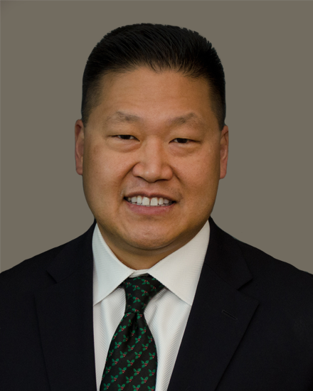 Michael Yung-Shun Wang合作论文数Department of Neurological Surgery, Miller School of Medicine, University of Miami;University of Miami Hospital20Gum Jeffrey L合作论文数Northwestern University, Duke University Medical Center18
 Pierce Nunley合作论文数18




