Background Traumatic brain injury (TBI) is a leading cause of death and disability worldwide, with secondary brain injury significantly contributing to poor outcomes. Early identification of patients at risk remains challenging in the prehospital environment due to limited diagnostic capability. Portable neuromonitoring technologies may enhance early assessment, however, their feasibility and impact on prehospital workflow are not well established. This study aimed to evaluate the integration of novel neuromonitoring devices into standard Helicopter Emergency Medical Service (HEMS) TBI care within a simulated environment. Methods A high-fidelity simulation study was conducted between August 2024 and May 2025 at a UK HEMS base. Twenty-four clinician pairs (critical care paramedics and doctors) participated in simulated TBI scenarios. Six portable neuromonitoring devices were introduced across 16 simulations, with 8 control simulations conducted without devices. The primary outcomes extracted from video recordings were phase of care for device deployment, time to data acquisition, and interruptions encountered. Secondary outcomes included the temporal impact on prehospital phases of care and standard clinical interventions. Results Neuromonitoring devices were successfully deployed in all 16 simulations and used almost exclusively during consecutive prehospital activity. Devices were predominantly used either prior to prehospital emergency anaesthesia or during transport. Data acquisition was achieved in all but three instances of device use, and time to data acquisition varied by device between 1.4–5.3 minutes. Interruptions occurred across all devices, most commonly due to user-related factors. There were no significant differences observed in the duration of prehospital phases of care between device and control groups including elapsed scene time until anaesthesia (device: 25.7mins [IQR 22.0-29.6], control: 25.4mins [IQR 22.8–28.1], p > 0.99). Conclusion Portable neuromonitoring devices can be integrated into simulated prehospital TBI care without delaying critical interventions or prolonging scene time. While deployment is generally successful, variability in duration of device use and associated interruptions highlight the importance of targeted training for optimisation of user–device integration. These findings support further investigation into real-world application and the potential of neuromonitoring to improve early TBI assessment and management.
Background:Traumatic acute subdural haematomas often require surgical evacuation via craniotomy or decompressive craniectomy. Decompressive craniectomy may prevent intracranial hypertension; however, it is unclear whether it is associated with better outcomes. Objective:Multicentre, pragmatic, parallel-group randomised trial to compare the clinical and cost-effectiveness of decompressive craniectomy versus craniotomy for evacuation of acute subdural haematomas. Design:International, multicentre, pragmatic, parallel-group randomised trial with additional observational arm. Setting:Hospitals with neurosurgical services in the UK and internationally. Participants:Patients aged ≥ 16 years, with a diagnosis of acute subdural haematomas on a computed tomography scan that required evacuation with a large bone flap either by craniotomy or decompressive craniectomy according to the opinion of the admitting neurosurgeon. Interventions:The enrolled patients underwent acute subdural haematoma evacuation in the operating room under general anaesthesia. A large bone flap ipsilateral to the haematoma was raised, the dura opened and the haematoma evacuated. Other haematomas, such as contusions, were evacuated at the discretion of the surgeon. If clinically appropriate randomisation occurred, the bone flap was either replaced (craniotomy) or not replaced (decompressive craniectomy). Patients who could not be randomised were followed up in the observational arm. Main outcome measures:Primary outcome measure was the extended Glasgow Outcome Scale assessed 12 months post injury. An economic evaluation (based on UK participants) was undertaken to estimate the cost-effectiveness of craniotomy compared to decompressive craniectomy. Results:Four hundred and fifty patients were randomised: 228 to craniotomy and 222 to craniectomy - with the common odds ratio for the differences across the Glasgow Outcome Scale scores of 0.85 [95% confidence interval (0.6 to 1.18; p = 0.324)]. The results were similar at 6 months. At 12 months, death occurred in 30.2% of the craniotomy group versus 32.2% of the decompressive craniectomy group, vegetative state occurred in 2.3% versus 2.8%, and good recovery occurred in 25.6% and 19.9%, respectively. In the observed cohort, those who had a decompressive craniectomy had significantly worse outcomes at 6 and 12 months, but their baseline characteristics were different. Limitations:Clinicians were not blinded to the trial groups. Decompressive craniectomy was performed in 8.8% of patients allocated to the craniotomy group, and 5.4% of patients allocated to the decompressive craniectomy group underwent craniotomy. Intraoperative non-adherence with allocation did not influence the primary analysis, which was based on the intention-to-treat principle. Conclusions:Among patients undergoing evacuation of acute subdural haematomas, the outcomes were similar in both groups. Additional surgery was required in a higher proportion of patients in the craniotomy group, but more wound complications occurred in the decompressive craniectomy group. Future work:Long-term outcomes of patients following decompressive craniectomy, timing and impact of cranial reconstruction on a patient's rehabilitation. Trial registration:This trial is registered as ISRCTN87370545. Funding:This award was funded by the National Institute for Health and Care Research (NIHR) Health Technology Assessment programme (NIHR award ref: 12/35/57) and is published in full in Health Technology Assessment; Vol. 30, No. 66. See the NIHR Funding and Awards website for further award information.
Traumatic brain injury (TBI) affects approximately 70 million individuals annually worldwide, often leading to significant morbidity and mortality. Intracranial pressure (ICP) monitoring plays a vital role in managing severe TBI and other conditions associated with intracranial hypertension by guiding timely medical interventions. However, invasive ICP monitoring requires drilling the skull to place a sensor transducer inside the head, also called a probe. Noninvasive ICP monitoring offers the potential to monitor patients during the critical "golden hour," when timely interventions can significantly influence outcomes. Additionally, it enables continuous surveillance for patients who are not candidates for invasive bolt placement. However, a reliable, continuous, noninvasive ICP measurement remains aspirational despite extensive research efforts. Crainio has developed a noninvasive ICP monitoring system utilising an optical sensor adhered to the forehead. This monitor collects cerebral photoplethysmography signals to estimate ICP changes and detect hypertension, enabling early management and improved patient outcomes. This feasibility study aims to improve the diagnostic precision of Crainio’s algorithm by augmenting the existing dataset, which currently comprises data from 40 TBI patients. A total of 54 participants will be enrolled over one year. Data from simultaneous recordings from the optical sensor and invasive ICP monitors create a more robust dataset to refine and optimise the algorithm for greater clinical accuracy. Additionally, the study will investigate the influence of potential confounders. The ultimate objective is to establish a robust noninvasive monitoring tool to optimize the management of TBI patients, advancing both safety and efficacy in ICP monitoring.
ObjectiveTo estimate the cost-effectiveness of craniotomy, compared with decompressive craniectomy (DC) in UK patients undergoing evacuation of acute subdural haematoma (ASDH).DesignEconomic evaluation undertaken using health resource use and outcome data from the 12-month multicentre, pragmatic, parallel-group, randomised, Randomised Evaluation of Surgery with Craniectomy for Patients Undergoing Evacuation-ASDH trial.SettingUK secondary care.Participants248 UK patients undergoing surgery for traumatic ASDH were randomised to craniotomy (N=126) or DC (N=122).InterventionsSurgical evacuation via craniotomy (bone flap replaced) or DC (bone flap left out with a view to replace later: cranioplasty surgery).Main outcome measuresIn the base-case analysis, costs were estimated from a National Health Service and Personal Social Services perspective. Outcomes were assessed via the quality-adjusted life-years (QALY) derived from the EuroQoL 5-Dimension 5-Level questionnaire (cost-utility analysis) and the Extended Glasgow Outcome Scale (GOSE) (cost-effectiveness analysis). Multiple imputation and regression analyses were conducted to estimate the mean incremental cost and effect of craniotomy compared with DC. The most cost-effective option was selected, irrespective of the level of statistical significance as is argued by economists.ResultsIn the cost-utility analysis, the mean incremental cost of craniotomy compared with DC was estimated to be −£5520 (95% CI −£18 060 to £7020) with a mean QALY gain of 0.093 (95% CI 0.029 to 0.156). In the cost-effectiveness analysis, the mean incremental cost was estimated to be −£4536 (95% CI −£17 374 to £8301) with an OR of 1.682 (95% CI 0.995 to 2.842) for a favourable outcome on the GOSE.ConclusionsIn a UK population with traumatic ASDH, craniotomy was estimated to be cost-effective compared with DC: craniotomy was estimated to have a lower mean cost, higher mean QALY gain and higher probability of a more favourable outcome on the GOSE (though not all estimated differences between the two approaches were statistically significant).EthicsEthical approval for the trial was obtained from the North West—Haydock Research Ethics Committee in the UK on 17 July 2014 (14/NW/1076).Trial registration numberISRCTN87370545.
Purpose In this research, a non-invasive intracranial pressure (nICP) optical sensor was developed and evaluated in a clinical pilot study. The technology relied on infrared light to probe brain tissue, using photodetectors to capture backscattered light modulated by vascular pulsations within the brain’s vascular tissue. The underlying hypothesis was that changes in extramural arterial pressure could affect the morphology of recorded optical signals (photoplethysmograms, or PPGs), and analysing these signals with a custom algorithm could enable the non-invasive calculation of intracranial pressure (nICP). Methods This pilot study was the first to evaluate the nICP probe alongside invasive ICP monitoring as a gold standard. nICP monitoring occurred in 40 patients undergoing invasive ICP monitoring, with data randomly split for machine learning. Quality PPG signals were extracted and analysed for time-based features. The study employed Bland–Altman analysis and ROC curve calculations to assess nICP accuracy compared to invasive ICP data. Results Successful acquisition of cerebral PPG signals from traumatic brain injury (TBI) patients allowed for the development of a bagging tree model to estimate nICP non-invasively. The nICP estimation exhibited 95% limits of agreement of 3.8 mmHg with minimal bias and a correlation of 0.8254 with invasive ICP monitoring. ROC curve analysis showed strong diagnostic capability with 80% sensitivity and 89% specificity. Conclusion The clinical evaluation of this innovative optical nICP sensor revealed its ability to estimate ICP non-invasively with acceptable and clinically useful accuracy. This breakthrough opens the door to further technological refinement and larger-scale clinical studies in the future. Trial registration NCT05632302, 11th November 2022, retrospectively registered.
Severe traumatic brain injury (TBI) is a complex disease, and understanding its injury-induced cellular pathobiology is vital to predicting outcomes and providing effective treatment and precision healthcare [...]
SFX-01 is a novel drug for clinical delivery of sulforaphane (SFN). SFN is a potent nuclear factor erythroid 2-related factor 2 activator that reduces inflammation and oxidation, improving outcomes after subarachnoid haemorrhage (SAH) in animal models. This was a multi-centre, double-blind, placebo-controlled, parallel-group randomised clinical trial to evaluate the safety, pharmacokinetics and efficacy of 28 days of SFX-01 300 mg BD in patients aged 18–80 with spontaneous SAH and high blood load on CT. Primary outcomes were (1) safety, (2) plasma and CSF SFN and metabolite levels and (3) vasospasm on transcranial doppler ultrasound. Secondary outcomes included CSF haptoglobin and malondialdehyde and clinical outcome on the modified Rankin Scale (mRS) and SAH outcome tool (SAHOT). A total of 105 patients were randomised (54 SFX-01, 51 placebo). There were no differences in adverse events other than nausea (9 SFX-01 (16.7
INTRODUCTION:The top research priority for cavernoma, identified by a James Lind Alliance Priority setting partnership was 'Does treatment (with neurosurgery or stereotactic radiosurgery) or no treatment improve outcome for people diagnosed with a cavernoma?' This pilot randomised controlled trial (RCT) aims to determine the feasibility of answering this question in a main phase RCT. METHODS AND ANALYSIS:We will perform a pilot phase, parallel group, pragmatic RCT involving approximately 60 children or adults with mental capacity, resident in the UK or Ireland, with an unresected symptomatic brain cavernoma. Participants will be randomised by web-based randomisation 1:1 to treatment with medical management and with surgery (neurosurgery or stereotactic radiosurgery) versus medical management alone, stratified by prerandomisation preference for type of surgery. In addition to 13 feasibility outcomes, the primary clinical outcome is symptomatic intracranial haemorrhage or new persistent/progressive focal neurological deficit measured at 6 monthly intervals. An integrated QuinteT Recruitment Intervention (QRI) evaluates screening logs, audio recordings of recruitment discussions, and interviews with recruiters and patients/parents/carers to identify and address barriers to participation. A Patient Advisory Group has codesigned the study and will oversee its progress. ETHICS AND DISSEMINATION:This study was approved by the Yorkshire and The Humber-Leeds East Research Ethics Committee (21/YH/0046). We will submit manuscripts to peer-reviewed journals, describing the findings of the QRI and the Cavernomas: A Randomised Evaluation (CARE) pilot trial. We will present at national specialty meetings. We will disseminate a plain English summary of the findings of the CARE pilot trial to participants and public audiences with input from, and acknowledgement of, the Patient Advisory Group. TRIAL REGISTRATION NUMBER:ISRCTN41647111.
The mechanisms underlying neurodegenerative sequelae of traumatic brain injury (TBI) are poorly understood. The normal plasma protein, serum amyloid P component (SAP), which is normally rigorously excluded from the brain, is directly neurocytotoxic for cerebral neurones and also binds to Aβ amyloid fibrils and neurofibrillary tangles, promoting formation and persistence of Aβ fibrils. Increased brain exposure to SAP is common to many risk factors for dementia, including TBI, and dementia at death in the elderly is significantly associated with neocortical SAP content. Here, in 18 of 30 severe TBI cases, we report immunohistochemical staining for SAP in contused brain tissue with blood-brain barrier disruption. The SAP was localized to neurofilaments in a subset of neurones and their processes, particularly damaged axons and cell bodies, and was present regardless of the time after injury. No SAP was detected on astrocytes, microglia, cerebral capillaries or serotoninergic neurones and was absent from undamaged brain. C-reactive protein, the control plasma protein most closely similar to SAP, was only detected within capillary lumina. The appearance of neurocytotoxic SAP in the brain after TBI, and its persistent, selective deposition in cerebral neurones, are consistent with a potential contribution to subsequent neurodegeneration.
INTRODUCTION:Unruptured intracranial aneurysms (UIA) are common in the adult population, but only a relatively small proportion will rupture. It is therefore essential to have accurate estimates of rupture risk to target treatment towards those who stand to benefit and avoid exposing patients to the risks of unnecessary treatment. The best available UIA natural history data are the PHASES study. However, this has never been validated and given the known heterogeneity in the populations, methods and biases of the constituent studies, there is a need to do so. There are also many potential predictors not considered in PHASES that require evaluation, and the estimated rupture risk is largely based on short-term follow-up (mostly 1 year). The aims of this study are to: (1) test the accuracy of PHASES in a UK population, (2) evaluate additional predictors of rupture and (3) assess long-term UIA rupture rates. METHODS AND ANALYSIS:The Risk of Aneurysm Rupture study is a longitudinal multicentre study that will identify patients with known UIA seen in neurosurgery units. Patients will have baseline demographics and aneurysm characteristics collected by their neurosurgery unit and then a single aggregated national cohort will be linked to databases of hospital admissions and deaths to identify all patients who may have subsequently suffered a subarachnoid haemorrhage. All matched admissions and deaths will be checked against medical records to confirm the diagnosis of aneurysmal subarachnoid haemorrhage. The target sample size is 20 000 patients. The primary outcome will be aneurysm rupture resulting in hospital admission or death. Cox regression models will be built to test each of the study's aims. ETHICS AND DISSEMINATION:Ethical approval has been given by South Central Hampshire A Research Ethics Committee (21SC0064) and Confidentiality Advisory Group support (21CAG0033) provided under Section 251 of the NHS Act 2006. The results will be disseminated in peer-reviewed journals. TRIAL REGISTRATION NUMBER:ISRCTN17658526.
Traumatic Brain Injury (TBI) can alter the brain’s ability to maintain an adequate supply of oxygen and metabolites to brain tissue by disrupting the autoregulatory mechanisms that maintain constant cerebral blood flow. Impaired cerebral autoregulation can result in brain hypoxia leading to morbidity and mortality so maintenance of cerebral blood flow after injury is of paramount importance. Currently, this is managed using limited information and various assumptions, hence there is significant interest in developing better correlates for establishing whether cerebral autoregulation is impaired or intact. In this study we simultaneously measure cerebral blood flow (CBF) using a non-invasive optical approach, intracranial pressure (ICP, measured invasively) and arterial blood pressure (ABP) with the aim of investigating the relationships between these signals over multiple timescales, and ultimately assessing how these measurements may best be combined and interpreted to aid the treatment of TBI.
Intracranial pressure (ICP) is an important measurement in the treatment of Traumatic Brain Injury (TBI). Currently, ICP can only be measured invasively, which exposes patients to operative risk and can only be performed by neurosurgeons. Hence, there is a significant need for a non-invasive ICP technology. This paper describes the evaluation of a novel non-invasive intracranial pressure (nICP) monitor which uses the Photoplethysmogram (PPG) to measure the ICP. The monitor was evaluated in an in vitro model that simulated cerebral haemodynamics and allowed the controlled manipulation of ICP. A number of features from the PPG were extracted and utilised in a machine learning model to estimate ICP. Three separate measurements in which the ICP was varied were performed, and the estimated ICP (nICP) was compared with reference (invasive) ICP measurements. The ICP estimated by the nICP monitor was highly correlated with reference ICP measurements (Pearson's correlation coefficient between 0.95 and 0.98). The nICP monitor also showed a low Root Mean Square Error from the reference ICP measure (3.12, 1.48, and 1.45 mmHg). Analysis of agreement by Bland and Altman also revealed good agreement between the two techniques. The optical nICP monitor was able to estimate the ICP non-invasively from an in vitro model simulating intracranial hypertension. The non-invasive ICP monitor showed very promising results which can set the base for further investigations. This work contributes significantly to the quest for non-invasive ICP monitoring in Traumatic Brain Injury (TBI), and paves the way for further research in this field.
BACKGROUND:Traumatic acute subdural hematomas frequently warrant surgical evacuation by means of a craniotomy (bone flap replaced) or decompressive craniectomy (bone flap not replaced). Craniectomy may prevent intracranial hypertension, but whether it is associated with better outcomes is unclear. METHODS:We conducted a trial in which patients undergoing surgery for traumatic acute subdural hematoma were randomly assigned to undergo craniotomy or decompressive craniectomy. An inclusion criterion was a bone flap with an anteroposterior diameter of 11 cm or more. The primary outcome was the rating on the Extended Glasgow Outcome Scale (GOSE) (an 8-point scale, ranging from death to "upper good recovery" [no injury-related problems]) at 12 months. Secondary outcomes included the GOSE rating at 6 months and quality of life as assessed by the EuroQol Group 5-Dimension 5-Level questionnaire (EQ-5D-5L). RESULTS:A total of 228 patients were assigned to the craniotomy group and 222 to the decompressive craniectomy group. The median diameter of the bone flap was 13 cm (interquartile range, 12 to 14) in both groups. The common odds ratio for the differences across GOSE ratings at 12 months was 0.85 (95% confidence interval, 0.60 to 1.18; P = 0.32). Results were similar at 6 months. At 12 months, death had occurred in 30.2% of the patients in the craniotomy group and in 32.2% of those in the craniectomy group; a vegetative state occurred in 2.3% and 2.8%, respectively, and a lower or upper good recovery occurred in 25.6% and 19.9%. EQ-5D-5L scores were similar in the two groups at 12 months. Additional cranial surgery within 2 weeks after randomization was performed in 14.6% of the craniotomy group and in 6.9% of the craniectomy group. Wound complications occurred in 3.9% of the craniotomy group and in 12.2% of the craniectomy group. CONCLUSIONS:Among patients with traumatic acute subdural hematoma who underwent craniotomy or decompressive craniectomy, disability and quality-of-life outcomes were similar with the two approaches. Additional surgery was performed in a higher proportion of the craniotomy group, but more wound complications occurred in the craniectomy group. (Funded by the National Institute for Health and Care Research; RESCUE-ASDH ISRCTN Registry number, ISRCTN87370545.).
ObjectiveVagus Nerve Stimulation (VNS) paired with rehabilitation delivered by the Vivistim® Paired VNS™ System was approved by the FDA in 2021 to improve motor deficits in chronic ischemic stroke survivors with moderate to severe arm and hand impairment. Vagus nerve stimulators have previously been implanted in over 125,000 patients for treatment-resistant epilepsy and the surgical procedure is generally well-tolerated and safe. In this report, we describe the Vivistim implantation procedure, perioperative management, and complications for chronic stroke survivors enrolled in the pivotal trial.MethodsThe pivotal, multisite, randomized, triple-blind, sham-controlled trial (VNS-REHAB) enrolled 108 participants. All participants were implanted with the VNS device in an outpatient procedure. Thrombolytic agents were temporarily discontinued during the perioperative period. Participants were discharged within 48 hrs and started rehabilitation therapy approximately 10 days after the Procedure.ResultsThe rate of surgery-related adverse events was lower than previously reported for VNS implantation for epilepsy and depression. One participant had vocal cord paresis that eventually resolved. There were no serious adverse events related to device stimulation. Over 90% of participants were taking antiplatelet drugs (APD) or anticoagulants and no adverse events or serious adverse events were reported as a result of withholding these medications during the perioperative period.ConclusionsThis study is the largest, randomized, controlled trial in which a VNS device was implanted in chronic stroke survivors. Results support the use of the Vivistim System in chronic stroke survivors, with a safety profile similar to VNS implantations for epilepsy and depression.
The distinguishing pathogenic features of neurodegenerative diseases include mitochondrial dysfunction and derived reactive oxygen species generation. The neural tissue is highly sensitive to oxidative stress and this is a prominent factor in both chronic and acute neurodegeneration. Based on this, therapeutic strategies using antioxidant molecules towards redox equilibrium have been widely used for the treatment of several brain pathologies. Globally, polyphenols, carotenes and vitamins are among the most typical exogenous antioxidant agents that have been tested in neurodegeneration as adjunctive therapies. However, other types of antioxidants, including hormones, such as the widely used melatonin, are also considered neuroprotective agents and have been used in different neurodegenerative contexts. This review highlights the most relevant mitochondrial antioxidant targets in the main neurodegenerative disorders including Alzheimer’s disease, Parkinson’s disease, and Huntington’s disease and also in the less represented amyotrophic lateral sclerosis, as well as traumatic brain injury, while summarizing the latest randomized placebo-controlled trials.
Traumatic brain injury (TBI) is an extremely complex disease and current systems classifying TBI as mild, moderate, and severe often fail to capture this complexity. Neuroimaging cannot resolve the cellular and molecular changes due to lack of resolution, and post-mortem tissue examination may not adequately represent acute disease. Therefore, we examined the cellular and molecular sequelae of TBI in fresh brain samples and related these to clinical outcomes. Brain biopsies, obtained shortly after injury from 25 living adult patients suffering severe TBI, underwent immunohistochemical analysis. There were no adverse events. Immunostaining revealed various qualitative cellular and biomolecular changes relating to neuronal injury, dendritic injury, neurovascular injury, and neuroinflammation, which we classified into 4 subgroups for each injury type using the newly devised Yip, Hasan and Uff (YHU) grading system. Based on the Glasgow Outcome Scale-Extended, a total YHU grade of ≤8 or ≥11 had a favourable and unfavourable outcome, respectively. Biomolecular changes observed in fresh brain samples enabled classification of this heterogeneous patient population into various injury severity categories based on the cellular and molecular pathophysiology according to the YHU grading system, which correlated with outcome. This is the first study investigating the acute biomolecular response to TBI.
Neuroinflammation has recently been identified as a fundamentally important pathological process in most, if not all, CNS diseases. The main contributor to neuroinflammation is the microglia, which constitute the innate immune response system. Accurate identification of microglia and their reactivity state is therefore essential to further our understanding of CNS pathophysiology. Many staining techniques have been used to visualise microglia in rodent and human tissue, and immunostaining is currently the most frequently used. Historically, identification of microglia was predominantly based on morphological structure, however, recently there has been a reliance on selective antigen expression, and microglia-specific markers have been identified providing increased certainty that the cells observed are in fact microglia, rather than the similar yet distinct macrophages. To date, the most microglia-specific markers are P2Y12 and TMEM119. However, other microglia-related markers can also be useful for demonstrating activation state, phagocytic state, and for neuroimaging purposes in longitudinal studies. Overall, it is important to be aware of the microglia-selectivity issues of the various stains and immunomarkers used by researchers to distinguish microglia in CNS tissue to avoid misinterpretation.
In this chapter, we describe the process of obtaining medical imaging data and its storage protocol. The authors also explain in a step-by-step approach how to extract and prepare the medical imaging data for machine learning algorithms. And finally, the process of building and assessing a convolutional neural network for medical imaging data is illustrated.
 Jeff Bamber合作论文数Department of Physics, The Institute of Cancer Research and Royal Marsden NHS Foundation Trust6
 Neil L Dorward合作论文数5Hasan Shumaila合作论文数5
 Neil Kitchen合作论文数National Hospital for Neurology and Neurosurgery;University College London Hospitals NHS Foundation Trust5
 Diederik O. Bulters合作论文数Department of Neurosurgery, Addenbrooke’s Hospital5
 Jérémie Fromageau合作论文数4Aabir Chakraborty合作论文数4
 Ping Yip合作论文数Blizard Institute, Faculty of Medicine and Dentistry, Queen Mary University of London4
 Adel Helmy合作论文数Division of Neurosurgery, University of Cambridge4
 Maria Roldan合作论文数3Huan Wee Chan合作论文数3
 Ardalan Zolnourian合作论文数3
 Peter J A Hutchinson合作论文数Department of Clinical Neurosciences, Robinson College, University of Cambridge3
 Graham Treece合作论文数Department of Engineering, University of Cambridge3
 Carole L Turner合作论文数Department of Neurosurgery, Addenbrooke’s Hospital3Leo Garcia合作论文数3Sarah Pyne合作论文数2Dev Bhattacharyya合作论文数2Tony Whitworth合作论文数2
 Patrick Grover合作论文数2
 Rushna Ali合作论文数2Andrew Bacon合作论文数2Alistair Jenkins合作论文数2Nihal Gurusinghe合作论文数2O. Hilmi合作论文数2
 Pragnesh Bhatt合作论文数2
 Harry Mee合作论文数2
 Matt Stovell合作论文数2
 Edoardo Viaroli合作论文数2James Housden合作论文数2Robert Bradford合作论文数Natl Hosp Neurol & Neurosurg, Dept Neurosurg, London, England2
 Midhun Mohan合作论文数2Barbara A Gregson合作论文数2
 Caitlin Hoffman合作论文数Neurological Surgery, Weill Cornell Medicine, Cornell University2
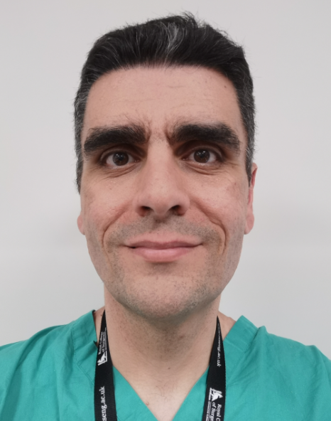 Angelos Kolias合作论文数Department of Clinical Neuroscience, Addenbrooke's Hospital;CRUK Cambridge Centre, Cancer Research UK Cambridge Institute, University of Cambridge2
 Maria Thom合作论文数Department of Neuropathology, University College London;Institute of Neurology, University College London2Adams Hadie合作论文数2
 Jonathan J. Russin合作论文数Department of Neurological Surgery, Keck Medicine of USC 2
 Sebastian Brandner合作论文数Division of Neuropathology, Queen Square Institute of Neurology, University College London2Alamri A合作论文数2
 Tomas Ysehak Abay合作论文数Sch. of Eng. & Math. Sci., City Univ. London;c;Sch. of Eng. & Math. Sci., City Univ. London2Camp Sophie合作论文数2Chavredakis Emmanuel合作论文数2
 Pervinder Bhogal合作论文数Department of Neuroradiology, The Karolinska Hospital2
 Tamas Revesz合作论文数Department of Neurodegenerative Diseases, Queen Square Institute of Neurology, Faculty of Brain Sciences, University College London2
 Charles Liu合作论文数Clinical Neurological Surgery;Director, USC Neurorestoration Center2
 Mario Teo合作论文数2
 Antonio Belli合作论文数Institute of Inflammation and Ageing, College of Medical and Dental Sciences, University of Birmingham2
 David Menon合作论文数Division of Anaesthesia, Department of Medicine, Queens' College, University of Cambridge2




