PURPOSE:This study describes the structure and timing of the clinical education components of MD-PhD programs to illustrate how variations in preclerkship curriculum correlate with the opportunity for early clinical exposure and other key program characteristics. METHOD:A survey was disseminated to U.S. MD-PhD programs on May 25, 2022, asking about the preclerkship curriculum length (long [> 18 months], medium [13-18 months], or short [12 months]), United States Medical Licensing Examination Step 1 timing (relative to the PhD training and clerkships), and opportunity for clerkships before the PhD phase. This survey was supplemented with data from publicly available sources to include 92 MD-PhD programs. RESULTS:This study found a wide range of MD-PhD clinical curricula. A strong association was found between shorter preclerkship curriculum length and the opportunity for clerkships before the PhD (10 of 50 programs [20%] with long preclerkship curriculum, 19 of 35 programs [54%] with medium preclerkship curriculum, and 7 of 7 programs [100%] with short preclerkship curriculum; P < .001). Variations in United States Medical Licensing Examination Step 1 timing also exist based on preclerkship curriculum length and the opportunity for clerkships before the PhD. Shorter preclerkship curriculum length was associated with National Institutes of Health funding of the MD-PhD program (20 [40%] of long, 25 [69%] of medium, and 6 [86%] of short preclerkship curricula; P = .006) and larger MD-PhD program size (35 students with long, 70 with medium, and 86 with short preclerkship curricula; P < .001). Preclerkship curriculum length was not associated with public vs private medical schools, although the West had shorter preclerkship curricula. CONCLUSIONS:This study underscores the need for collaborative efforts to gain insights into the effectiveness and implications of educational interventions in MD-PhD programs, ultimately informing future training strategies and policies.
The Eight-Twenty-One (ETO)/Myeloid Translocation Gene (MTG) family of transcriptional corepressors play a key role in adult stem cell function across multiple tissues and may be affected by mutation, deletion, or translocation in solid tumors and leukemia. Structural studies of the first conserved domain identified residues that make specific contacts with E proteins, such as HEB and E2A. We generated mice with a mutation in a critical phenylalanine (F210A) in Mtg16 to test the physiological significance of Mtg16 association with E proteins and compared these mice to mice containing a nearby cancer-associated mutation (P209T). We found that Mtg16-/- and Mtg16F210A/F210A mice showed impaired lymphopoiesis following competitive bone marrow transplant, suggesting that the repression of E protein-dependent transcription is critical for B- and T-cell development. Although Mtg16-/-, Mtg16P209T/P209T, and Mtg16F210A/F210A animals showed significant defects in burst forming potential (BFU-E) after phenylhydrazine treatment, only Mtg16-/- mice showed overt signs of anemia. Thus, we propose that, although Mtg16 is a critical regulator of hematopoietic stem and progenitor cell (HSPC) function, response to hemolytic anemia, and lymphoid development, the interaction between Mtg16 and E proteins is particularly important for lymphopoiesis.
The Alliance for Academic Internal Medicine (AAIM) first convened a workshop in 2015 that brought a small group of internal medicine program directors together who recognized the growing success of early-phase physician-scientist training programs but the unclear path afterward for these trainees. The meeting subsequently evolved into what is now the annual American Society for Clinical Investigation/AAIM/Burroughs Wellcome Fund (ASCI/AAIM/BWF) Physician-Scientist Pathways Workshop, which continues to bring stakeholders together to discuss the obstacles to success that physician-scientists face at all stages of their careers. This perspective presents the history and goals of the workshop, with an emphasis on the most recent meeting in 2024, and looks ahead to the work that still needs to be done to ensure a robust physician-scientist workforce.
Physician-scientists in academic medical centers require extramural grant support to launch and maintain their research careers. In order to cultivate the next generation of biomedical researchers, including physician-scientists, the NIH supports multiple career development (K series) awards. For many, their first experience in grant writing is composing a career development award (CDA) application. From the applicant's perspective, this process can be difficult. For one, NIH institute-specific differences between the same K mechanism can be confusing. Additionally, the importance of the various elements that make up the K application are frequently misunderstood. Furthermore, many K applications will not be funded on the initial submission; therefore, the need to resubmit an application should not be viewed as a sign of failure, but rather can be viewed as an element of resilience in biomedical research. In this piece, we aim to provide guidance for aspiring K applicants - in particular, from the reviewer perspective - with the intent of making the application process more understandable. We offer dos and don'ts on different components of the K application, advice on when to reach out to a program officer, and tips on resubmission. Our overarching goal is to provide support for prospective K applicants in their effort to obtain a K award. While targeted to K applications, most of the contents of this summary apply to any CDA.
Interleukin-10 (IL-10) is an immunomodulatory cytokine critical for intestinal immune homeostasis. IL-10 is produced by various immune cells but IL-10 receptor signaling in intestinal CX3CR1+ mononuclear phagocytes is necessary to prevent spontaneous colitis in mice. Here, we utilized fluorescent protein reporters and cell-specific targeting and found that Rorc-expressing innate lymphoid cells (ILCs) produce IL-10 in response to anti-CD40-mediated intestinal inflammation. Deletion of Il10 specifically in Rorc-expressing ILCs led to phenotypic changes in intestinal macrophages and exacerbated both innate and adaptive immune-mediated models of experimental colitis. The population of IL-10+ producing ILCs shared markers with both ILC2 and ILC3 with nearly all ILC3s being of the NCR+ subtype. Interestingly, Ccl26 was enriched in IL-10+ ILCs and was markedly reduced in IL-10-deficient ILC3s. Since CCL26 is a ligand for CX3CR1, we employed RNA in situ hybridization and observed increased numbers of ILCs in close proximity to Cx3cr1-expressing cells under inflammatory conditions. Finally, we generated transgenic RorctdTomato reporter mice that faithfully marked RORγt+ cells that could rescue disease pathology and aberrant macrophage phenotype following adoptive transfer into mice with selective Il10 deficiency in ILC3s. These results demonstrate that IL-10 production by a population of ILCs functions to promote immune homeostasis in the intestine possibly via direct effects on intestinal macrophages.
Selenoproteins are involved in immune cell metabolism, yet the roles of these proteins in T cell development and function remain largely unknown. The Trsp gene encodes the selenocysteine tRNA (tRNA Sec ) required for translation of all selenoproteins. In this study, we found that Trsp was required for thymopoiesis, with the majority of tRNA Sec -deficient T cells not progressing beyond double negative 3 stage, with egressed thymocytes undergoing peripheral homeostatic expansion. Trsp- deficient CD4 + T cells exhibited impairments in TCR and IL-2 signaling and did not cause inflammation in experimental models. On the other hand, Trsp -deficient regulatory T (Treg) cells exhibited defects in suppressive function ex vivo and Treg-specific Trsp deletion using Trsp fl/fl Foxp3 YFP-Cre ( Trsp !ιTreg ) mice caused fatal autoimmunity similar to FOXP3-deficient mice. Reducing oxidative stress via 2-HOBA administration prolonged survival in these Trsp !ιTreg mice. These findings indicate that tRNA Sec is required for T cell homeostasis and may be therapeutic targets in inflammation. One sentence summary:Trsp , a gene required for translation of all selenoproteins, is essential for all T cell development and function, especially regulatory T cells.
Vertebrate retinas share a basic blueprint comprising 5 neuronal classes arranged according to a common wiring diagram. Yet, vision is aligned with species differences in behavior and ecology, raising the question of how evolution acts on this circuit to adjust its computational characteristics. We address that problem by comparing the thalamic visual code and retinal cell composition in closely related species occupying different niches: Rhabdomys pumilio, which are day-active murid rodents, and nocturnal laboratory mice (Mus musculus). Using high-density electrophysiological recordings, we compare visual responses at both single-unit and population levels in the thalamus of these two species. We find that Rhabdomys achieves a higher spatiotemporal resolution visual code through the selective expansion of information channels characterized by non-linear spatiotemporal summation. Comparative analysis of single-cell transcriptomic atlases reveals that this difference originates with the increased relative abundance of retinal bipolar and ganglion cell types supporting OFF and ON-OFF responses. These findings demonstrate that evolution may drive changes in neural computation by adjusting the proportions of shared cell types rather than inventing new types and show the power of matching high-density physiological recordings with transcriptomic cell atlases to study evolution in the brain.
IntroductionThe pro-inflammatory cytokine interleukin-23 (IL-23) has been implicated in colorectal cancer (CRC). Yet, the cell-specific contributions of IL-23 receptor (IL-23R) signaling in CRC remain unknown. One of the cell types that highly expresses IL-23R are colonic regulatory T cells (Treg cells). The aim of this study was to define the contribution of Treg cell-specific IL-23R signaling in sporadic and inflammation-associated CRC.MethodsIn mice, the role of IL-23R in Treg cells in colitis-associated cancer (CAC) was investigated using azoxymethane/dextran sodium sulphate in wild-type Treg cell reporter mice (WT, Foxp3YFP-iCre), and mice harboring a Treg cell-specific deletion of IL-23 (Il23rΔTreg). The role of IL-23R signaling in Treg cells in sporadic CRC was examined utilizing orthotopic injection of the syngeneic colon cancer cell line MC-38 submucosally into the colon/rectum of mice. The function of macrophages was studied using clodronate. Finally, single-cell RNA-seq of a previously published dataset in human sporadic cancer was reanalyzed to corroborate these findings.ResultsIn CAC, Il23rΔTreg mice had increased tumor size and increased dysplasia compared to WT mice that was associated with decreased tumor-infiltrating macrophages. In the sporadic cancer model, Il23rΔTreg mice had increased survival and decreased tumor size compared to WT mice. Additionally, MC-38 tumors of Il23rΔTreg mice exhibited a higher frequency of pro-inflammatory macrophages and IL-17 producing CD4+ T cells. The decreased tumor size in Il23rΔTreg mice was macrophage-dependent. These data suggest that loss of IL-23R signaling in Treg cells permits IL-17 production by CD4+ T cells that in turn promotes pro-inflammatory macrophages to clear tumors. Finally, analysis of TCGA data and single-cell RNA-seq analysis of a previously published dataset in human sporadic cancer, revealed that IL23R was highly expressed in CRC compared to other cancers and specifically in tumor-associated Treg cells.ConclusionInflammation in colorectal carcinogenesis differs with respect to the contribution of IL-23R signaling in regulatory T cells.
Physician -scientists play a crucial role in advancing medical knowledge and patient care, yet the long periods of time required to complete training may impede expansion of this workforce. We examined the relationship between postgraduate training and time to receipt of NIH or Veterans Affairs career development awards (CDAs) for physician -scientists in internal medicine. Data from NIH RePORTER were analyzed for internal medicine residency graduates who received specific CDAs (K08, K23, K99, or IK2) in 2022. Additionally, information on degrees and training duration was collected. Internal medicine residency graduates constituted 19% of K awardees and 28% of IK2 awardees. Of MD -PhD internal medicine-trained graduates who received a K award, 92% received a K08 award; of MD -only graduates who received a K award, a majority received a K23 award. The median time from medical school graduation to CDA was 9.6 years for K awardees and 10.2 years for IK2 awardees. The time from medical school graduation to K or IK2 award was shorter for US MD -PhD graduates than US MD -only graduates. We propose that the time from medical school graduation to receipt of CDAs must be shortened to accelerate training and retention of physician -scientists.
How does evolution act on neuronal populations to match computational characteristics to functional demands? We address this problem by comparing visual code and retinal cell composition in closely related murid species with different behaviours. Rhabdomys pumilio are diurnal and have substantially thicker inner retina and larger visual thalamus than nocturnal Mus musculus. High-density electrophysiological recordings of visual response features in the dorsal lateral geniculate nucleus (dLGN) reveals that Rhabdomys attains higher spatiotemporal acuity both by denser coverage of the visual scene and a selective expansion of elements of the code characterised by non-linear spatiotemporal summation. Comparative analysis of single cell transcriptomic cell atlases reveals that realignment of the visual code is associated with increased relative abundance of bipolar and ganglion cell types supporting OFF and ON-OFF responses. These findings demonstrate how changes in retinal cell complement can reconfigure the coding of visual information to match changes in visual needs.
Undifferentiated intestinal stem cells (ISCs), particularly those marked by Lgr5, are crucial for maintaining homeostasis and resolving injury. Lgr5+ cells in the crypt base constantly divide, pushing daughter cells upward along the crypt axis, where they differentiate into a variety of specialized cell types. This process requires coordinated execution of complex transcriptional programs, which allow for the maintenance of undifferentiated stem cells while permitting differentiation of the wide array of intestinal cells necessary for homeostasis. Thus, disrupting these programs may negatively impact homeostasis and response to injury. Previously, members of the myeloid translocation gene (MTG) family have been identified as transcriptional co-repressors that regulate stem cell maintenance and differentiation programs in multiple organ systems, including the intestine. One MTG family member, myeloid translocation gene related 1 (MTGR1), has been recognized as a crucial regulator of secretory cell differentiation and response to injury. However, whether MTGR1 contributes to the function of ISCs has not yet been examined. Here, using Mtgr1-/- mice, we have assessed the effects of MTGR1 loss on ISC biology and differentiation programs. Interestingly, loss of MTGR1 increased the total number of cells expressing Lgr5, the canonical marker of cycling ISCs, suggesting higher overall stem cell numbers. However, expanded transcriptomic analyses revealed MTGR1 loss may instead promote stem cell differentiation into transit-amplifying cells at the expense of cycling ISC populations. Furthermore, ex vivo intestinal organoids established from Mtgr1 null were found nearly completely unable to survive and expand, likely due to aberrant ISC differentiation, suggesting that Mtgr1 null ISCs were functionally deficient as compared to WT ISCs. Together, these results identify a novel role for MTGR1 in ISC function and suggest that MTGR1 is required to maintain the undifferentiated state.
Although selenium deficiency correlates with colorectal cancer (CRC) risk, the roles of the selenium-rich antioxidant selenoprotein P (SELENOP) in CRC remain unclear. In this study, we defined SELENOP's contributions to sporadic CRC. In human single-cell cRNA-Seq (scRNA-Seq) data sets, we discovered that SELENOP expression rose as normal colon stem cells transformed into adenomas that progressed into carcinomas. We next examined the effects of Selenop KO in a mouse adenoma model that involved conditional, intestinal epithelium-specific deletion of the tumor suppressor adenomatous polyposis coli (Apc) and found that Selenop KO decreased colon tumor incidence and size. We mechanistically interrogated SELENOP-driven phenotypes in tumor organoids as well as in CRC and noncancer cell lines. Selenop-KO tumor organoids demonstrated defects in organoid formation and decreases in WNT target gene expression, which could be reversed by SELENOP restoration. Moreover, SELENOP increased canonical WNT signaling activity in noncancer and CRC cell lines. In defining the mechanism of action of SELENOP, we mapped protein-protein interactions between SELENOP and the WNT coreceptors low-density lipoprotein receptor-related proteins 5 and 6 (LRP5/6). Last, we confirmed that SELENOP-LRP5/6 interactions contributed to the effects of SELENOP on WNT activity. Overall, our results position SELENOP as a modulator of the WNT signaling pathway in sporadic CRC.
Supporting the career pathways of clinician-investigators (CIs) during residency and fellowship training presents a complex, multifaceted issue—a “wicked problem” that encompasses numerous stakeholders, with diverse priorities, values, competing interests, and agendas. Consensus on solutions may be elusive due to many intractable issues, such as human capital for mentorship, funding for protected research time during residency, and lack of research infrastructure. These factors collectively hinder CIs’ development during graduate medical education (GME). Since the COVID-19 pandemic, financial constraints have further exacerbated these limitations. How can we best support residents and fellows pursuing CI careers in this challenging environment?CIs play an essential role in translating scientific discoveries into medical practice; thus, sustaining this workforce is vital. Despite specialty-specific accreditation and board certification requirements, support for GME CIs predominantly occurs at individual residency or fellowship program levels. Given the complex and interconnected nature of CI training pathways, one solution lies in uniting and igniting these siloed communities for collective impacts, through the establishment of a CI community of practice (CoP). A CoP incorporates 3 key features—“mutual engagement, joint enterprise, and shared repertoire”1—which shift the focus from valuing individual achievements to collective experiences of clinicians and scholars. This can enhance and sustain successes across many programs.To form and sustain a CoP, 3 modes of belonging are essential: engagement, imagination, and alignment.2Engagement promotes collaboration, imagination stimulates self-reflection and new possibilities, and alignment ensures open communication and consistency in community values and goals. As a CoP evolves in a rapidly changing and complex environment, it would benefit from interacting with other CoPs beyond its local sphere of influence to survive and thrive.1,3This Rip Out provides practical recommendations for building a local CoP, expanding to a network of practice (NoP), and legitimizing the landscape of practice (LoP) for CIs. An LoP is an interconnected, complex social learning system where various CoPs coexist.4 GME research leaders, program directors, and designated institutional officials can strengthen and sustain their CoPs by extending collaborative networks across programs and institutions. Conceptually, the 3-Phase Evolution of Clinician Investigators CoPs focuses on the “collaboration continuum” (x-axis) to extend beyond boundaries (y-axis). This boundary crossing can amplify learning, collaboration, and innovation within and across communities.
Supplementary Legends for Figures and Tables from MTGR1 Is Required for Tumorigenesis in the Murine AOM/DSS Colitis-Associated Carcinoma Model
PDF file - 1856K, Intratumoral apoptosis is not altered in Gpx3-/- mice. TUNEL immunohistochemistry was performed to identify apoptotic cells in WT (N=13) and Gpx3-/- (N=14) tumors. TUNEL+ cells were counted within each high-powered field (HPF) and then averaged for each mouse. P=n.s.
PDF file - 2063K, Gpx3 is decreased in plasma of mice subjected to the AOM/DSS protocol. Western blot of Gpx3 protein expression in plasma from WT water treated and AOM/DSS treated mice (top). Quantification is presented as fold change intensity and the graph (bottom) displays plasma Gpx3 protein in the mice shown in the western blot above. P=0.046.
 Mary Kay Washington合作论文数Department of Pathology, Microbiology and Immunology, Vanderbilt University Medical Center;School of Medicine, Vanderbilt University80
 Sarah Short合作论文数70
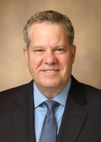 Keith T Wilson合作论文数Division of Gastroenterology, Hepatology and Nutrition, Department of Medicine, Vanderbilt University Medical Center;Vanderbilt Center for Mucosal Inflammation and Cancer, Vanderbilt University Medical Center;School of Medicine, Vanderbilt University52
 Yash Choksi合作论文数43
 Caitlyn W Barrett合作论文数40Lori A. Coburn合作论文数Vanderbilt University Medical Center Inflammatory Bowel Disease Center32
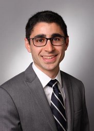 Parang Bobak合作论文数Departments of Cancer Biology, Pathology, Microbiology, and Immunology, and Biochemistry, Vanderbilt University;Department of Biology, Lipscomb University;Division of Pediatrics-Gastroenterology, Baylor University School of Medicine;Division of Pediatric Hematology/Oncology, University of Utah;and **Vanderbilt Ingram Cancer Center, Tennessee Valley Health Care System;Lipscomb University, Vanderbilt University31
 Rupesh Chaturvedi合作论文数Veterans Affairs Tennessee Valley Healthcare System31
 Scott W. Hiebert合作论文数Department of Biochemistry, Vanderbilt University30Wei Ning合作论文数29
 Vishruth K Reddy合作论文数26
 Raymond N. DuBois合作论文数Departments of Biochemistry and Molecular Biology, College of Medicine, Medical University of South Carolina;Hollings Cancer Center, Medical University of South Carolina;The Mark Foundation for Cancer Research;American Association for Cancer Research24Jennifer M. Pilat合作论文数24Frank Revetta合作论文数24
 Rachel Brown合作论文数21
 Joshua Thompson合作论文数21
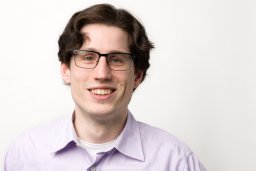 Justin Jacobse合作论文数Med Ctr, Leiden Univ20
 Jeremy Goettel合作论文数Monroe Carell Jr Children's Hospital at Vanderbilt, Vanderbilt University School of Medicine19
 Amber Bradley合作论文数17
 Mukul Mittal合作论文数Sanofi17
 Raymond F. Burk合作论文数Vanderbilt University16Poindexter Shenika V合作论文数15Margaret M Allaman合作论文数14
 Xi Chen (陈曦)合作论文数Department of Systems Biology, School of Life Sciences, Southern University of Science and Technology14Kristina E. Hill合作论文数14Kshipra Singh合作论文数13
 R. Daniel Beauchamp合作论文数School of Medicine, Vanderbilt University12
 Maria Blanca Piazuelo合作论文数Department of Medicine, Vanderbilt University Medical Center;Ingram Cancer Center, Vanderbilt University Medical Center12
 David Schwartz合作论文数Center for Genes, Environment, & Health, National Jewish Health;Department of Medicine, School of Medicine, University of Colorado;Department of Medicine-Pulmonary Sciences & Critical Care, School of Medicine, University of Colorado12Elizabeth McDonough合作论文数11
 Andrew Chan合作论文数Clinical and Translational Epidemiology Unit, Massachusetts General Hospital;Mass General Cancer Center, Massachusetts General Hospital11Horst Sara合作论文数11
 David W. Hill合作论文数Applied Physiology Laboratory, Department of Kinesiology, Health Promotion, and Recreation, College of Education, University of North Texas11Dawn B. Beaulieu合作论文数11
 Amanda Williams合作论文数10
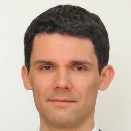 Thomas Brand合作论文数10
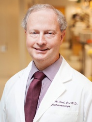 Richard M. Peek合作论文数Department of Pathology, Microbiology and Immunology, Vanderbilt University Medical Center;Division of Gastroenterology, Hepatology, and Nutrition, Vanderbilt University Medical Center10
 Larry W. Burggraf合作论文数AFIT/ENP, Air Force Institute of Technology10Cody E Keating合作论文数9
 J. Joshua Smith合作论文数Univ Washington, Dept Genome Sci, Seattle, WA 98195 USA9
 Hiremath Girish合作论文数Texas Children's Hospital, Baylor College of Medicine9Noah F. Shroyer合作论文数MLC, Cincinnati Children’s Hospital9Koral M Blunt合作论文数8
 Anthony Bilotta合作论文数Univ Texas Med Branch, Dept Microbiol & Immunol, 4-142C Med Res Bldg,301 Univ Blvd, Galveston, TX 77555 USA8Brown Caroline T合作论文数8Amy K. Motley合作论文数8
 Michael J Rosen合作论文数Division of Pediatric Gastroenterology, Hepatology & Nutrition, Department of Pediatrics, Stanford School of Medicine, Stanford University;Stanford Medicine Children's Health Center for IBD and Celiac Disease, Stanford University8




