In current clinical practice, Tumor Treating Fields (TTFields) are delivered through insulated ceramic electrode arrays via capacitive coupling, which limits the efficiency of electric field energy transfer. In this study, we propose a new TTFields delivery mode based on conductive electrodes, termed conductive TTFields (Ce-TTFields), to enhance energy delivery efficiency. Electromagnetic-field and lumped-circuit analysis was conducted to understand the underlying mechanisms of TTFields delivery and proposed the novel Ce-TTFields concept. We designed and fabricated a Ce-TTFields culture dish and conducted electromagnetic simulations, in vitro electric-field measurements, and U-87 glioma cell proliferation assays to validate this novel concept. Simulation and test experimental results demonstrate that Ce-TTFields produce stronger electric field intensities in the cell culture and the simulated human brain model compared with conventional insulated electrodes under the same driving voltage. U-87 glioma cell proliferation assays consistently confirmed that the U-87 glioma inhibition efficiency is enhanced by Ce-TTFields, indicating significantly improved energy-delivery efficiency. These findings suggest that Ce-TTFields may help optimize TTFields treatment protocols and offer a promising direction for developing more efficient, lightweight, and cost-effective TTFields therapeutic systems.
Irreversible electroporation (IRE) is a promising non-thermal tumor ablation method, but achieving optimal outcomes is challenging due to complex patient-specific 3D anatomies. Effective treatment requires synergistic optimization of both electrode placement and electrical parameters. To address this, this study proposes a multi-objective sparrow search algorithm based on evolutionary reference-points (ERM-SSA), which utilizes a 3D electric field distribution model incorporating heterogeneous tissue conductivity. Key optimization variables include the number of electrode needles, needles’ position and voltage. ERM-SSA establishes a multi-objective optimization model aiming to maximize tumor ablation rate while minimizing damage to the surrounding healthy tissues. To improve the optimization performance, ERM-SSA introduces several key improvements based on the standard sparrow search algorithm (SSA). First, Poisson disk sampling is used for population initialization to enhance the diversity of solutions. Secondly, an adaptive reference point strategy based on the electrode needle position is designed to improve the search guidance ability in high-dimensional space. Furthermore, its position update formula is optimized to make it more suitable for solving multi-objective problems. Finally, the improved sinusoidal perturbation strategy is integrated to effectively enhance its ability to jump out of the local optimum. Experimental results demonstrate that ERM-SSA achieves remarkable optimization performance across various tumor sizes and clinical constraints. This study introduces a fully automated, high-precision 3D optimization tool, offering a significant advancement for clinical IRE treatment planning. Moreover, its dynamic search mechanism provides novel and valuable insights for the clinical application implementation of multi-objective optimization algorithms.
Taste perception is central to flavor experiences. Electroencephalography (EEG) signals carry rich information about taste perception. Because these neural data are independent of verbal reports and bypassing conscious filtering, EEG holds promise as a neural-sensing platform for objective taste representation. However, taste-evoked EEG signals exhibit complex spatiotemporal and spectral dynamics that require advanced computational approaches to decode effectively. This study developed an EEG-based Multi-Scale Hybrid Attention and Squeeze Network (MHASNet) for taste evaluation. EEG signals evoked by distinct taste stimuli were recorded, a dedicated taste-EEG dataset was compiled, and a novel deep learning architecture, MHASNet, was designed to classify these signals. MHASNet synergistically integrates multi-scale convolutions to capture temporal dynamics across different time scales, dual-attention mechanisms to localize discriminative brain regions and electrode positions, and a squeeze-and-excitation module to optimize frequency-band contributions—collectively enabling precise extraction of taste-specific neural signatures. Results showed that the proposed model achieved superior performance across five taste categories (sweet, sour, salty, bitter, and tasteless), with 94.33% accuracy, 91.37% F1-score, 91.89% precision, and 92.07% recall. These results surpass benchmark models while maintaining millisecond-level inference latency suitable for real-time applications. By complementing subjective evaluations and instrumental analyses, the model offers an objective, neurophysiology-based solution for taste evaluation.
Objective: Tumor Treating Fields (TTFields) therapy, a clinically established modality that disrupts cancer cell mitosis through biophysical mechanisms, presents a unique paradigm in oncology. Despite its proven efficacy, its broad application is hindered by significant challenges in optimizing treatment delivery for individual patients. Methods: This review synthesizes the landscape of advanced computational strategies designed to overcome these barriers. We argue that personalizing TTFields therapy requires tackling three interdependent obstacles: achieving accurate electric field dosimetry, ensuring thermal safety, and enabling adaptive treatment planning. Results: this review systematically analyzes the state-of-the-art computational solutions corresponding to each challenge. We first examine patient-specific electric field modeling, emphasizing the critical roles of high-fidelity segmentation and quantitative dosimetric criteria. We then delve into thermal safety analysis, focusing on coupled electro-thermal simulations for predicting and mitigating thermal risks. Finally, we explore the multifaceted approaches to personalization, reviewing the convergence of algorithmic array layout optimization, real-time monitoring systems, and synergistic surgical interventions. Significance: By structuring the current body of research within this “problem-solution” framework, this review provides a clear and cohesive synthesis of how computational engineering is paving the way for a new era of precise, safe, and adaptive TTFields therapy.
Objective: Asymmetric pulse waveforms represent an optimized treatment protocol for high-frequency irreversible electroporation (H-FIRE), offering a lower ablation threshold compared to symmetric waveforms. However, the impact of waveform asymmetry has not been considered in current numerical models aimed at predicting tissue ablation zones. This study aims to develop an asymmetric waveform ablation area prediction model incorporating dynamic conductivity and dielectric dispersion effects (AW-DCDE) to predict ablation zones under varying asymmetric conditions. Methods: A linear scaling parameter relating pulse asymmetry to the ablation threshold was integrated into a modified Heaviside conductivity function. Dielectric dispersion was captured via a fourth-order Debye model. Variations in electric field intensity and dielectric dispersion were analyzed at pulse widths of 5 μs and 10 μs across different asymmetry levels. Finally, potato tissue experiments were conducted to validate the AW-DCDE model. Results: Increasing waveform asymmetry leads to a reduction in electric field intensity and polarization current, but a significant expansion of the ablation area. Statistical analysis confirmed no significant difference (p > 0.05) between AW-DCDE predictions and experimental results. Dynamic conductivity was found to have a greater influence on the predicted ablation area than dielectric dispersion. Conclusion: Waveform asymmetry significantly affects ablation area and electrical parameters. Significance: The AW DCDE model provides a theoretical framework for optimizing clinical H-FIRE treatment protocols.
OBJECTIVES:Micro-computed tomography (Micro-CT) is renowned for its high resolution, holding a pivotal role in advancing medical science research. However, compared to CT medical imaging datasets, there are fewer publicly available Micro-CT datasets, especially those annotated for multiple objects, leading to segmentation models with limited generalization abilities. METHODS:In order to improve the accuracy of multi-organ segmentation in Micro-CT, we developed a novel segmentation model called MOSnet which can utilize annotations from different datasets to enhance the whole segmentation performance. The proposed MOSnet includes a control module coupled with a reconstruction block that forms a multi-task structure, effectively addressing the absence of complete annotations. RESULTS:Experiments on 85 contrast-enhanced micro-CTscans and 140 native micro-CTscans for mice demonstrate that MOSnet is superior to the most of advanced segmentation networks. Compared to the best results of ResUnet, Unet3+, DAVnet3+ and AIMOS, our method improved dice similarity coefficient by 4.1 and 2.4 %, increased jaccard similarity coefficient by 4.1 and 3.1 %, and reduced HD95 by 16.3 and 19.3 % on the two datasets respectively at least. CONCLUSIONS:Our proposed model proves to be a robust and effective method for multi-organ segmentation in micro-CT, especially in situations where comprehensive annotations are lacking within a dataset.
To develop a deep learning (DL) model based on MRI to predict muscle-invasive bladder cancer (MIBC). A total of 559 patients, including 521 patients in our center and 38 patients in external centers were collected from 2012 to 2023 to construct the DL model. In this study, the DL model was utilized to differentiate between MIBC and NMIBC based on three-channel image inputs, including original T2WI images, segmented bladder, and regions of interest. Inception V3 was employed for model construction. The accuracy, sensitivity (SN), specificity (SP), positive predictive value (PPV) and negative predictive value (NPV) for predicting MIBC by DL model were 92.4%, 94.7%, 91.5%, 81.8% and 97.7% in the validation set and 92.1%, 86.8%, 94.6%, 88.5% and 93.8% in the internal test set. In the external test set, these values were 81.6%, 57.1%, 87.1%, 50.0% and 90.0%. Additionally, the accuracy, SN, SP, PPV, and NPV for predicting MIBC were 93.5%, 100%, 93.4%, 11.1%, and 100% in VI-RADS 2; 80.0%, 66.7%, 87.2%, 73.7% and 82.9% in VI-RADS 3; 90.3%, 91.7%, 85.7%, 95.7%, 75.0% in VI-RADS 4. The accuracy, SN, and PPV were 93.9%, 93.9%, and 100% in VI-RADS 5. The DL model based on T2WI can effectively predict MIBC and serve as a valuable complement to VI-RADS 3.
Accurate segmentation of glioblastoma (GBM), including the whole tumor (WT), tumor core (TC), and enhancing tumor (ET), from multi-modal magnetic resonance images (MRI) is essential for precise Tumor Treating Fields (TTFields) simulation. This study aims to address the challenges of this segmentation task to improve the accuracy of TTFields simulation results. We propose enhanced nnUnet (EnnUnet), a novel framework for multi-modal MRI segmentation that enhances the robust and widely-used nnUnet architecture. This advanced architecture integrates three key innovations: (1) Generalized Multi-kernel Convolution blocks are incorporated to capture multi-scale features and long-range dependencies. (2) A dual attention mechanism is employed at skip connections to refine feature fusion. (3) A novel boundary and Top-K loss is implemented for boundary-based refinement and to focus the training process on hard-to-segment pixels. The effectiveness of each enhancement was systematically evaluated through an ablation study on the BraTS 2023 dataset. The final EnnUnet model achieved superior performance, with average Dice scores of 93.52%, 92.07%, and 87.60% for the WT, TC, and ET, respectively, consistently outperforming other state-of-the-art methods. Furthermore, TTFields simulations on real patient data demonstrated that our precise segmentations yield more realistic electric field distributions compared to simplified homogeneous tumor models. The proposed EnnUnet architecture showcases promising potential for highly accurate and robust glioma segmentation. It offers a more reliable foundation for computational modeling, which is essential for enhancing the precision of TTFields treatment planning and advancing personalized therapeutic strategies for GBM patients.
Tumor treating fields (TTFields) is a promising non-invasive cancer treatment that uses alternating electric fields to disrupt tumor cell division. Despite its potential, there is a significant lack of precise and reliable methods for evaluating the efficacy of TTFields in clinical settings. The aim of this study is to develop and validate a new method for real-time assessment of the efficacy of TTFields. We proposed a novel neural network based on a collaborative fusion strategy of dual-branch (CFS-DB) to reconstruct the conductivity of tumor region for real-time assessment of the efficacy of TTFields. The proposed CFS-DB includes two independent branches: a conductivity branch and a structure branch. The conductivity branch employs FC-UNet to learn the mapping from measured boundary voltages to conductivity. The structural branch uses the results reconstructed by Gaussian-Newton method as the input for image-to-image training. Finally, the features from both branches are fused for coordinated end-to-end training. The simulation and experimental results show that the proposed CFS-DB has superior performance compared to five state-of-the-art deep learning networks. The CFS-DB method offers a novel and precise approach for evaluating the efficacy of TTFields, providing a new paradigm for clinical assessment.
A robust and efficient two-dimensional/three-dimensional (2D/3D) registration algorithm is critical to imageguided interventions, as it allows for intuitive, reproducible, and high-accuracy robot-assisted surgical procedures at competitive costs. Our approach adopts a multi-stage, self-supervised framework tailored to patientspecific contexts to tackle the 2D/3D registration problem. Preoperatively, a regression neural network is trained using synthesized X-rays to achieve robust initialization of rigid-body poses from the Special Euclidean group SE(3). However, SE(3) lacks a bi-invariant metric for measuring the distance between poses as it is not a direct product of compact and abelian groups. At the same time, existing left-invariant metrics fail to sufficiently account for the consistency and symmetry of spatial displacements under Lie group operations, which may hinder the network from correctly comprehending the 2D/3D projective geometry. To address these limitations, we propose a practical pose parameterization approach that embeds na & iuml;ve SE(3) pose elements into the fourdimensional Special Orthogonal group SO(4), thereby deriving an approximate bi-invariant metric for network training. Additionally, we present a cross-stage partial style, lightweight network CSP-ConvNeXt towards low-cost systematic solutions. Intraoperatively, we perform gradient-based optimization for real-time pose refinement. We report mean target registration error, network registration success rate, and sub-millimeter registration success rate for stage-dependent evaluations. Experimental results demonstrate that our method achieves state-of-the-art registration performance on two public datasets and one in-house dataset across all registration stages.
Medical image segmentation plays a crucial role in accurate disease diagnosis and effective treatment planning. However, cross-modality medical image segmentation poses significant challenges due to inter-domain differences. This study proposes a novel unsupervised domain adaptive method, UDAS2S, which integrates adversarial learning and semi-supervised learning for unpaired cross-modality image segmentation. UDAS2S comprises two main components: a synthetic network based on patch contrastive learning (SPCL) and a semi-supervised segmentation network (SSN). The SPCL is based on the Cycle-Consistent Generative Adversarial Networks (CycleGAN) to implement the transformation from the source images to target images, and we introduced patch comparison learning to facilitate more accurate image synthesis by comparing the local patches of the source and target images. Additionally, a semi-supervised segmentation model based on cross-pseudo-supervised is used to fully employ the information from unlabeled target images and enhance the segmentation performance of the model. This method enhances the segmentation performance through the interaction between the source and target images, which allows us to achieve more accurate cross-modality segmentation. Experimental evaluations on two publicly available datasets—Multi-Modality Whole Heart Segmentation and Combined Healthy Abdominal Organ Segmentation—demonstrate that UDAS2S outperforms state-of-the-art unsupervised domain adaptive methods in terms of Average Symmetric Surface Distance, Dice Similarity Coefficient, and 95 https://github.com/XYY311/UDAS2S .
This study investigates how Tumor Treating Fields (TTFields) disrupt tumor cell division by exerting dielectrophoretic (DEP) forces on septins, key proteins in cytokinesis. Computational models show that TTFields induce negative DEP forces, displacing septins away from the cleavage furrow. A three-phase electrode configuration was 11.2% more effective at displacing septins than an orthogonal setup. The findings suggest TTFields interfere with cytokinesis by causing septin mislocalization, which likely disrupts the contractile ring and midbody. This offers a novel mechanism and suggests that enhancing these nDEP forces by tailoring electric field configurations could improve future cancer therapies.
Purpose To assist doctors in clinical diagnosis, we propose a multipath deep learning (MP-DL) model to distinguish between muscle-invasive bladder cancer (MIBC) and non-muscle-invasive bladder cancer (NMIBC) using multiparametric magnetic resonance imaging (mp-MRI) and synthesized samples. Methods The proposed MP-DL model integrates T2-weighted image (T2WI), diffusion-weighted imaging (DWI), and dynamic contrast-enhanced imaging (DCE) branches, combining high-level features from these sequences. Multichannel inputs, including the original image, segmented bladder, and region of interest from the T2WI branch, enhance the focus on the tumor region. InceptionV3 served as the model backbone for feature extraction with a multitask framework in the DWI and DCE branches. Synthesized samples were generated to supplement missing DWI and DCE sequences, enlarging the sample size and boosting model performance. Evaluation through five-fold cross-validation and internal-external testing demonstrated the model's robustness and generalization capabilities. Results The dataset consisted of 401 cases (287 NMIBC and 114 MIBC cases), which included a training set of 313 cases (containing partly synthesized DWI and DCE samples), validation of 26 cases, internal testing of 34 cases, and external testing of 28 cases. In the internal testing, the model achieved area under curve, accuracy, sensitivity, specificity, and F1 scores of 0.914, 0.835, 0.880, 0.817, and 0.761, respectively. In the external testing, the results were 0.821, 0.814, 0.730, 0.845, and 0.619, respectively. The model performance surpasses that of medical professionals in internal testing while outperforming urologists and approaching senior radiologists in external testing. Conclusions The MP-DL model, incorporating synthesized samples, shows promise in preoperative MIBC prediction, potentially aiding primary urologists and radiologists.
Electrical impedance tomography (EIT), a non-invasive, real-time, and cost-effective imaging technique, is widely studied in medical diagnostics for lung diseases. However, the severely ill-posed nonlinear inverse problem in EIT leads to reconstructed images being susceptible to noise-induced artifacts. This study aims to advance a deep learning technique to reconstruct high-resolution conductivity distributions using voltages measured by EIT sensors. We proposed a novel reconstruction algorithm called generative adversarial network based on conditional variational autoencoder (CVAE-GAN). We incorporated the true conductivity as a conditional variable into the latent representation of the variational autoencoder (VAE) decoder and encoder to form a conditional variational autoencoder (CVAE). A residual module was introduced into the CVAE decoder and encoder to facilitate the network in learning deeper feature representations, which improves the performance of the model. The adversarial learning strategy leverages the improved CVAE as the generator in a GAN framework, substantially enhancing the accuracy and robustness of the reconstructed images. Experimental results demonstrate that CVAE-GAN outperforms five state-of-the-art deep learning methods. Compared to the best alternative model, it achieves an 8.9% improvement in peak signal-to-noise ratio (PSNR) and a 3.2% improvement in structural similarity index (SSIM), while reducing mean squared error (MSE) by 33.33% and relative error (RE) by 24.57%. These results highlight the significant performance gains in terms of both accuracy and robustness for EIT image reconstruction. The proposed CVAE-GAN framework represents a significant advancement in EIT image reconstruction. By addressing key challenges such as noise-induced artifacts and achieving robust reconstructions, it provides a generalizable approach with transformative potential for real-world applications in medical imaging, particularly in the diagnostics and monitoring of lung diseases.
Tumor treating fields (TTFields) have emerged as an innovative therapeutic strategy for glioblastoma. The effectiveness of TTFields depends on factors such as electric field intensity, direction, and treatment duration. MRI slices from two glioblastoma patients are segmented and meshed to obtain discrete nodes representing different tissues. Then, we employ the multi- objective node adaptive optimization (MONAO) algorithm to optimize the positions of the electrode arrays and the phase differences between the voltages of different arrays, aiming to enhance the therapeutic efficacy of TTFields. The algorithm serializes the discrete mesh nodes and incorporates a dual-population mechanism to ensure diversity among individuals. To enhance exploration capabilities, it employs an adaptive position update strategy and a dynamic mutation mechanism. Additionally, a binary encoding system is used to represent the positions of the electrode arrays and the phase differences, aiming to maximize both the average and minimum electric field intensity within the tumor throughout the treatment cycle. Experimental results show that MONAO delivers the highest therapeutic doses, reaching 32.1022 V & sdot; h/cm for patient1 and 31.1646 V & sdot; h/cm for patient2, outperforming other algorithms. Additionally, MONAO achieves these results using only three electrode arrays and 28V input, providing more electric field directions while reducing thermal side effects on the scalp, whereas other methods require four electrode arrays and 40V input, offering a more economical and efficient approach for personalized TTFields therapy.
This study presents an innovative KAN-UKAN framework that integrates the Kolmogorov-Arnold network (KAN) in the UNet architecture, aiming at achieving high-resolution 3D electrical impedance tomography (EIT) image reconstruction for accurate assessment of the efficacy of tumor treating fields (TTFields). In this framework, KAN replaces the traditional fully connected layers in reconstruction networks, thereby improving the precision of mapping boundary voltages to conductivity distributions. Additionally, we strategically incorporate the KAN modules between the encoder and decoder of the UNet, referred to as UKAN, which strengthens the model’s ability to capture and learn complex nonlinear relationships in EIT data. Finally, the model’s overall performance and robustness were enhanced by combining the loss functions of the initial KAN predictions and the UKAN predictions with the ground truth. Simulation results on the lung cancer dataset and phantom experimental results show that the proposed KAN-UKAN framework significantly outperforms the four state-of-the-art reconstruction models. This superiority is evident in both quantitative metrics and qualitative analysis, demonstrating the accuracy and robustness of the framework in 3D EIT image reconstruction. The methodology proposed in this study provides a novel and accurate way to assess the efficacy of TTFields and provides a new paradigm for clinical evaluation.
BackgroundTumor treating fields (TTFields) is an innovative cancer therapy utilizing electric fields to disrupt cell division, where stronger fields are more effective at inhibiting tumor cells. This study seeks to enhance TTFields’ effectiveness by optimizing electrode array’s position using an intelligent algorithm, thus increasing electric field intensity within the tumor region.MethodThe study employed two representative real abdominal models of liver cancer patients. We utilized an improved white shark optimization (IWSO) algorithm to optimize the positions of four electrode arrays on the abdominal skin. The IWSO algorithm was enhanced through the integration of sinusoidal chaotic mapping, Levy flight strategy, and differential evolution. Furthermore, we utilized the finite element method to compute electric field intensity within tumor region as the objective function for the IWSO algorithm.ResultsExperiments on two real abdominal modes demonstrate that the electrode arrays by our approach is superior to the default layout in terms of average electric field intensity (Eave), average power loss density (Pave), average current density (Cave), and the average treatment coverage of the tumor ATV1 to ATV3. For Patient 1, the Eave, Pave, and Cave of the tumor area were improved by 46.71%, 106.33%, and 48.14%. For Patient 2, the Eave, Pave, and Cave of the tumor area increased by 11.86%, 22.09%, and 50.25%.ConclusionThis study demonstrates that optimizing the position of TTFields electrode arrays using the IWSO algorithm can effectively enhance the electric field intensity and treatment coverage of tumors, providing a more effective method for personalized TTFields treatment.
In this paper, the multi-task dense-feature-fusion survival prediction (DFFSP) model is proposed to predict the three-year survival for glioblastoma (GBM) patients based on radiogenomics data. The contrast-enhanced T1-weighted (T1w) image, T2-weighted (T2w) image and copy number variation (CNV) is used as the input of the three branches of the DFFSP model. This model uses two image extraction modules consisting of residual blocks and one dense feature fusion module to make multi-scale fusion of T1w and T2w image features as backbone. Also, a gene feature extraction module is used to adaptively weight CNV fragments. Besides, a transfer learning module is introduced to solve the small sample problem and an image reconstruction module is adopted to make the model anatomy-aware under a multi-task framework. 256 sample pairs (T1w and corresponding T2w MRI slices) and 187 CNVs of 74 patients were used. The experimental results show that the proposed model can predict the three-year survival of GBM patients with the accuracy of 89.1 %, which is improved by 3.2 and 4.7 % compared with the model without genes and the model using last fusion strategy, respectively. This model could also classify the patients into high-risk and low-risk groups, which will effectively assist doctors in diagnosing GBM patients.
The multifaceted nature of photodynamic therapy (PDT) requires a throughout evaluation of a multitude of parameters when devising preclinical protocols. In this study, we constructed MCF-7 human breast tumor spheroid assays to infer PDT irradiation doses at four gradient levels for violet light at 408 nm and red light at 625 nm under normal and hypoxic oxygen conditions. The compacted three-dimensional (3D) tumor models conferred PDT resistance as compared to monolayer cultures due to heterogenous distribution of photosensitizers along with the presence of internal hypoxic region. Cell viability results indicated that the violet light was more efficient to kill cells in the spheroids under normal oxygen conditions, while cells exposed to the hypoxic microenvironment exhibited minimal PDT-induced death. The combination of 3D tumor spheroid assays and the multiparametric screening platform presented a solid framework for assessing PDT efficacy across a wide range of different physiological conditions and therapeutic regimes.
Tumor treating fields (TTFields) is a novel therapeutic approach for the treatment of glioblastoma. The electric field intensity is a critical factor in the therapeutic efficacy of TTFields, as stronger electric field can more effectively impede the proliferation and survival of tumor cells. In this study, we aimed to improve the therapeutic effectiveness of TTFields by optimizing the position of electrode arrays, resulting in an increased electric field intensity at the tumor. Three representative head models of real glioblastoma patients were used as the research subjects in this study. The improved subtraction-average-based optimization (ISABO) algorithm based on circle chaos mapping, opposition-based learning and golden sine strategy, was employed to optimize the positions of the four sets of electrode arrays on the scalp. The electrode positions are dynamically adjusted through iterative search to maximize the electric field intensity at the tumor. The experimental results indicate that, in comparison to the conventional layout, the positions of the electrode arrays obtained by the ISABO algorithm can achieve average electric field intensity of 1.7887, 2.0058, and 1.3497 V/cm at the tumor of three glioblastoma patients, which are 23.6%, 29.4%, and 8.5% higher than the conventional layout, respectively. This study demonstrates that optimizing the location of the TTFields electrode array using the ISABO algorithm can effectively enhance the electric field intensity and treatment coverage in the tumor area, offering a more effective approach for personalized TTFields treatment.
 Zhiyu Qian (钱志余)合作论文数College of Automation Engineering, Nanjing University of Aeronautics and Astronautics23
 Xue Fu合作论文数19
 Liang Wang (汪良)合作论文数Union Hospital, Tongji Medical College, Huazhong University of Science and Technology19
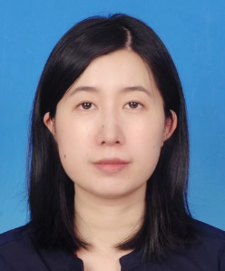 Yamin Yang (杨雅敏)合作论文数College of Automation Engineering, Nanjing University of Aeronautics and Astronautics10
 Ling Tao (陶玲)合作论文数College of Automation Engineering, Nanjing University of Aeronautics and Astronautics9
 Weitao Li (李韪韬)合作论文数College of Automation Engineering, Nanjing University of Aeronautics and Astronautics8
 Jie Yu合作论文数7Sijia Liu合作论文数7
 Yuan Zou (邹媛)合作论文数School of Ecology, Sun Yat-sen University7Xiao Yang合作论文数7CAI Lingkai (蔡令凯)合作论文数6Yueyue Xiao合作论文数5Yuewu Li合作论文数5Baorui Yuan合作论文数5
 Xin Chen合作论文数5Peng Wu (吴芃)合作论文数5Hua Tao (陶华)合作论文数5
 Xiong Lu (陆熊)合作论文数College of Automation Engineering, Nanjing University of Aeronautics and Astronautics5Jing Wang合作论文数5Jie Zhao合作论文数5
 Shijie Wang (王世杰)合作论文数School of Artificial Intelligence, Southeast University5
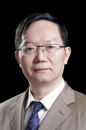 Guangming Lu (卢光明)合作论文数State Key Laboratory of Analytical Chemistry for Life Science, Nanjing University;Medical School of Nanjing University5
 Dongsheng Li (李东胜)合作论文数Microsoft Research Asia;School of Computer Science, Fudan University5
 Limin Luo (罗立民)合作论文数School of Computer Science and Engineering, Southeast University5
 Jianye Liu (刘建业)合作论文数College of Automation Engineering, Nanjing University of Aeronautics and Astronautics5
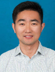 Rongfang Gong (龚荣芳)合作论文数Nanjing University of Aeronautics and Astronautics5Kun Li合作论文数4Yuehui Wen合作论文数4Qiuyi Wu合作论文数4Jiarong Zeng合作论文数4CHEN Zhiying (陈志颖)合作论文数4Peng Xu合作论文数4Junhao YANG (杨俊豪)合作论文数4Wen-qing LIU (刘雯卿)合作论文数4Jianfei Li合作论文数4
 Qiang Lv (吕强)合作论文数南京医科大学第一附属医院泌尿外科4
 Abai Xu (徐啊白)合作论文数Southern Medical University Zhujiang Hospital4Chen Binshen合作论文数4Jiani Wu (吴佳妮)合作论文数4Jian Li合作论文数3Qingyang Zang (臧青杨)合作论文数3Zhiying Chen合作论文数3WANG Hui-nan合作论文数3XU Junqi (徐俊琪)合作论文数3Rui Dong合作论文数3Peikun Liu合作论文数3Qiang Shao合作论文数3CUI Jianliang (崔建良)合作论文数3JIANG Ruilin (姜睿林)合作论文数3




