Suppressor/enhancer of Lin-12-like (SEL1L) is a component of the endoplasmic reticulum-associated degradation (ERAD) pathway which is part of the unfolded protein response (UPR). SEL1L may exert pro-tumorigenic or oncosuppressive functions in different tumor types, but its role in human cutaneous malignant melanoma (cMM) remains largely unexplored. Here, in silico analysis revealed that SEL1L is upregulated in cMM compared to normal skin. In addition, SEL1L expression in cMM positively correlated with Clark level at diagnosis, higher expression being associated with poorer patient survival. SEL1L co-expression with ERAD- and UPR-related genes, along with Gene Ontology enrichment analysis, supported its role in ER-stress responses in cMM. Immunohistochemistry and quantitative digital morphometric analysis of SEL1L protein levels in 60 human samples of benign melanocytic tumors and cMMs at different Breslow T category showed that SEL1L levels in cMM progressively increase in association with T category, tumor thickness, and the presence of ulceration. In addition, total and tumor-associated endothelial SEL1L expression correlates with microvessel density in the corresponding tumor specimens. In keeping with a non-redundant role of SEL1L in cMM, grafting of SEL1L-silenced A2058 human melanoma cells in immunodeficient mice yielded tumors with reduced cell proliferation, enhanced apoptosis, and increased necrosis, accompanied by heightened hypoxia and reduced vascularization. Accordingly, SEL1L-silenced cells showed upregulation of the angiosuppressive thrombospondin-encoding genes THSP1 and THSP2 and a reduced angiogenic potential in an endothelial sprouting assay in vitro and in the chick embryo chorionallantoic membrane assay in vivo. Together, these findings indicate that SEL1L may exert a pro-tumorigenic/pro-angiogenic function in human melanoma and pave the way to further studies aimed at investigating its potential as a therapeutic target in cMM.
PURPOSE:TMED10 is involved in unconventional protein secretion and ER-Golgi trafficking. TMED10 may exert protumorigenic or oncosuppressive functions in different tumor types, but its role in human cutaneous melanoma has never been explored. Here, TMED10 expression has been investigated in human benign melanocytic tumors and cutaneous malignant melanoma (cMM). METHODS:This study utilized in silico analysis on various publicly available web platforms to investigate TMED10 gene expression in normal skin and human melanoma at different Clark levels, and immunohistochemistry and digital morphometric analysis of TMED10 protein levels in 60 human samples of benign melanocytic tumors and cMMs at different Breslow T category. RESULTS:In silico analysis revealed that TMED10 is upregulated in cMM compared to normal skin. TMED10 expression in cMM positively correlated with Clark level at diagnosis, and higher expression was associated with the BRAFV600E mutation and poorer patient survival. TMED10 co-expression with TMED2, TMED8, HSP90AA1 and HSP90B1, along with Gene Ontology enrichment analysis, supported its role in ER-Golgi trafficking and unconventional protein secretion in human melanoma. Digital morphometric analysis of immunohistochemical investigation of 60 human specimens confirmed a significant increase in TMED10 protein levels with Breslow thickness-based T category in tumor cells and tumor-associated blood vessels. At variance, TMED10 immunoreactivity in infiltrating lymphocytes was reduced in T3 and T4 cMM. CONCLUSION:These findings point to a pro-tumorigenic function of TMED10 in human cMM and pave the way to further studies aimed at identifying its contribution to tumor progression, neovascularization, and immune escape, and its potential as a therapeutic target in cMM.
Supplementary Data - Single file with supplemental methods, tables, figures, legends. Supplemental Materials and Methods; Table S1: Primers used for RT-qPCR analysis; Table S2: GALC downregulation affects the lipid profile of murine B16-F10 melanoma cells; Table S3: Expression levels of enzymes involved in sphingolipid metabolism in B16-F10 cells; Table S4: RT-qPCR analysis of the expression of tumor infiltrate markers and immune checkpoints in B16-F10 tumor grafts; Figure S1: Galca does not affect melanocyte differentiation in zebrafish; Figure S2: Galc downregulation does not affect cell cycle distribution and cell death in B16-F10 cells; Figure S3: The neutral sphingomyelinase inhibitor GW4869 increases the tumorigenic activity of shGALC-B16-F10 cells; Figure S4: Inhibition of the tumorigenic activity in shGALC-bis-B16-F10 cells; Figure S5: Public data mining of GALC expression in human melanoma; Figure S6: Public data mining of SMPD3 expression in human melanoma.
<p>Time courses of pH and oxygen in presence of enzalutamide continuous treatment</p>
In spite of the huge advancements in both diagnosis and interventions, hormone refractory prostate cancer (HRPC) remains a major hurdle in prostate cancer (PCa). Metabolic reprogramming plays a key role in PCa oncogenesis and resistance. However, the dynamics between metabolism and oncogenesis are not fully understood. Here, we demonstrate that two multi-target natural products, cannabidiol (CBD) and cannabigerol (CBG), suppress HRPC development in the TRansgenic Adenocarcinoma of the Mouse Prostate (TRAMP) model by reprogramming metabolic and oncogenic signaling. Mechanistically, CBD increases glycolytic capacity and inhibits oxidative phosphorylation in enzalutamide-resistant HRPC cells. This action of CBD originates from its effect on metabolic plasticity via modulation of VDAC1 and hexokinase II (HKII) coupling on the outer mitochondrial membrane, which leads to strong shifts of mitochondrial functions and oncogenic signaling pathways. The effect of CBG on enzalutamide-resistant HRPC cells was less pronounced than CBD and only partially attributable to its action on mitochondria. However, when optimally combined, these two cannabinoids exhibited strong anti-tumor effects in TRAMP mice, even when these had become refractory to enzalutamide, thus pointing to their therapeutical potential against PCa.
Prostate cancer (PCa) is a leading cause of death in the male population commonly treated with androgen deprivation therapy that often relapses as androgen-independent and aggressive castration-resistant prostate cancer (CRPC). Ferroptosis is a recently described form of cell death that requires abundant cytosolic labile iron to promote membrane lipid peroxidation and which can be induced by agents that inhibit the glutathione peroxidase-4 activity such as RSL3. Exploiting in vitro and in vivo human and murine PCa models and the multistage transgenic TRAMP model of PCa we show that RSL3 induces ferroptosis in PCa cells and demonstrate for the first time that iron supplementation significantly increases the effect of RSL3 triggering lipid peroxidation, enhanced intracellular stress and leading to cancer cell death. Moreover, the combination with the second generation anti-androgen drug enzalutamide potentiates the effect of the RSL3 + iron combination leading to superior inhibition of PCa and preventing the onset of CRPC in the TRAMP mouse model. These data open new perspectives in the use of pro-ferroptotic approaches alone or in combination with enzalutamide for the treatment of PCa.
The Figure shows the long term behavior of sensitive and resistant cells (in absence of enzalutamide
Supplementary Fig. S1 from αvβ3 Integrin-dependent antiangiogenic activity of resveratrol stereoisomers
<p>Simulation and treatment of TRAMP mice treated with enzalutamide, cabazitaxel and the combination</p>
Globoid cell leukodystrophy (GLD), or Krabbe disease, is a neurodegenerative sphingolipidosis caused by genetic deficiency of lysosomal β-galactosylceramidase (GALC), characterized by neuroinflammation and demyelination of the central (CNS) and peripheral nervous system. The acute phase protein long pentraxin-3 (PTX3) is a soluble pattern recognition receptor and a regulator of innate immunity. Growing evidence points to the involvement of PTX3 in neurodegeneration. However, the expression and role of PTX3 in the neurodegenerative/neuroinflammatory processes that characterize GLD remain unexplored. Here, immunohistochemical analysis of brain samples from Krabbe patients showed that macrophages and globoid cells are intensely immunoreactive for PTX3. Accordingly, Ptx3 expression increases throughout the course of the disease in the cerebrum, cerebellum, and spinal cord of GALC-deficient twitcher (Galctwi/twi) mice, an authentic animal model of GLD. This was paralleled by the upregulation of proinflammatory genes and M1-polarized macrophage/microglia markers and of the levels of PTX3 protein in CNS and plasma of twitcher animals. Crossing of Galctwi/twi mice with transgenic PTX3 overexpressing animals (hPTX3 mice) demonstrated that constitutive PTX3 overexpression reduced the severity of clinical signs and the upregulation of proinflammatory genes in the spinal cord of P35 hPTX3/Galctwi/twi mice when compared to Galctwi/twi littermates, leading to a limited increase of their life span. However, this occurred in the absence of a significant impact on the histopathological findings and on the accumulation of the neurotoxic metabolite psychosine when evaluated at this late time point of the disease. In conclusion, our results provide the first evidence that PTX3 is produced in the CNS of GALC-deficient Krabbe patients and twitcher mice. PTX3 may exert a protective role by reducing the neuroinflammatory response that occurs in the spinal cord of GALC-deficient animals.
 Marco Presta合作论文数49
 Roberto Ronca合作论文数Department of Biomedical Sciences and Biotechnology, University of Brescia34Belleri Mirella合作论文数26
 Arianna Giacomini合作论文数25
 Domenico Ribatti合作论文数Department of Human Anatomy and Histology University of Bari Medical School17
 Sara Rezzola合作论文数Department of Molecular and Translational Medicine, University of Brescia15Alessi Patrizia合作论文数14Daria Leali合作论文数14Di Salle Emanuela合作论文数13Corsini Michela合作论文数Department of Molecular and Translational Medicine, University of Brescia11
 Federica Maccarinelli合作论文数Department of Molecular and Translational Medicine, University of Brescia Brescia10
 Stefania Mitola合作论文数Department of Biomedical Sciences and Biotechnology, University of Brescia10
 Alessia Ligresti合作论文数Endocannabinoid Research Group, CNR10Ravelli Cosetta合作论文数9
 Marianna Cerasuolo合作论文数Dipartimento di Matematica e Applicazioni “R. Caccioppoli”,, Università degli Studi di Napoli Federico II9Viviana Marolda合作论文数8Magdalena Kostrzewa合作论文数Institute of Biomolecular Chemistry, National Research Council of Italy8
 Debora Paris合作论文数8
 Ali Mokhtar Mahmoud合作论文数National Cancer Institute8
 Sara Matarazzo合作论文数8
 William Vermi合作论文数Department of Pathology;University of Brescia;Department of Pathology, University of Brescia8Gaia C. Ghedini合作论文数7Triggiani Luca合作论文数7Roberta Verde合作论文数7
 Dominique Melck合作论文数7
 Stefano Calza合作论文数Department of Molecular and Translational Medicine, University of Brescia6Francesco Semeraro合作论文数5Paola Chiodelli合作论文数School of Medicine, University of Brescia5Patrizi Ornella合作论文数Department of Molecular and Translational Medicine, University of Brescia4
 Elisabetta Grillo合作论文数4
 Silvia Parolini合作论文数University of Brescia4
 Tiziana Annese合作论文数Department of Human Anatomy and Histology, University of Bari Medical School4Giovanna Tabellini合作论文数4Adwaid Manu Krishna Chandran合作论文数3
 Stefania Nicoli合作论文数Molecular Medicine, Pharmacology, and Physiology, Biological & Biomedical Sciences, Yale University3
 Mohd Imtiaz Nawaz合作论文数Dept Ophthalmol, King Saud Univ3
 Chiara Tobia合作论文数3Rampinelli Fabio合作论文数3
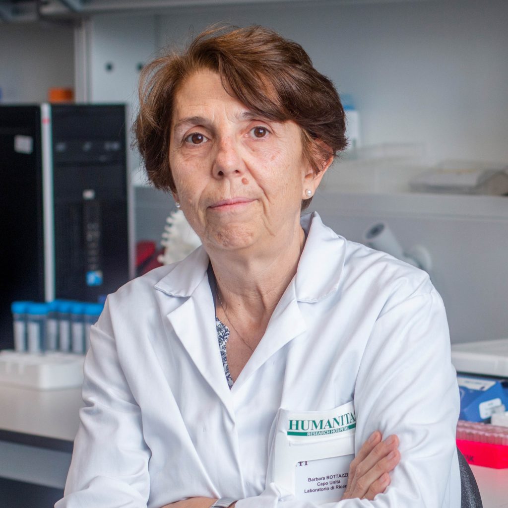 Barbara Bottazzi合作论文数Humanitas Research Hospital3
 Cecilia Garlanda合作论文数Humanitas University;Laboratory of Experimental Immunopathology, Dept. of Research in Immunology, Humanitas Clinical and Research Hospital3Stefano Indraccolo合作论文数Immunology and Molecular Oncology Unit, Veneto Institute of Oncology IOV -IRCCS3
 Emanuela Marcenaro合作论文数Dipartimento di Medicina Sperimentale, Università degli Studi di Genova3
 Josefina Casas合作论文数2Giuseppe Paganini合作论文数2
 Marco Righi合作论文数2Chiara Urbinati合作论文数2Liliana Lista合作论文数2
 Andrea Barbieri合作论文数2
 Luca Forti合作论文数2




