Purpose/Objective(s) To assess the anatomic conformity of custom thermoplastic boluses generated based on patient-specific 3D-printed molds. Materials/Methods A retrospective review of consecutive cases of 3D printer-assisted custom thermoplastic bolus was performed. For each case, target volume-specific contours (molds) were generated using computed tomography-based (CT) simulation scans in a treatment planning system. Molds were exported into a standard tessellation language (STL) file for rapid manufacturing on a 3D Printer using polylactic acid (PLA) filament. STL files were converted into G-code script and printed on slicer settings of 220°C nozzle temperature, 0.3 mm layer height, and 20% infill. The thickness of the thermoplastic bolus utilized was based on the treating radiation oncologist's prescription and ranged between 5.0 and 10.0 mm. Once heated, thermoplastic boluses were shaped using the 3D printed target volume-specific PLA molds. The quality of 3D printer-assisted thermoplastic boluses was confirmed on CT scanners before clinical use. Anatomic conformity of the boluses was quantified as mean/standard deviation (SD) of bolus-to-body air gap measured at five points equidistant from the center of the target volume. These isocentric points were assessed on first-fraction cone-beam CT scans. Results From September 2018 to January 2022, 13 3D printer-assisted thermoplastic boluses were generated for treatment to the scalp (4), nose (6), anus (2), and vulva (1) with a median dose of 50.0 Gy (range 24-70) and median of 20.0 fx prescribed (range 5-35). All treatments were 6MV/MeV with a median bolus thickness of 5.0 mm (range 5.0-10.0). The mean air gap visualized was 2.0 mm (range 0.0-6.8) ± 0.16 SD. Based on Malone Bolus Fit Classification System (excellent: no gap; good: <5 mm; tolerable: 5-10mm; poor: >10 mm), 12 cases were considered "good fit" with one case categorized as "excellent fit" with no notable air gaps. Conclusion This novel approach in generating 3D printer-assisted thermoplastic boluses allows for rapid offline manufacturing of patient-specific boluses without sacrificing quality in anatomic conformity.
Abstract Purpose: Breast reirradiation (reRT) after breast conserving surgery (BCS) has emerged as a viable alternative to mastectomy for women presenting with recurrent or new primary breast cancer. There are limited data on safety of different fractionation regimens. This multi-institutional study reports safety and efficacy among women treated with repeat BCS and reRT. Methods and Materials: Patients who underwent repeat BCS followed by RT from 2015–2021 at 2 institutions were analyzed. Univariate logistic regression models were used to identify predictors of acute and late toxicities. Kaplan Meier estimates were used to evaluate overall survival (OS), distant metastasis free survival (DMFS) and locoregional recurrence-free survival (LR-RFS). Results: Sixty-six patients were reviewed with median follow-up of 16 months (range: 3–60 months). At time of first recurrence, 41% had invasive carcinoma with a ductal carcinoma in situ (DCIS) component, 41% had invasive carcinoma alone and 18% had DCIS alone. All were clinically node negative. For the reirradiation course, 95% received partial breast irradiation (PBI) (57.5% with 1.5Gy BID; 27% with 1.8Gy daily; 10.5% with hypofractionation), and 5% received whole breast irradiation (1.8-2Gy/fx), all of whom had received PBI for initial course. One patient experienced grade 3 fibrosis, and one patient experienced grade 3 telangiectasia. None had grade 4 or higher late adverse events. We found no association between the fractionation of the second course of RT or the cumulative dose (measured as EQD2) with acute or late toxicity. At 2 years, OS was 100%, DMFS was 91.6%, and LR-RFS was 100%. Conclusion: In this multi-institutional series of patients with recurrent or new primary breast cancer, a second breast conservation surgery followed by reirradiation was effective with no local recurrences and an acceptable toxicity profile across a range of available fractionation regimens.
In this population of pts with unresected nodal disease, boost RT to radiographically positive LN regions can be safely delivered with low rates of grade 3+ toxicity. The majority of failures were distant with no isolated LRFs. Failures were highest in the IMN/axSCV group (∼40%). Further treatment escalation is necessary for these pts.
In this hypothesis-generating retrospective cohort study, patients with oligoprogressive malignancy treated with SBRT have similar freedom from new systemic therapy to patients with oligometastatic malignancy, strengthening the rationale for treating oligoprogressive malignancy with SBRT.
 P. Galavis合作论文数19J. Teruel合作论文数18
 Hesheng Wang合作论文数Department of Radiation Oncology, Grossman School of Medicine, New York University18
 Robert Jeraj合作论文数Department of Medical Physics, School of Medicine and Public Health, University of Wisconsin-Madison17J. Xue合作论文数16T. Chen合作论文数14
 Anna Mccarthy合作论文数Tisch School of the Arts, New York University14
 Samir Taneja合作论文数Northwell Health;Department of Urology, Donald & Barbara Zucker School of Medicine13M. Malin合作论文数9
 Indra J Das合作论文数Radiation Oncology, Feinberg School of Medicine, Northwestern University9A. Rea合作论文数8C. Hitchen合作论文数8K. Osterman合作论文数6N. Gerber合作论文数6
 Cooper Benjamin T合作论文数Perlmutter Cancer Center, New York University School of Medicine6Gerber Naamit K合作论文数6
 N. Bice合作论文数5T. Qu合作论文数5J. Nickles合作论文数5L. Forrest合作论文数5J. Xiao合作论文数4D Kondziolka合作论文数4Carmen A Perez合作论文数4
 Ryan A. Flynn合作论文数Department of Stem Cell and Regenerative Biology, Harvard University;Boston Children’s Hospital4J.R. Teruel合作论文数3J. Zhang合作论文数3
 J. Rembish合作论文数3K. Hu合作论文数3I. Duarte合作论文数3
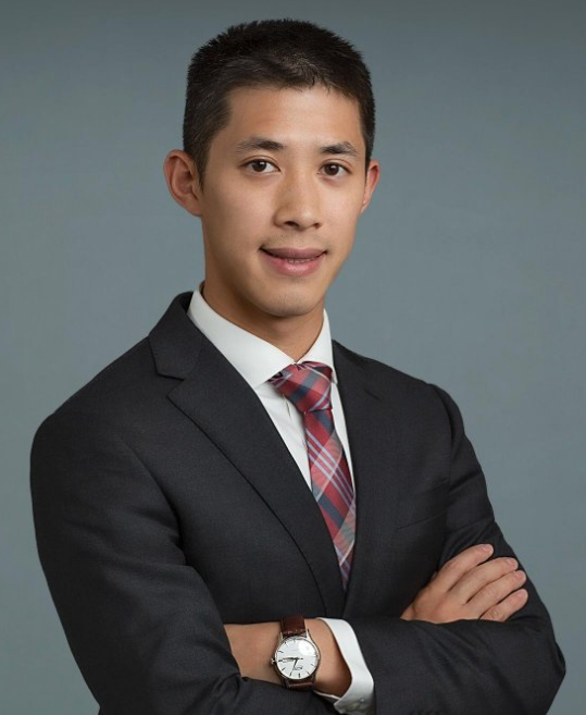 Moses M. Tam合作论文数Department of Radiation Oncology, Grossman School of Medicine, New York University3
 David W Dick合作论文数3
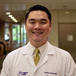 Byun David J合作论文数Center for Brachial Plexus and Traumatic Nerve Injury, Hospital for Special Surgery, New York, New York.3Lei Hu合作论文数3M. Vanderhoek合作论文数3Keisha C Mccall合作论文数3S. Osterman合作论文数2Ea Smith合作论文数2O. Dejesus合作论文数2Kenneth Bernstein合作论文数Departments of1Neurological Surgery.2C. Oh合作论文数2
 Karl Spuhler合作论文数Department of Radiation Oncology, New York University Grossman School of Medicine2S. Bowen合作论文数2S. Ayyalasomayajula合作论文数2W. Wang合作论文数2
 Adam S Jacobson合作论文数2
 Amber A. Guth合作论文数Division of Surgical Oncology, Department of Surgery, Grossman School of Medicine, New York University2
 Deborah M. Axelrod合作论文数Division of Surgical Oncology, Department of Surgery, Grossman School of Medicine, New York University2
 Fauzia Shaikh合作论文数Department of Radiation Oncology, NYU Grossman School of Medicine2
 Elizabeth A Ballegeer合作论文数2




