Objective-Leukocyte-associated immunoglobulin-like receptor-1 (LAIR-1) is a collagen receptor that belongs to the inhibitory immunoreceptor tyrosine-based inhibition motif-containing receptor family. It is an inhibitor of signaling via the immunoreceptor tyrosine-based activation motif-containing collagen receptor complex, glycoprotein VI-FcR gamma-chain. It is expressed on hematopoietic cells, including immature megakaryocytes, but is not detectable on platelets. Although the inhibitory function of LAIR-1 has been described in leukocytes, its physiological role in megakaryocytes and in particular in platelet formation has not been explored. In this study, we investigate the role of LAIR-1 in megakaryocyte development and platelet production by generating LAIR-1-deficient mice.Approach and Results-Mice lacking LAIR-1 exhibit a significant increase in platelet counts, a prolonged platelet half-life in vivo, and increased proplatelet formation in vitro. Interestingly, platelets from LAIR-1-deficient mice exhibit an enhanced reactivity to collagen and the glycoprotein VI-specific agonist collagen-related peptide despite not expressing LAIR-1, and mice showed enhanced thrombus formation in the carotid artery after ferric chloride injury. Targeted deletion of LAIR-1 in mice results in an increase in signaling downstream of the glycoprotein VI-FcR gamma-chain and integrin alpha IIb beta 3 in megakaryocytes because of enhanced Src family kinase activity.Conclusions-Findings from this study demonstrate that ablation of LAIR-1 in megakaryocytes leads to increased Src family kinase activity and downstream signaling in response to collagen that is transmitted to platelets, rendering them hyper-reactive specifically to agonists that signal through Syk tyrosine kinases, but not to G-protein-coupled receptors.
BackgroundInherited platelet function disorders (PFDs) are heterogeneous, and identification of the underlying genetic defects is difficult when based solely on phenotypic and clinical features of the patient.ObjectiveTo analyze 329 genes regulating platelet function, number, and size in order to identify candidate gene defects in patients with PFDs.Patients/methodsTargeted analysis of candidate PFD genes was undertaken after next-generation sequencing of exomic DNA from 18 unrelated index cases with PFDs who were recruited into the UK Genotyping and Phenotyping of Platelets (GAPP) study and diagnosed with platelet abnormalities affecting either Gi signaling (n=12) or secretion (n=6). The potential pathogenicity of candidate gene defects was assessed using computational predictive algorithms.ResultsAnalysis of the 329 candidate PFD genes identified 63 candidate defects, affecting 40 genes, among index cases with Gi signaling abnormalities, while 53 defects, within 49 genes, were identified among patients with secretion abnormalities. Homozygous gene defects were more commonly associated with secretion abnormalities. Functional annotation analysis identified distinct gene clusters in the two patient subgroups. Thirteen genes with significant annotation enrichment for intracellular signaling' harbored 16 of the candidate gene defects identified in nine index cases with Gi signaling abnormalities. Four gene clusters, representing 14 genes, with significantly associated gene ontology annotations were identified among the cases with secretion abnormalities, the most significant association being with establishment of protein localization.'ConclusionOur findings demonstrate the genetic complexity of PFDs and highlight plausible candidate genes for targeted analysis in patients with platelet secretion and Gi signaling abnormalities.
M. L . JONES* , S . L . MURDEN* , D . BEM , S . J . MU N DELL , P . G I SSEN§ , M. E . DALY– , S . P . WATSO N , and A . D . MUMFORD, * ON BEHALF O F T HE UK GAPP STUDY G ROUP *Bristol Heart Institute & School of Cellular and Molecular Medicine, University of Bristol, Bristol; Centre for Cardiovascular Sciences, College of Medical and Dental Sciences, University of Birmingham, Birmingham; School of Physiology and Pharmacology, University of Bristol, Bristol; §Institute of Child Health and MRC Laboratory for Molecular Cell Biology, University College London, London; and –Department of Cardiovascular Science, University of Sheffield, Sheffield, UK
The activation of NF-κB and phosphatidylinositol-3 (PI3) kinase by TNF-α and TRAIL overrides the pro-apoptotic effects of these ligands in carcinoma cells and hinders their therapeutic application. In this report we show that CD40 ligand, another member of the TNF superfamily, also triggers the activation of these signalling pathways but, importantly, utilises only the PI3 kinase cascade for anti-apoptotic responses, inasmuch as suppression of PI3 kinase but not NF-κB sensitises carcinoma cells to CD40L-induced apoptosis. Therefore, NF-κB activation does not always confer anti-apoptotic effects. Moreover, no cross-talk between the two pathways was observed, as the specific suppression of PI3 kinase with chemical inhibitors did not influence CD40-mediated IκBα phosphorylation and degradation or NF-κB binding and transactivation. Similarly, whilst suppression of Akt expression by RNA interference sensitised tumour cells to CD40L-induced apoptosis, it had no effect on CD40-mediated IκBα degradation. These data provide new evidence for the role of NF-κB and PI3 kinase/Akt in phenotypic effects mediated by CD40 ligation and highlight differences in the mechanisms by which TNF family members regulate apoptosis in carcinoma cells.
 Steve P. Watson合作论文数Institute of Cardiovascular Sciences, University of Birmingham;Department of Cardiovascular Sciences, College of Medicine and Health, University of Birmingham5
 Paul Gissen合作论文数4
 Marie Lordkipanidze合作论文数Department of Pharmacy, Hôpital du Sacré-Cœur de Montréal3Martina Daly合作论文数Department of Infection, Immunity and Cardiovascular Disease, The Medical School, Faculty of Medicine, Dentistry & Health, The University of Sheffield3Andrew D. Mumford合作论文数Clinical Sciences Centre;Faculty of Medicine;Imperial College;Hammersmith Hospital;Imperial College, Hammersmith Hospital Campus3Jones M L合作论文数2
 Drake Sian合作论文数2
 Neil Morgan合作论文数Institute of Cardiovascular Sciences, College of Medical and Dental Sciences, University of Birmingham2J Rivera合作论文数1A. Mazharian合作论文数1G. C. Lowe合作论文数1B. Dawood合作论文数1MA Kurian合作论文数1I A Aligianis合作论文数1R Nunes-Bastos合作论文数1Fc Bond合作论文数1M. Jones合作论文数1Michael A. Simpson合作论文数1Ga Kirby合作论文数1Imran Masood合作论文数1Jm Graham合作论文数1Ss Pasha合作论文数1Ar Cullinane合作论文数1Aa Straatman-Iwanowska合作论文数1Mtw Handley合作论文数1Lydie Burglen合作论文数1S. Fletcher合作论文数1Z Ahmed合作论文数1
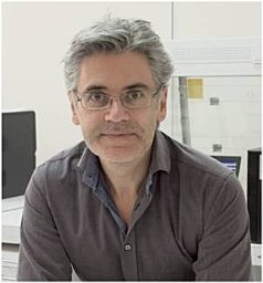 Eliopoulos Aristides G合作论文数Mol & Cellular Biol Lab, Univ Crete1
 Stuart Mundell合作论文数1
 Jayashree Motwani合作论文数1
 Stockley Jacqueline合作论文数1
 Lawrence Young合作论文数Department of Biomedical Sciences, Warwick Medical School, University of Warwick1
 Gillian C Lowe合作论文数1
 Isabel Sanchez-Guiu合作论文数Universidad de Murcia, Hospital Universitario Morales Meseguer1
 David L. Williams合作论文数Department of Surgery, Quillen College of Medicine, East Tennessee State University1
 Michael Makris合作论文数Department of Infection, Immunity and Cardiovascular Disease, The Medical School, The University of Sheffield1
 Francis Barr合作论文数Department of Biochemistry, Medical Sciences Division, University of Oxford1Heising Silke合作论文数1
 William Dobyns合作论文数Department of Pediatrics, Medical School, University of Minnesota;Division of Genetics and Metabolism, University of Minnesota1
 Keith Foster合作论文数1
 Peter Collins合作论文数School of Medicine, Cardiff University1Stuart J. Mundell合作论文数1
 Alisdair McNeill合作论文数Department of Neuroscience, The Medical School, Faculty of Medicine, Dentistry & Health, The University of Sheffield1Yotis A. Senis合作论文数Institute of Biomedical Research, University of Birmingham1Yavor Hadzhiev合作论文数1Linde Meyaard合作论文数1Clare C. Davies合作论文数Institute of Cancer and Genomic Sciences, College of Medical and Dental Sciences, University of
Birmingham, B15 2TT, United Kingdom1
Alisdair McNeill合作论文数Department of Neuroscience, The Medical School, Faculty of Medicine, Dentistry & Health, The University of Sheffield1Yotis A. Senis合作论文数Institute of Biomedical Research, University of Birmingham1Yavor Hadzhiev合作论文数1Linde Meyaard合作论文数1Clare C. Davies合作论文数Institute of Cancer and Genomic Sciences, College of Medical and Dental Sciences, University of
Birmingham, B15 2TT, United Kingdom1 Ben Johnson合作论文数1




