Telehealth and telemedicine have encountered explosive growth since the beginning of the COVID-19 pandemic, resulting in increased access to care for patients located far from medical centers and clinics. Subspecialty clinicians in behavioral neurology & neuropsychiatry (BNNP) have implemented the use of telemedicine platforms to perform cognitive examinations that were previously office based. In this perspective article, BNNP clinicians at Massachusetts General Hospital (MGH) describe their experience performing cognitive examinations via telemedicine. The article reviews the goals, prerequisites, advantages, and potential limitations of performing a video- or telephone-based telemedicine cognitive examination. The article shares the approaches used by MGH BNNP clinicians to examine cognitive and behavioral areas, such as orientation, attention and executive functions, language, verbal learning and memory, visual learning and memory, visuospatial function, praxis, and abstract abilities, as well as to survey for neuropsychiatric symptoms and assess activities of daily living. Limitations of telemedicine-based cognitive examinations include limited access to and familiarity with telecommunication technologies on the patient side, limitations of the technology itself on the clinician side, and the limited psychometric validation of virtual assessments. Therefore, an in-person examination with a BNNP clinician or a formal in-person neuropsychological examination with a neuropsychologist may be recommended. Overall, this article emphasizes the use of standardized cognitive and behavioral assessment instruments that are either in the public domain or, if copyrighted, are nonproprietary and do not require a fee to be used by the practicing BNNP clinician.
Studies investigating the effects of language intervention on the re-organization of language networks in chronic aphasia have resulted in mixed findings, likely related to-among other factors-the language function targeted during treatment. The present study investigated the effects of the type of treatment provided on neural reorganization. Seventy individuals with chronic stroke-induced aphasia, recruited from three research laboratories and meeting criteria for agrammatism, anomia or dysgraphia were assigned to either treatment (N = 51) or control (N = 19) groups. Participants in the treatment group received 12-weeks of language intervention targeting sentence comprehension/production, naming, or spelling. At baseline and post-testing, all participants performed an fMRI story comprehension task, with blocks of auditorily-presented stories alternated with blocks of reversed speech. Participants in the treatment, but not control, group significantly improved in the treated language domain. FMRI region-of-interest (ROI) analyses, conducted within regions that were either active (or homologous to active) regions in a group of 22 healthy participants on the story comprehension task, revealed a significant increase in activation from pre-to post-treatment in right-hemisphere homologues of these regions for participants in the sentence and spelling, but not naming, treatment groups, not predicted by left-hemisphere lesion size. For the sentence (but not the spelling) treatment group, activation changes within right-hemisphere homologues of language regions were positively associated with changes in measures of verb and sentence comprehension. These findings support previous research pointing to recruitment of right hemisphere tissue as a viable route for language recovery and suggest that sentence-level treatment may promote greater neuroplasticity on naturalistic, language comprehension tasks, compared to word-level treatment.
Inconsistent findings have been reported about the impact of structural disconnections on language function in post-stroke aphasia. This study investigated patterns of structural disconnections associated with chronic language impairments using disconnectome maps.Seventy-six individuals with post-stroke aphasia underwent a battery of language assessments and a structural MRI scan. Support-vector regression disconnectome-symptom mapping analyses were performed to examine the correlations between disconnectome maps, representing the probability of disconnection at each white matter voxel and different language scores. To further understand whether significant disconnections were primarily representing focal damage or a more extended network of seemingly preserved but disconnected areas beyond the lesion site, results were qualitatively compared to support-vector regression lesion-symptom mapping analyses.Part of the left white matter perisylvian network was similarly disconnected in 90% of the individuals with aphasia. Surrounding this common left perisylvian disconnectome, specific structural disconnections in the left fronto-temporo-parietal network were significantly associated with aphasia severity and with lower performance in auditory comprehension, syntactic comprehension, syntactic production, repetition and naming tasks. Auditory comprehension, repetition and syntactic processing deficits were related to disconnections in areas that overlapped with and extended beyond lesion sites significant in SVR-LSM analyses. In contrast, overall language abilities as measured by aphasia severity and naming seemed to be mostly explained by focal damage at the level of the insular and central opercular cortices, given the high overlap between SVR-DSM and SVR-LSM results for these scores.While focal damage seems to be sufficient to explain broad measures of language performance, the structural disconnections between language areas provide additional information on the neural basis of specific and persistent language impairments at the chronic stage beyond lesion volume. Leveraging routinely available clinical data, disconnectome mapping furthers our understanding of anatomical connectivity constraints that may limit the recovery of some language abilities in chronic post-stroke aphasia.
This study investigates the relationship between verb-related morphosyntactic production (VRMP) and locality (i.e., critical cue being adjacent to the target or not), verbal Working Memory (vWM), nonverbal/visuospatial WM (nvWM), verbal short-term memory (vSTM), nonverbal/visuospatial STM (nvSTM), speed of processing, and education. Eighty healthy middle-aged and older Greek-speaking participants were administered a sentence completion task tapping into production of subject–verb Agreement, Time Reference/Tense, and grammatical Aspect in local and nonlocal configurations, and cognitive tasks tapping into vSTM, nvSTM, vWM, nvWM, and speed of processing. Aspect elicited worse performance than Time Reference and Agreement, and Time Reference elicited worse performance than Agreement. There were main effects of vSTM, vWM, education, and locality: the greater the participants’ vSTM/vWM capacity, and the higher their educational level, the better their VRMP; nonlocal configurations elicited worse performance on VRMP than local configurations. Moreover, vWM affected Aspect and Time Reference/Tense more than Agreement, and education affected VRMP more in local than in nonlocal configurations. Lastly, locality affected Agreement and Aspect (with nonlocal configurations eliciting more agreement and aspect errors than local configurations) but not Time Reference. That vSTM/vWM (but not nvSTM/nvWM) were found to subserve VRMP suggests that VRMP is predominantly supported by domain-specific, not by domain-general, memory resources. The main effects of vWM and vSTM suggest that both the processing and storage components of WM are relevant to VRMP. That vWM (but not vSTM) interacts with production of Aspect, Time Reference, and Agreement suggests that Aspect and Time Reference are computationally more demanding than Agreement. These findings are consistent with earlier findings that, in individuals with aphasia, vWM interacts with production of Aspect, Time Reference, and Agreement. The differential effect of education on VRMP in local vs. nonlocal configurations could be accounted for by assuming that education is a proxy for an assumed procedural memory system that is sensitive to frequency patterns in language and better supports VRMP in more frequent than in less frequent configurations. In the same vein, the interaction between locality and the three morphosyntactic categories might reflect the statistical distribution of local vs. nonlocal Aspect, Agreement, and Time Reference/Tense in Greek.
Stroke-induced alterations in cerebral blood flow (perfusion) may contribute to functional language impairments in chronic aphasia, particularly in perilesional tissue. Abnormal perfusion in this region may also serve as a biomarker for predicting functional improvements with behavioral treatment interventions. Using pseudo-continuous arterial spin labeling in magnetic resonance imaging (MRI), we examined perfusion in chronic aphasia, in perilesional rings in the left hemisphere and their right hemisphere homologues. In the left hemisphere we found a gradient pattern of decreasing perfusion closer to the lesion. The opposite pattern was found in the right hemisphere, with significantly increased perfusion close to the lesion homologue. Perfusion was also increased in the right hemisphere lesion homologue region relative to the surrounding tissue. We next examined changes in perfusion in two groups: one group who underwent MRI scanning before and after three months of a behavioral treatment intervention that led to significant language gains, and a second group who was scanned twice at a three-month interval without a treatment intervention. For both groups, there was no difference in perfusion over time in either the left or the right hemisphere. Moreover, within the treatment group pre-treatment perfusion scores did not predict treatment response; neither did pre-treatment perfusion predict post-treatment language performance. These results indicate that perfusion is chronically abnormal in both hemispheres, but chronically abnormal perfusion did not change in response to our behavioral treatment interventions, and did not predict responsiveness to language treatment.
An 81-year-old right-handed White non-Hispanic previously healthy woman with glaucoma presented with acute weakness and slurred speech nearly 10 hours after last seen well. She was found to have new onset atrial fibrillation. Her NIH stroke scale (NIHSS) was 18 with points given for failure to answer questions, left lower facial paralysis, right gaze preference with ability to cross midline, left field cut, left hemiplegia, severe dysarthria, mild aphasia, and severe left sensory neglect.
Can sentence comprehension impairments in aphasia be explained by difficulties arising from dependency completion processes in parsing? Two distinct models of dependency completion difficulty are investigated, the Lewis and Vasishth (2005) activation-based model and the direct-access model (DA; McElree, 2000). These models' predictive performance is compared using data from individuals with aphasia (IWAs) and control participants. The data are from a self-paced listening task involving subject and object relative clauses. The relative predictive performance of the models is evaluated using k-fold cross-validation. For both IWAs and controls, the activation-based model furnishes a somewhat better quantitative fit to the data than the DA. Model comparisons using Bayes factors show that, assuming an activation-based model, intermittent deficiencies may be the best explanation for the cause of impairments in IWAs, although slowed syntax and lexical delayed access may also play a role. This is the first computational evaluation of different models of dependency completion using data from impaired and unimpaired individuals. This evaluation develops a systematic approach that can be used to quantitatively compare the predictions of competing models of language processing.
Predicting the potential recovery outcome of post-stroke aphasia remains a challenging task. Our previous work[10] applied machine learning algorithms to predict participant response to therapy using a complex set of brain and behavioral data in individuals with post-stroke aphasia. The present work explores the additional predictive value of cognitive composite scores (CS), which measure visuo-spatial processing and verbal working memory; high-dimensional resting-state (RS) functional magnetic resonance imaging (fMRI) data, which measures the functional connectivity between brain regions; and diffusion tensor imaging (DTI) data, which provides information related to microstructural integrity via fractional anisotropy (FA) values. We first perform feature selection on the RS data as it has about 5 times more features than than all the other feature-sets combined. Next, we append these RS features, CS scores, and FA values to our existing data set. Finally, we train Support Vector Machine (SVM) and Random Forest (RF) classifiers for various combinations of feature-sets and compare their performance in terms of accuracy, F1-score, sensitivity and selectivity. Results show that combinations of feature-sets outperform most individual feature-sets and whereas each feature-set is present among the top 20 combinations, many of them contain RS.
Background: Despite growing evidence that anxiety is critical in the development and maintenance of postconcussion symptoms after mild traumatic brain injury (mTBI), little is known about potential mechanisms through which anxiety may affect these symptoms. Objective: To test the strength and reliability of cognitive (pain catastrophizing) and behavioral (limiting behaviors) pathways mediating the relationship between anxiety and postconcussion symptoms among patients with mTBI. Method: Patients with mTBI (N = 57) completed self-report measures of anxiety, postconcussion symptoms, pain catastrophizing, and limiting behavior. After preliminary simple-mediation models (for pain catastrophizing and limiting behavior separately), we ran a multiplemediation model (pathways modeled simultaneously). Bootstrapping with 10,000 resampling iterations assessed mediation reliability. Results: In preliminary simple mediation models, both pain catastrophizing (beta = 0.24, 95% confidence interval [CI] = 0.03-0.44, P = 0.02) and limiting behaviors (beta = 0.14, 95% CI = 0.03-0.26, P = 0.01) partially mediated the relationship between anxiety and postconcussion symptoms. In the multiple mediation model, pain catastrophizing was a less reliable but numerically stronger mediator (beta = 0.19, 95% CI = -0.01 to 0.38; P = 0.05) and explained more variance in postconcussion symptoms (R-2 = 0.41) than limiting behavior (beta = 0.10, 95% CI = 0.02-0.21, P = 0.03; R-2 = 0.22), although mediators did not significantly differ in strength (beta = 0.08, 95% CI = -0.16 to 0.32; P = 0.49). Results provide novel evidence for the role of pain catastrophizing and limiting behaviors in explaining the association between anxiety and postconcussion symptoms. Addressing both factors may improve the recovery trajectory of individuals with mTBI. Emphasizing limiting behavior may yield more consistent and reliable effects. Conclusion: Results support developing interventions to directly target anxiety, for pain catastrophizing, and for activity engagement despite symptoms, to decrease symptom severity among patients with mTBI.
Background White matter hyperintensities (WMH) are a radiological marker of brain health that has been associated with language status in poststroke aphasia; however, its association with language treatment outcomes remains unknown. Objective To determine whether WMH in the right hemisphere (RH) predict response to language therapy independently from demographics and stroke lesion-related factors in poststroke aphasia. Methods We used the Fazekas scale to rate WMH in the RH in 30 patients with poststroke aphasia who received language treatment. We developed ordinal regression models to examine language treatment effects as a function of WMH severity after controlling for aphasia severity, stroke lesion volume, time post onset, age, and education level. We also evaluated associations between WMH severity and both pre-treatment naming ability and executive function. Results The severity of WMH in the RH predicted treatment response independently from demographic and stroke-related factors such that patients with less severe WMH exhibited better treatment outcome. WMH scores were not significantly correlated with pretreatment language scores, but they were significantly correlated with pretreatment scores of executive function. Conclusion We suggest that the severity of WMH in the RH is a clinically relevant predictor of treatment response in this population.
The potential recovery of post-stroke aphasia is highly variable and the rehabilitation outcomes are difficult to predict. This interdisciplinary collaboration builds on data collected as part of a large set of behavioral and brain variables in patients with post-stroke aphasia, charting the course of recovery associated with therapy across language domains and examining the basis of neuroplasticity. In this pilot study, we created and tested a predictive framework based on a subset of the data collected and developed machine-learning algorithms that take as input a complex set of brain and behavioral features to classify and predict the participants' responsiveness to therapy. We developed Random Forest models that enabled us to rank the importance of these features. We then compared the contributions of different feature sets and discussed their physiological implications. Our preliminary results suggest the potential of our framework, and, thus, this study takes an important first step towards predicting individualized rehabilitation outcomes.
Functional neuroimaging and lesion-symptom mapping investigations implicate a left frontal–temporal–parietal network for sentence processing. The majority of studies have focused on sentence comprehension, with fewer in the domain of sentence production, which have not fully elucidated overlapping and/or unique brain structures associated with the two domains, particularly for sentences with noncanonical word order. Using voxel-based lesion symptom mapping (VLSM) we examined the relationship between lesions within the left hemisphere language network and both sentence comprehension and production of simple and complex syntactic structures in 76 participants with chronic stroke-induced aphasia. Results revealed shared regions across domains in the anterior and posterior superior temporal gyri (aSTG, pSTG), and the temporal pole (adjusted for verb production/comprehension). Additionally, comprehension was associated with lesions in the anterior and posterior middle temporal gyri (aMTG, pMTG), the MTG temporooccipital regions, SMG/AG, central and parietal operculum, and the insula. Subsequent VLSM analyses (production versus comprehension) revealed critical regions associated with each domain: anterior temporal lesions were associated with production; posterior temporo-parietal lesions were associated with comprehension, implicating important roles for regions within the ventral and dorsal stream processing routes, respectively. Processing of syntactically complex, noncanonical (adjusted for canonical), sentences was associated with damage to the pSTG across domains, with additional damage to the pMTG and IPL associated with impaired sentence comprehension, suggesting that the pSTG is crucial for computing noncanonical sentences across domains and that the pMTG, and IPL are necessary for re-analysis of thematic roles as required for resolution of long-distance dependencies. These findings converge with previous studies and extend our knowledge of the neural mechanisms of sentence comprehension to production, highlighting critical regions associated with both domains, and further address the mechanism engaged for syntactic computation, controlled for the contribution of verb processing.
Coronavirus 2019 (COVID-19) has profoundly impacted the well-being of society and the practice of medicine across health care systems worldwide. As with many other subspecialties, the clinical paradigm in behavioral neurology and neuropsychiatry (BN-NP) was transformed abruptly, transitioning to real-time telemedicine for the assessment and management of the vast majorities of patient populations served by our subspecialty. In this commentary, we outline themes from the BN-NP perspective that reflect the emerging lessons we learned using telemedicine during the COVID-19 pandemic. Positive developments include the ability to extend consultations and management to patients in our high-demand field, maintenance of continuity of care, enhanced ecological validity, greater access to a variety of well-reimbursed telemedicine options (telephone and video) that help bridge the digital divide, and educational and research opportunities. Challenges include the need to adapt the mental state examination to the telemedicine environment, the ability to perform detailed motor neurologic examinations in patients where motor features are important diagnostic considerations, appreciating nonverbal cues, managing acute safety and behavioral concerns in less controlled environments, and navigating intervention-based (neuromodulation) clinics requiring in-person contact. We hope that our reflections help to catalyze discussions that should take place within the Society for Behavioral and Cognitive Neurology, the American Neuropsychiatric Association, and allied organizations regarding how to optimize real-time telemedicine practices for our subspecialty now and into the future.
A Woman with Type 1 Diabetes Mellitus and Obtundation A 59-year-old woman with type 1 diabetes and a 2-year history of cognitive decline presented with obtundation. There was diffuse, symmetric hyp...
Functional neurologic disorder (FND), although neglected for much of the 20th century, is among the most common conditions encountered by neurologists across clinical settings. High prevalence rates and limited provider expertise in FND have created a considerable need to develop educational initiatives and practical suggestions to guide neurologists in training working with this population. To help avoid diagnostic errors, trainees should keep in mind that (1) marginally positive functional examination signs have low specificity; (2) FND can coexist with other neurologic comorbidities; and (3) bizarre, not previously encountered, neurologic presentations should not be mistakenly diagnosed as FND. Furthermore, trainees should be encouraged to longitudinally follow in their clinics a subset of patients with FND to develop the interview, diagnostic, and neuropsychiatric skills needed to effectively care for this population. As the landscape of neurologic care evolves, neurologists with expertise in FND should advise on shaping elements of the educational curriculum for neurology residents.
Investigating the neurobiology of language impairment and treatment in chronic stroke aphasia using fMRI requires an understanding of measurement variability within and between participants. In this multicenter study, we evaluated the scan–rescan reliability of an auditory and visual (written) story comprehension paradigm in stroke participants with aphasia ( N = 65) and healthy controls ( N = 22). The multi‐modal task was conducted twice (~1 week apart) on separate visits upon study enrolment and twice again at completion three months later. A non‐language visuomotor task was studied in the aphasia group only, which was conducted once per time point (3 months apart). While participants were asked to make responses during the comprehension task, these in‐scanner responses were not recorded. Reliability was assessed using intraclass correlation coefficient (ICC) at both group and individual participant levels. The visual story comprehension condition had higher reliability than the auditory condition in both groups, with participants with aphasia exhibiting lower reliability than controls in both conditions (stroke ICC = .43, healthy ICC = .81). Differences in reliability within the group of participants with aphasia were found to be partially explained by overall language impairment as well as greater head motion. In the participants with aphasia, the visuomotor paradigm was found to have greater reliability than the story comprehension task at equivalent interscan intervals (visuomotor = 0.50, comprehension = 0.34), and its reliability was not associated with language impairment. This work highlights the importance of considering the reliability of fMRI tasks in aphasia research, provides strategies to improve reliability and has potential implications for the field of clinical neuroimaging in general.
Comprehending and producing sentences is a complex endeavor requiring the coordinated activity of multiple brain regions. We examined three issues related to the brain networks underlying sentence comprehension and production in healthy individuals: First, which regions are recruited for sentence comprehension and sentence production? Second, are there differences for auditory sentence comprehension vs. visual sentence comprehension? Third, which regions are specifically recruited for the comprehension of syntactically complex sentences? Results from activation likelihood estimation (ALE) analyses (from 45 studies) implicated a sentence comprehension network occupying bilateral frontal and temporal lobe regions. Regions implicated in production (from 15 studies) overlapped with the set of regions associated with sentence comprehension in the left hemisphere, but did not include inferior frontal cortex, and did not extend to the right hemisphere. Modality differences between auditory and visual sentence comprehension were found principally in the temporal lobes. Results from the analysis of complex syntax (from 37 studies) showed engagement of left inferior frontal and posterior temporal regions, as well as the right insula. The involvement of the right hemisphere in the comprehension of these structures has potentially important implications for language treatment and recovery in individuals with agrammatic aphasia following left hemisphere brain damage.
Event Abstract Back to Event Structural disconnection maps associated with language impairment in chronic aphasia Anne Billot1*, Michel Thiebaut De Schotten2, 3, 4, Todd Parrish5, 6, Cynthia K. Thompson5, 7*, Brenda Rapp8, David Caplan9 and Swathi Kiran10 1 Division of Graduate Medical Sciences, Boston University, United States 2 Sorbonne Universités, France 3 INSERM U1127 Institut du Cerveau et de la Moelle épinière (ICM), France 4 Université de Bordeaux, France 5 Feinberg School of Medicine, Northwestern University, United States 6 Department of Radiology, Feinberg School of Medicine, Northwestern University, United States 7 Center for the Neurobiology of Language Recovery, Northwestern University, United States 8 Department of Cognitive Science, Krieger School of Arts and Sciences, Johns Hopkins University, United States 9 Massachusetts General Hospital, Harvard Medical School, United States 10 Department of Speech, Language and Hearing Sciences, Boston University, United States The analysis of residual white matter connections between brain regions is essential to understand language impairment in post-stroke aphasia [1]. Tractography studies have found that white matter integrity is a good predictor of language outcomes after stroke [2]. However, few studies have investigated the relationship between seemingly spared but directly disconnected white matter pathways and language impairments in chronic aphasia. Probabilistic maps of disconnection based on anatomical connectomes of healthy controls are a complementary method to measure the impact of the lesion on neuronal circuits [3]. This project examined the relationship between language deficits and the degree of structural disconnections in the brain of people with chronic aphasia. This study includes 76 individuals with chronic aphasia resulting from a single left-hemisphere stroke. Participants were recruited from three sites (Boston, Johns Hopkins, and Northwestern Universities) and underwent an MRI and DTI imaging and an extensive language assessment (Western Aphasia Battery-Revised - WAB [4]; Northwestern Assessment of Verbs and Sentences - NAVS, [5]; Psycholinguistic Assessments of Language Processing - PALPA [6]). 75 individuals have completed NAVS (sentence production) and 74 have completed PALPA. Probability maps of white matter tracts' disconnection were computed for each participant with the “disconnectome map” tool of the BCBToolkit [3] using 178 healthy controls diffusion weighting imaging datasets (7T, www.humanconnectomeproject.org). For each individual with aphasia, the lesion mask was registered in MNI space and used as a seed to produce the tractography of healthy controls’ fibers passing through the lesion. Then, at each voxel, tractographies from healthy controls were binarized and summed to produce a percentage overlap map, and consequently, the probability of disconnection (range 0-100%). A threshold of 50% (50% of controls had tracts passing through the lesion) was applied for this study. Nonparametric permutation analyses and threshold-free cluster enhancement technique were used to evaluate the correlation between the probability of white matter disconnection and each language score (WAB: aphasia severity, auditory verbal comprehension, repetition, naming; NAVS: sentence comprehension, sentence production priming test; PALPA-40: spelling). Demographic variables and lesion size were included as covariates of no interest in the general linear model implemented in Randomise (FSL package, [7]). Family-wise error rate was controlled at p<0.001 (Figure 1). Atlasquery tool (FSL package, [7]) was used to identify disconnected tracts significantly correlated with each language score. Tracts with a probability of overlapping with more than 1% of the statistical mapping of the disconnections were selected. Table 1 shows that disconnected tracts contributing most to all language impairments included in this study were the left external capsule, the left posterior limb of the internal capsule, the left cerebral peduncle and the middle cerebellar peduncle (p=0.001). In addition, specific linguistic domain scores were correlated with different disconnected tracts including the Left IFOF, ILF and SLF (Table 1). This study identified white matter tracts’ disconnections associated with aphasia severity and the degree of impairment in verbal comprehension, repetition, naming, sentence processing, and spelling. Disconnectome mapping confirms previous findings showing that structural damage is related to language outcomes beyond cortical necrosis. Figure 1 Figure 2 Acknowledgements This work was supported by the NIH/NIDCD Clinical Research Center Grant, P50DC012283, and the Center for the Neurobiology of Language Recovery. References [1] Catani, M., & Mesulam, M. (2008). The arcuate fasciculus and the disconnection theme in language and aphasia: history and current state. cortex, 44(8), 953-961. [2] Kiran, S., & Thompson, C. K. (2019). Neuroplasticity of Language Networks in Aphasia: Advances, Updates and Future Challenges. Frontiers in neurology, 10, 295. [3] Foulon, C., Cerliani, L., Kinkingnehun, S., Levy, R., Rosso, C., Urbanski, M., ... & Thiebaut de Schotten, M. (2018). Advanced lesion symptom mapping analyses and implementation as BCBtoolkit. GigaScience, 7(3), giy004. [4] Kertesz, A. (2007). WAB-R: Western aphasia battery-revised. PsychCorp. [5] Thompson, C. K. (2012). Northwestern Assessment of Verbs and Sentences (NAVS). [6] Kay, J., Lesser, R., & Coltheart, M. (1996). Psycholinguistic assessments of language processing in aphasia (PALPA): An introduction. Aphasiology, 10(2), 159-180. [7] Winkler, A. M., Ridgway, G. R., Webster, M. A., Smith, S. M., & Nichols, T. E. (2014). Permutation inference for the general linear model. Neuroimage, 92, 381-397. Keywords: Aphasia, Stroke, White matter (WM), Language, Disconnection, Magnetic Resonance Image (MRI) Conference: Academy of Aphasia 57th Annual Meeting, Macau, Macao, SAR China, 27 Oct - 29 Oct, 2019. Presentation Type: Platform presentation Topic: Eligible for student award Citation: Billot A, Thiebaut De Schotten M, Parrish T, Thompson CK, Rapp B, Caplan D and Kiran S (2019). Structural disconnection maps associated with language impairment in chronic aphasia. Front. Hum. Neurosci. Conference Abstract: Academy of Aphasia 57th Annual Meeting. doi: 10.3389/conf.fnhum.2019.01.00040 Copyright: The abstracts in this collection have not been subject to any Frontiers peer review or checks, and are not endorsed by Frontiers. They are made available through the Frontiers publishing platform as a service to conference organizers and presenters. The copyright in the individual abstracts is owned by the author of each abstract or his/her employer unless otherwise stated. Each abstract, as well as the collection of abstracts, are published under a Creative Commons CC-BY 4.0 (attribution) licence (https://creativecommons.org/licenses/by/4.0/) and may thus be reproduced, translated, adapted and be the subject of derivative works provided the authors and Frontiers are attributed. For Frontiers’ terms and conditions please see https://www.frontiersin.org/legal/terms-and-conditions. Received: 06 May 2019; Published Online: 09 Oct 2019. * Correspondence: Mx. Anne Billot, Division of Graduate Medical Sciences, Boston University, Boston, Massachusetts, 02118, United States, abillot@bu.edu Prof. Cynthia K Thompson, Center for the Neurobiology of Language Recovery, Northwestern University, Evanston, Illinois, 60208, United States, ckthom@northwestern.edu Login Required This action requires you to be registered with Frontiers and logged in. To register or login click here. Abstract Info Abstract The Authors in Frontiers Anne Billot Michel Thiebaut De Schotten Todd Parrish Cynthia K Thompson Brenda Rapp David Caplan Swathi Kiran Google Anne Billot Michel Thiebaut De Schotten Todd Parrish Cynthia K Thompson Brenda Rapp David Caplan Swathi Kiran Google Scholar Anne Billot Michel Thiebaut De Schotten Todd Parrish Cynthia K Thompson Brenda Rapp David Caplan Swathi Kiran PubMed Anne Billot Michel Thiebaut De Schotten Todd Parrish Cynthia K Thompson Brenda Rapp David Caplan Swathi Kiran Related Article in Frontiers Google Scholar PubMed Abstract Close Back to top Javascript is disabled. Please enable Javascript in your browser settings in order to see all the content on this page.
Event Abstract Back to Event Effect of word- and sentence-level interventions on language network re-organization in chronic aphasia: an fMRI study. Elena Barbieri1, 2*, James Higgins1, 3, Kaitlyn Litcofsky1, 2, Kathy Xie1, 2, David Caplan1, 4, Brenda Rapp1, 5, Swathi Kiran1, 6, Todd Parrish1, 3 and Cynthia K. Thompson1, 2 1 Center for the Neurobiology of Language Recovery, Northwestern University, United States 2 Department of Communication Sciences and Disorders, Bouvé College of Health Sciences, Northeastern University, United States 3 Department of Radiology, Feinberg School of Medicine, Northwestern University, United States 4 Department of Neurology, Massachusetts General Hospital, Harvard Medical School, United States 5 Department of Cognitive Science, Krieger School of Arts and Sciences, Johns Hopkins University, United States 6 Department of Speech, Language and Hearing Sciences, Boston University, United States Studies investigating the effects of language intervention on the re-organization of language networks in chronic aphasia have resulted in mixed findings, likely related to – among other factors – the language function targeted during treatment and the language task used to elicit brain activation [1]. Most studies have focused on naming, reporting greater activation (post- vs. pre-treatment) within intact regions in the left hemisphere (LH); however, studies have also found recruitment of regions in the right hemisphere (RH), positively correlated with treatment outcome [2]. The present study investigated the effects of the type of treatment provided for patients with chronic aphasia on neural activation using an auditory story comprehension task that in healthy participants reliably recruits a left fronto-temporo-parietal network [3]. We hypothesized that behavioral improvements associated with sentence processing, but not naming or spelling, treatments would result in shifts in activation using this task. Eighty-five individuals with chronic LH stroke-induced aphasia, recruited from three research laboratories (Northwestern University, NU; Boston University, BU; Johns Hopkins University, JHU) were assigned to either a language treatment (N=61) or control group (N=24). Participants in the treatment group received approximately 12 weeks of language treatment targeting one of three language domains: sentence comprehension/production (NU), naming (BU) or spelling (JHU). At baseline and post-testing, participants in both groups underwent language testing and performed an fMRI story comprehension task, which included alternating blocks of auditorily-presented short stories and a control condition (reversed speech) and required participants to listen to the stories and answer comprehension questions. At all recruitment sites, participants in the treatment, but not control, group evinced significant behavioral gains in the treated language domains. Region-of-Interest analyses of the fMRI story comprehension data revealed a significant increase in activation from pre- to post-treatment for the NU treatment group, but no changes in activation were found in the BU or JHU treatment groups, or in the control group. Results for the NU treatment group showed post-treatment upregulation of regions within the language network and were restricted to the RH. When overlaid onto activation maps derived from a group of healthy individuals performing the same task (Figure 1), post-treatment activation maps for the NU treatment group showed post- (vs. pre-treatment) recruitment of RH regions that were either active (i.e., posterior middle temporal gyrus, inferior frontal gyrus) or homologous to active LH regions (i.e., superior frontal, precentral gyri) in healthy individuals. Increased activation was positively correlated with behavioral change (i.e., verb comprehension), derived from a Principal Component Analysis of the behavioral data. These findings indicate that the language domain targeted for treatment affects re-organization of the language network. Sentence-level language intervention impacted comprehension of naturalistic narrative language and recruited regions with the normal language network in the RH, whereas (spoken or written) word-level treatments did not, indicating that treatment that exploits specific language processes impacts brain mechanisms associated with those process. Figure 1 Acknowledgements This study is part of a larger, multi-site project examining the neurobiology of language recovery in people with aphasia (NIH-NIDCD Clinical Research Center grant P50DC012283, PI: Cynthia K. Thompson). References 1. Kiran, S., & Thompson, C. K. (2019). Neuroplasticity of Language Networks in Aphasia: Advances, Updates and Future Challenges. Frontiers in neurology, 10, 295. 2. Barbieri, E., Mack, J., Chiappetta, B., Europa, E., & Thompson, C.K. (under review). Recovery of offline and online sentence processing in aphasia: Language and domain-general network neuroplasticity. Cortex. 3. Wilson, S. M., Molnar-Szakacs, I., & Iacoboni, M. (2007). Beyond superior temporal cortex: intersubject correlations in narrative speech comprehension. Cerebral cortex, 18(1), 230-242. Keywords: neuroplasticity, sentence processing, Word Processing, Language treatment, fMRI, Aphasia Conference: Academy of Aphasia 57th Annual Meeting, Macau, Macao, SAR China, 27 Oct - 29 Oct, 2019. Presentation Type: Platform presentation Topic: Not eligible for student award Citation: Barbieri E, Higgins J, Litcofsky K, Xie K, Caplan D, Rapp B, Kiran S, Parrish T and Thompson CK (2019). Effect of word- and sentence-level interventions on language network re-organization in chronic aphasia: an fMRI study.. Front. Hum. Neurosci. Conference Abstract: Academy of Aphasia 57th Annual Meeting. doi: 10.3389/conf.fnhum.2019.01.00115 Copyright: The abstracts in this collection have not been subject to any Frontiers peer review or checks, and are not endorsed by Frontiers. They are made available through the Frontiers publishing platform as a service to conference organizers and presenters. The copyright in the individual abstracts is owned by the author of each abstract or his/her employer unless otherwise stated. Each abstract, as well as the collection of abstracts, are published under a Creative Commons CC-BY 4.0 (attribution) licence (https://creativecommons.org/licenses/by/4.0/) and may thus be reproduced, translated, adapted and be the subject of derivative works provided the authors and Frontiers are attributed. For Frontiers’ terms and conditions please see https://www.frontiersin.org/legal/terms-and-conditions. Received: 05 May 2019; Published Online: 09 Oct 2019. * Correspondence: Dr. Elena Barbieri, Center for the Neurobiology of Language Recovery, Northwestern University, Evanston, United States, elena.barbieri@northwestern.edu Login Required This action requires you to be registered with Frontiers and logged in. To register or login click here. Abstract Info Abstract The Authors in Frontiers Elena Barbieri James Higgins Kaitlyn Litcofsky Kathy Xie David Caplan Brenda Rapp Swathi Kiran Todd Parrish Cynthia K Thompson Google Elena Barbieri James Higgins Kaitlyn Litcofsky Kathy Xie David Caplan Brenda Rapp Swathi Kiran Todd Parrish Cynthia K Thompson Google Scholar Elena Barbieri James Higgins Kaitlyn Litcofsky Kathy Xie David Caplan Brenda Rapp Swathi Kiran Todd Parrish Cynthia K Thompson PubMed Elena Barbieri James Higgins Kaitlyn Litcofsky Kathy Xie David Caplan Brenda Rapp Swathi Kiran Todd Parrish Cynthia K Thompson Related Article in Frontiers Google Scholar PubMed Abstract Close Back to top Javascript is disabled. Please enable Javascript in your browser settings in order to see all the content on this page.
Purpose The present work investigated whether verbal working memory (WM) affects morphosyntactic production in configurations that do not involve or favor similarity-based interference and whether WM interacts with verb-related morphosyntactic categories and/or cue–target distance (locality). It also explored whether the findings related to the questions above lend support to a recent account of agrammatic morphosyntactic production: Interpretable Features' Impairment Hypothesis (Fyndanis, Varlokosta, & Tsapkini, 2012). Method A sentence completion task testing production of subject–verb agreement, tense/time reference, and aspect in local and nonlocal conditions and two verbal WM tasks were administered to 8 Greek-speaking persons with agrammatic aphasia (PWA) and 103 healthy participants. Results The 3 morphosyntactic categories dissociated in both groups (agreement > tense > aspect). A significant interaction emerged in both groups between the 3 morphosyntactic categories and WM. There was no main effect of locality in either of the 2 groups. At the individual level, all 8 PWA exhibited dissociations between agreement, tense, and aspect, and effects of locality were contradictory. Conclusions Results suggest that individuals with WM limitations (both PWA and healthy older speakers) show dissociations between the production of verb-related morphosyntactic categories. WM affects performance shaping the pattern of morphosyntactic production (in Greek: subject–verb agreement > tense > aspect). The absence of an effect of locality suggests that executive capacities tapped by WM tasks are involved in morphosyntactic processing of demanding categories even when the cue is adjacent to the target. Results are consistent with the Interpretable Features' Impairment Hypothesis (Fyndanis et al., 2012). Supplemental Material https://doi.org/10.23641/asha.6024428
 Gloria Sydna Waters合作论文数Department of Speech, Language & Hearing Sciences, College of Health & Rehabilitation Sciences, Boston University103
 Jennifer Michaud合作论文数24
 Swathi Kiran合作论文数Aphasia Research Laboratory, Sargent College, Boston University;Department of Speech Language and Hearing Sciences, Sargent College, Boston University;Aphasia Resource Center, Boston University;Center for Systems Neuroscience, Boston University;Center For Brain Recovery, Boston University19Gayle Dede合作论文数Sargent College, Boston University17
 Gina Kuperberg合作论文数Department of Psychology, School of Arts and Sciences, Tufts University;Department of Psychiatry, Massachusetts General Hospital16
 Cynthia K. Thompson合作论文数Department of Communication Sciences and Disorders, School of Communication, Northwestern University14
 Nancy Hildebrandt合作论文数13
 Todd Parrish合作论文数Biomedical Engineering, McCormick School of Engineering and Applied Science, Northwestern University12
 Brenda C. Rapp合作论文数Department of Psychological & Brain Sciences, Johns Hopkins University/Department of Cognitive Science, Krieger College of Arts & Sciences, Johns Hopkins University11
 Phillip J Holcomb合作论文数Department of Psychology, San Diego State University11Bradford C. Dickerson合作论文数9Nathaniel Alpert合作论文数9Hufford Rebecca合作论文数7
 Nikos Makris合作论文数Departments of Psychiatry, Massachusetts General Hospital;Departments of Neurology, Harvard Medical School7Louise Stanczak合作论文数6
 Amanda Reddy合作论文数6
 David N Kennedy合作论文数Department of Psychiatry, T.H. Chan School of Medicine, University of Massachusetts;Department of Radiology, T.H. Chan School of Medicine, University of Massachusetts;NeuroNexus Institute, T.H. Chan School of Medicine, University of Massachusetts;Department of Neuroscience, Morningside Graduate School of Biomedical Sciences, University of Massachusetts6
 Elizabeth Rochon合作论文数Rehabilitation Sciences Institute, Temerty Faculty of Medicine, University of Toronto6
 Joshua Levy合作论文数6
 David L Perez合作论文数Department of Neurology, Massachusetts General Hospital6T Sitnikova合作论文数5
 Elena Barbieri合作论文数5
 Caroline West合作论文数Department of Philosophy, School of Philosophical and Historical Inquiry, Faculty of Arts and Social Sciences, The University of Sydney5Hoover Elizabeth合作论文数5Chemali Zeina N合作论文数5
 James C. Higgins合作论文数Electronic Systems Center5
 Douglas Greve合作论文数Massachusetts General Hospital;Laboratory for Computational Neuroimaging, Athinoula A. Martinos Center for Biomedical Imaging, Mass General Research Institute;Harvard Medical School5
 Anne Billot合作论文数Sargent College, Boston University4
 Alexander Berardino合作论文数NYU, Ctr Neural Sci, New York, NY 10003 USA4
 Chaleece Wyatt Sandberg合作论文数Department of Speech and Hearing Sciences, Boston University4
 Varkanitsa Maria合作论文数Department of Linguistics, University College London (UCL),4Vanessa Baudiffier合作论文数4J. F. Bates合作论文数3A M Dale合作论文数3WC West合作论文数3MWL Chee合作论文数3Vs Caviness合作论文数3CL Leonard合作论文数3
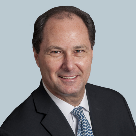 Jeremy Schmahmann合作论文数Massachusetts General Hospital/Harvard Medical School3MM Mesulam合作论文数3
 Debra Titone合作论文数Department of Psychology, Faculty of Science, McGill University3
 Roee Gutman合作论文数Department of Biostatistics, Brown University3Rigalleau F合作论文数3
 Arthur Wingfield合作论文数Department of Psychology, Brandeis University/School of Life Sciences, Brandeis University3E Chen合作论文数3
 Lauren R Moo合作论文数Department of Neurology, Johns Hopkins University School of Medicine3Evan Chen合作论文数3Donna A. Kreher合作论文数3
 Nathaniel M. Alpert合作论文数Department of Radiology, Massachusetts General Hospital3




