The sections in this article are: 1 General Considerations 1.1 Passive Changes in Splanchnic Blood Volume 1.2 Active Constriction of Resistance and Capacitance Elements 1.2.1 Direct 1.2.2 Reflex 1.3 Active and Passive Components of Sympathetically Induced Change in Splanchnic Blood Volume 2 Spleen 2.1 Innervation 2.2 Arterial Inflow 2.3 Reflexly Mediated Changes in Splenic Blood Volume 2.4 Conscious Dogs 2.5 Humans 3 Intestine 3.1 Blood Flow and Blood Volume 3.2 Sympathetic Adrenergic Nerve Activity 3.2.1 Direct Excitation of Sympathetic Nerves 3.2.2 Reflex Control of Intestinal Blood Flow and Blood Volume 3.2.3 Autoregulation of Flow and Escape from Sympathetic Adrenergic Vasoconstriction 3.3 Stress (Exercise, Excitement, and Body Heating) 3.4 Effect of Food Intake 4 Liver 4.1 Hepatic Microcirculation 4.2 Studies in Conscious Animals 4.3 Hepatic Blood Flow 4.4 Uptake and Release of Blood by the Liver 4.4.1 Increase in Hepatic Venous Pressure, Volume Expansion, and Hemorrhage 4.4.2 Excitation of Hepatic Sympathetic Nerves 4.4.3 Effect of Vasoactive Substances on Hepatic Blood Flow and Blood Volume 5 conclusion5 Conclusion
Widely distributed throughout the heart is a network of fibers connected to the medullary cardiovascular centers by nonmedullated vagal afferent fibers. When the traffic in these fibers is interrupted by vagal cooling, and the input from the arterial baroreceptors is prevented, the arterial blood pressure increases. Thus, these receptors act to inhibit tonically the vasomotor center. The receptors in the atria alter their rate of discharge with changes in atrial transmural pressure and contractility and are most active during end-inspiration and early expiration when the transmural pressure is maximal. The receptors in the ventricles respond to changes in ventricular end-diastolic pressure (preload), to the pressure generated during systole (afterload) and to changes in ventricular contractility. The cardiac mechanoreceptors have an equal or greater effect on the renal bed than the arterial mechanoreceptors and this effect is enhanced by hypercapnia. In animals, the cardiac mechanoreceptors have less control of the muscle vessels than the arterial mechanoreceptors, but the reverse is true in man. Both the cardiac and arterial mechanoreceptors can modulate the output of renin from the kidney, but the cardiac mechanoreceptors are more sensitive to small changes in blood volume. During coronary occlusion, in association with the bulging of the ischemic myocardium, the rate of discharge of these cardiac receptors is greatly increased.
Regional patterns of sequential contraction of the right and left ventricles under physiological conditions, have been described (1,2) and the important detrimental role of abnormal asynchronous contraction patterns in the heart due to aberrant electrical activation or ischemic regions has also been recognized. Such data have indicated the necessity for measurements of regional myocardial function over a major portion of the ventricular wall but for practical technological reasons most geometric measurements made directly on the dynamic intact heart involve a small number of regions of the ventricle. Simplifying shape assumptions such as those commonly used in calculations of ventricular volumes (3,4) or myo-cardial wall mass (5), as well as the arbitrary selection of frames of reference in the quantitation of regional dynamics all affect the validity of these indices to an unknown extent.
Summary o.1.Twenty-six dogs were cooled by a whole body, closed chest perfusion technique to end perfusion brain temperatures of 14.1° to 0.2° C. inclusive. 2.There were no deaths in the group of six dogs in which the end perfusion brain temperature was 10.0° C. or higher. 3.In the 17 dogs cooled to brain temperatures of 5.0° C. or less there were nine deaths, of which five occurred quickly from cardiovascular failure, while four resulted from gross neurologic disturbances. 4.There was no evidence of aortic incompetence during whole body, closed chest hypothermic perfusion. 5.The lower the temperature of the brain at the end of the cooling perfusion, the greater was the passive transfer of heat from the environment. Summary o.1.Twenty-six dogs were cooled by a whole body, closed chest perfusion technique to end perfusion brain temperatures of 14.1° to 0.2° C. inclusive. 2.There were no deaths in the group of six dogs in which the end perfusion brain temperature was 10.0° C. or higher. 3.In the 17 dogs cooled to brain temperatures of 5.0° C. or less there were nine deaths, of which five occurred quickly from cardiovascular failure, while four resulted from gross neurologic disturbances. 4.There was no evidence of aortic incompetence during whole body, closed chest hypothermic perfusion. 5.The lower the temperature of the brain at the end of the cooling perfusion, the greater was the passive transfer of heat from the environment.
 JULIUS H. COMROE合作论文数3LEONARD T. SKEGGS合作论文数3JAMES R. NEELY合作论文数3JEAN M. MARSHALL合作论文数3THOMAS C. LLOYD合作论文数3W. RAY GIBBONS合作论文数3ALEXANDRE FABIATO合作论文数3DIRK DURRER合作论文数3Robert J. White合作论文数3ROBERT B. CASE合作论文数3Fj Haddy合作论文数3James Scheuer合作论文数3MICHAEL R. ROSEN合作论文数3Pj Martin合作论文数3
 Robert Burgess Jennings合作论文数Department of Pathology, Duke University School of Medicine3
 John West合作论文数School of Medicine, University of California, San Diego3
 Madison Stockton Spach合作论文数Department of Pediatrics, School of Medicine, Duke University3
 Rick White合作论文数Mayo Clinic3
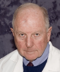 Bertram Pitt合作论文数Samuel and Jean Frankel Cardiovascular Center, University of Michigan;School of Medicine, University of Michigan3Edward Carmeliet合作论文数3
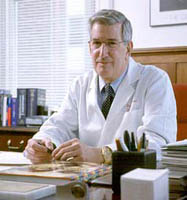 Burton E. Sobel合作论文数University of Vermont Colchester Research Facility3Brian F. Hoffman合作论文数3Shu Chien合作论文数3Ray A. Olsson合作论文数3
 Eveline E. Schneeberger合作论文数Department of Pathology, Medical School, Harvard3
 Matthew Nathan Levy合作论文数Case Western Reserve University3
 John C. McGiff合作论文数Department of Pharmacology, New York Medical College3Colin M. Bloor合作论文数3
 Mario Vassalle合作论文数3Merrill Tarr合作论文数3Lloyd L. Hefner合作论文数3Arthur L. Bassett合作论文数3Michael Heymann合作论文数University of California San Francisco3
 Eugene M. Renkin合作论文数University of California at Davis3Robert M. Berne合作论文数3
 John T. Shepherd合作论文数Mayo Clinic3Derek Bergel合作论文数3Philip A. Khairallah合作论文数3S. P. Sutera合作论文数3
 Andrew Wit合作论文数Department of Molecular Pharmacology and Therapeutics, Columbia University3George I. Drummond合作论文数3Kiichi Sagawa合作论文数School of Medicine, John Hopkins University3
 Roger C. Barr合作论文数Department of Biomedical Engineering, Pratt School of Engineering, Duke University;Duke Institute for Brain Sciences, Duke University3
 Aubrey Taylor合作论文数College of Medicine, University of South Alabama3MARK NOBLE合作论文数2
 Saul Winegrad合作论文数Department of Physiology, Perelman School of Medicine, University of Pennsylvania2
 Ray Fuller合作论文数Eli Lilly and Company2
 William Watts Parmley合作论文数University of California, San Francisco2
 Philip D. Henry合作论文数Internal Medicine, James H. Quillen College of Medicine, East Tennessee State University2




