Background Leprosy relapse is defined as patients presenting new clinical signs and symptoms of active disease after they were adequately treated with a standard therapeutic regimen and discharged as cured. The trend of relapse over the years after completion of MDT is a crucial epidemiological indicator, which can help in designing an appropriate follow up strategy. Objective To study the profile of leprosy relapse patients (MB and PB) after treatment with WHO-MDT at a tertiary Institution of South India. Material and methods A hospital record based retrospective follow up study was conducted in the Dermatology, Venereology and Leprosy department of Schieffelin Institute of Health-Research Leprosy centre, Karigiri, Vellore, Tamilnadu by reviewing the 5-year medical records of leprosy patients from March 2008 to 2013. A total of 33 (25 males and 8 females) leprosy relapse patients were enrolled in this study. Histopathological findings were used to classify the patient according to the Ridley- Jopling classification. Data were entered into Microsoft Excel 2007 and were analyzed using software Statistical Package for Social Sciences (SPSS) version 22.0. Results Multibacillary relapse occurred in 32 cases (97%) and paucibacillary relapse in 1 case (3%), according to WHO criteria. Among 26 patients previously treated with MBMDT, 14 relapsed as BT, 7 as BL, 4 as LL and 1 as histoid LL, while 7 patients who had received PBMDT (including one patient who took single-dose ROM) relapsed as 3 BT, 2 BL, 1 LL and 1 TT leprosy. Conclusion WHO MDT is highly effective against all the relapse cases but "Risk of relapse" is associated with every leprosy patient category.
Background: Smartphone usage has become increasingly popular in recent decade. Though it’s of great utility in many aspects, excessive usage among youth has shown increased restlessness, careless lifestyles and greater susceptibility to stress. Aim & Objective: To assess the pattern and purpose of smartphone usage and its dependence among medical students. Methods & Material: A cross-sectional, descriptive study was conducted among 363 MBBS students of 1st, IInd and IIIrd professional year between October 2018 to December 2018 at a government medical college. A 20-item self-administrated questionnaire, focusing on purpose and pattern of use of smart phone and its dependence was filled by all the students. Data was collected, entered and analysed using SPSS Software. Result: Among 363 students, 53% participants were males and rest were females. 80% students were using smartphone for more than 2 years, while 60% were having daily usage of more than 3 hours. Significant association between severity of smartphone dependence and variables like MBBS professional year, daily phone call made and received, daily SMS received, daily WhatsApp message sent and received were observed. Dependence was mild in 8.8% of the students; Moderate in 72.2% while 19% were suffering from severe smartphone dependence. Conclusion: Smart phone dependence is an established and emerging psychological issue which needs attention and intervention. It is of serious concern that all medical students were suffering from smartphone dependence with varying grades of severity. Increased awareness regarding the harmful effects of smartphone addiction is the need of hour.
BACKGROUND AND AIMS Medical thoracoscopy, also called pleuroscopy, has received renewed interest in the recent past for diagnostic as well as therapeutic uses. In this study, we describe our experience with thoracoscopy for undiagnosed pleural effusions. METHODS In a retrospective analysis of thoracoscopic procedures we performed between January 2007 and December 2008, yield of thoracoscopic pleural biopsy for achieving a diagnosis in undiagnosed pleural effusions, defined as pleural effusions with adenosine deaminase (ADA) levels less than 70 IU/L and negative pleural fluid cytology for malignancy on three occasions was evaluated. Complications of thoracoscopy were also analysed. RESULTS Overall diagnostic yield of thoracoscopic pleural biopsy was 74.3% in patients with undiagnosed pleural effusions. Pleural malignancy was diagnosed in 48.6% of patients. There was only one case of mesothelioma and the rest were due to pleural metastasis. Lung cancer and breast cancer were the most common sites of primary malignancy. Tuberculosis was diagnosed with pleural biopsy in 22.8% of patients. We had low complication rate after thoracoscopy. Only two cases of empyema were observed. CONCLUSION Medical thoracoscopy is a safe procedure and has good diagnostic yield in patients with undiagnosed pleural effusions.
Background: Medical Education curriculum is considered to be one of the toughest across the world.The long duration of course along with frequent assessments done make it stressful journey for medicalstudents. Assessment of stress and its determinants among medical students is of utmost importance so thatcorrectable actions can be taken.Objective: Assessment of stress and its underlying causes among Indian Medical Undergraduate Students.Material and Method: A descriptive cross-sectional study carried among 380 MBBS (Undergraduate)students of Government Medical College of North India to explore various stress determinants (Stressors)which were categorized into 6 domains viz. Academic Related Stressors (ARS), Intrapersonal andInterpersonal Related Stressors (IRS),Teaching and Learning-Related Stressors (TLRS), Social RelatedStressors (SRS), Drive and Desire Related Stressors (DRS) and Group Activities Related Stressors (GARS).Data collection was carried out for a period of 3 month and self-administrated validated MSSQ questionnairewas used as a Data tool. Data entered and analyzed with SPSS software.Result: 31% medical students suffers from Severe and 56% medical students suffers from moderateAcademic Stress. 40% medical students have documented that Teaching and Learning Method as a sourceof Moderate stress. Academic related stress and Social related Stress has significant association among bothgender when compared with 1st, 3rd and 5th Semester curriculum. Father’s Education status of a FemaleStudent and their Residence has significant association with Academic related stress. Mother occupation and1st, 3rd and 5th Semester Curriculum has significant association with Teaching Related stress.Conclusion: Stress exists in various domains and with variable intensity during whole MBBS course.
Background Primitive neuroectodermal tumor commonly occurs in bones and is equivalent to Ewing’s sarcoma. Very few cases have been reported in the literature and they had a very different presentation and very aggressive behavior. Case presentation We present a case of a young 23-year-old male who presented with complaints of pain in left lumbar region of abdomen since 8 months and hematuria off and on. CT scan of the abdomen was done which revealed a large heterogeneously enhancing mass lesion in the left lumbar region arising from the superior and mid-pole of left kidney showing multiple non-enhancing necrotic areas. A diagnosis of left renal cell carcinoma was kept. Through the transperitoneal approach, the left kidney was approached and a left radical nephrectomy was done. The histopathology report revealed the tumor cells to be positive for CD99 and focally positive for Vimentin and negative for cytokeratin thereby making a diagnosis of primary neuroectodermal tumor (PNET). Conclusions Renal PNET is a rare renal malignancy that should be kept in the differential diagnosis of a renal SOL especially when it is a presenting feature in adolescent and young adult. It has a very aggressive course and multimodal therapy has to be considered in its treatment
Perception of medical student regarding factors influencing the decision to serve in rural health services of Uttarakhand - JPMHH- Print ISSN No: - 2454-6704 Online ISSN No:- 2454-6712 Article DOI No:- 10.18231/j.jpmhh.2020.007, Journal of Preventive Medicine and Holistic Health-J Prev Med Holist Health
Assessment of attitude and perception of medical students towards rural health services in hilly region of Uttarakhand - IJFCM- Print ISSN No: - 2394-6768 Online ISSN No:- 2394-6776 Article DOI No:- 10.18231/j.ijfcm.2019.014, Indian Journal of Forensic and Community Medicine-Indian J Forensic Community Med
Introduction: According to WHO, schizophrenia is a severe mental disorder affecting about 23 million people worldwide. Psychoses, including schizophrenia, are characterized by distortions in thinking, perception, emotions, language, sense of self and behavior. The impacts of these disorders are severe, with approximately 1 million people committing suicide annually. There is also an increase in co morbidity of these different conditions. So, this study was carried out to assess the symptom profile of patients with schizophrenia and other psychotic disorders (excluding mood disorders, substance induced psychotic disorders, organic psychotic disorders). Material and Method: Discharge record sheets of all cases admitted (Total 86) to the psychiatric ward over the study period were reviewed. Cases diagnosed as schizophrenia and other psychotic disorders were enrolled. Major symptoms were listed from the discharge papers. Results: Out of the total number of patients enrolled in the research, 68.6% were male while 31.4% were female. The mean age of the patients was found to be 31.03 years with a minimum age of 16 years and maximum 63 years. Most were from lower middle class family, Hindu by religion and unmarried. Among 86 cases, 26(30.2%) were diagnosed as Unspecified nonorganic psychosis, 24(27.9%) as Schizophrenia, 19(22.1%) as Schizoaffective disorder, 14(16.3%) as Acute & Transient Psychotic Disorder, 3(3.5%) as Persistent delusional disorder. Delusion of persecution was present in 46 cases (53.5%), Delusion of reference in 33 cases (38.4%) and Hallucinations in 32 cases (37.2%). Out of the 86 patients enrolled in the study, 15 (17.4%) had at least one suicidal attempt. Conclusion: Significant number of patients were diagnosed as Schizophrenia which is itself a chronic, debilitating illness. Hallucinations and delusions were present in most of the cases.
BACKGROUND:Breast cancer (BC) continues to be a major cause of morbidity and mortality throughout the world. Early detection of BC and early treatment increases the chance of survival. According to Breast Health Global Initiative guidelines for low and middle income countries, diagnosing BCs early by promoting breast self-awareness; clinical breast examination (CBE) and resource adapted mammographic screening will reduce BC mortality. There is a paucity of data on the knowledge and awareness of BC and self-breast examination in India. We designed this hospital based cross sectional descriptive study to evaluate the current status of knowledge, awareness and practices related to BC and breast self-examination in the female rural population attending a teaching hospital.MATERIALS AND METHODS:We did a random sampling to identify and enroll 360 women and their female relatives. We excluded a participant from the study if she had already undergone a screening mammography or had had a BC. The data was collected by a self-administered questionnaire in vernacular language.RESULTS:Our study population included 360 women with a mean age of 45.81 (±10.9) years. Only 5 (1.38%) females had a family history of BC. A whopping 81% of women did not have any knowledge about BC. All the women thought that CBE by doctors was the only way for screening BC.CONCLUSIONS:We concluded that with the results of this study, it is imperative to increase awareness about BC and its detection methods in the community through health education campaigns. We should have major policy changes to increase future screening programs and health education programs which would have an overall positive impact on reducing the disease burden.
Background: Monitoring disease activity in sarcoidosis remains a clinical challenge as there is no gold standard. Positron emission tomography (PET) imaging is a novel tool to assess the metabolic activity. There is limited data on the role of serial PET scans in monitoring the disease activity. Methods: This is a prospective study of 27 sarcoidosis patients treated with systemic corticosteroids. Patients underwent two serial PET/CT scans: one before initiating therapy and the follow up scan at end of therapy. The metabolic response on PET scan was classified as: (a) complete metabolic response (CMR); (b) partial metabolic response (PMR); (c) stable metabolic disease (SMD); and, (d) progressive metabolic disease (PMD). Patients with either CMR or PMR were classified as PET responders while those with SMD or PMD were considered as PET non-responders. All patients were followed at 3, 6 and 12 months after completion of therapy. Relapse rates and relapse-free survival was compared between the various groups. Results: There was significant decline in the median SUVmax of the mediastinal lymph nodes, peripheral lymph nodes and the lung parenchyma in the follow up PET scan. Eight patients achieved CMR, 6 patients achieved PMR while 13 patients were PET non-responders. There was no difference in the clinical remission rates between the responders and non-responders. However, the relapse rate was significantly higher in non-responders vs. responders (61.5% vs. 14.2%, p=0.018). None of the patients who achieved a CMR relapsed during the study period. Conclusions: Patients with metabolic response on PET scan have significantly fewer relapses as compared to those with no response on PET scan.
Background and Aims. The combination of inhaled corticosteroids (ICS) and long-acting β2 agonists (LABA) has been used as a single inhaler both for maintenance and reliever therapy in asthma, the SMART approach. The administration of additional CS with each reliever inhalation in response to symptoms is expected to provide better control of airway inflammation. The aim of this meta-analysis was to evaluate the efficacy and safety of the SMART approach versus other approaches in the management of asthma in preventing asthma exacerbations. Methods. We searched the MEDLINE and EMBASE databases for studies that have reported exacerbations in the SMART group versus the control group. We calculated the odds ratio (OR) and 95% confidence intervals (CI) to assess the exacerbations in the two groups and pooled the results using a random-effects model. Results. Our search yielded eight studies. The use of SMART approach compared to fixed-dose ICS-LABA combination significantly decreased the odds of a severe exacerbation (OR 0.65; 95% CI, 0.53-0.80) and severe exacerbation requiring hospitalization/ER treatment (OR 0.69; 95% CI, 058- 0.83). The use of SMART approach compared to fixed-dose ICS also significantly decreased the odds of a severe exacerbation (OR 0.52; 95% CI, 0.45-0.61) and severe exacerbation requiring medical intervention (OR 0.52; 95% CI, 0.42-0.65). The occurrence of adverse events was similar in the two groups. There was some evidence of statistical heterogeneity. Conclusions. The SMART approach using formoterol- budesonide is superior in preventing exacerbations when compared to traditional therapy with fixed dose ICS or ICS-LABA combination without any increase in adverse events.
BACKGROUND & OBJECTIVES:Several patients with cancer in India are not aware of their diagnosis. We evaluated the impact of awareness of cancer diagnosis on health-related quality of life (HRQL) in newly diagnosed patients with lung cancer. METHODS:A total of 391 treatment-naïve patients with lung cancer, seen at the Lung Cancer Clinic of a tertiary care hospital in north India, were categorized into those aware of their diagnosis (group A) and those not aware (group B). All patients answered Hindi versions of abbreviated World Health Organization Quality of Life questionnaire (WHOQOL-Bref) and European Organization for Research and Treatment of Cancer Quality of Life questionnaire (EORTC QLQ-C30), and its lung cancer module, EORTC QLQ-LC13. Various domain scores were computed and compared between the two groups. Analysis of covariance was used to determine significance of differences after adjustment for potential confounding factors. RESULTS:Only 117 (29.9%) patients were aware of their diagnosis. Of all, 302 (77.2%) patients had non-small cell lung cancer, and 301 (77.0%) had advanced disease. All HRQL domain scores were similar between the two groups, except that group B patients had significantly poorer median (interquartile range) Physical [39.3 (28.6-50.0) vs 46.4 (28.6-57.1)] and Environment [46.9 (40.6-56.3) vs 53.1 (0.6-65.6)] domain scores of WHOQOL-Bref, and p0 hysical function [60.0 (40.0-73.3) vs 66.7 (46.7-80.0)] and Fatigue [66.7 (55.6-77.8) vs 66.7 (44.4-66.7)] scores of QLQ-C30. After adjusting for gender, age, education, family income, and tumour extent, these differences were not significant. INTERPRETATION & CONCLUSIONS:Disclosure of cancer diagnosis, or lack of it, had no significant impact on HRQL in patients with lung cancer after adjustment of potential confounders.
the diagnostic role of endobronchial ultrasoundguided transbronchial needle aspiration (EBUSTBNA) compared to conventional TBNA (cTBNA) in stage I and stage II sarcoidosis (1). The authors describe the yield of EBUS-TBNA to be significantly higher than cTBNA overall but was similar in the patients with lymph node station (4R and 7: group A) while it was higher in other lymph node stations (group B). While intuitive, that the yield was significantly higher in the other lymph node stations compared to stations 4R and 7, it would be of further interest to know the overall diagnostic yield of TBNA when combined with endobronchial biopsy (EBB) and transbronchial lung biopsy (TBLB). In a recent trial comprising of 117 patients with stage I and stage II sarcoidosis, combining EBB and TBLB with cTBNA provided yields comparable to EBUS-TBNA (cTBNA: 53/62 [85.5%] vs. EBUSTBNA: 51/55 [92.7%]; p=0.34) (2). In this study, the diagnostic yield of EBUS-TBNA increased from 74.5% to 91% when combined with TBLB. Although the diagnostic yield of EBUS-TBNA alone is superior to cTBNA, this advantage is balanced if cTBNA is combined with EBB and TBLB. In a real life scenario, sarcoidosis is generally associated with symmetrical mediastinal lymph node enlargement and the relevance of targeting other lymph node stations (2R, 2L, 4L, 11R, 11L and others) by cTBNA is not important especially when cTBNA is combined with EBB and TBLB. This is also reflected in the current study where the numbers of patients with other lymph node stations formed only a small subset. This is an important point for centers that do not have the facility of EBUS-TBNA because the diagnosis of stage I and stage II sarcoidosis can be safely made in almost 86% of patients by cTBNA plus EBB and TBLB (3).
Background: It is not clear how well sarcoidosis-associated fatigue improves with definitive therapy. Objective: To evaluate improvement in fatigue in patients of sarcoidosis receiving oral corticosteroid therapy, and correlate it with clinical recovery and change in health-related quality of life (HRQL). Methods: We studied 51 newly diagnosed adult patients of pulmonary sarcoidosis. Sarcoidosis Health questionnaire (SHQ) was used to assess HRQL. Fatigue was evaluated using Fatigue Assessment Scale (FAS). All patients received six month treatment with oral prednisolone, and SHQ and FAS were administered before initiation and after completion of treatment. Baseline and post-treatment scores were compared and a 4-point change in FAS scores considered clinically significant. Results: All patients improved clinically and radiologically with therapy. Body aches, however, persisted in 12 of 28 (42.9%) patients having this symptom at baseline. 33 patients (64.7%) had pretreatment fatigue (FAS >=22), of which seven (13.7%) were severely fatigued (FAS score >=35). Of them 21 (63.6%) and 4 (12.1%) patients respectively showed improvement and deterioration in FAS score by >4 points. Only five of 18 patients with baseline FAS score <22 also showed >4 point score increment after treatment, and four of them developed fatigue during therapy. All patients with baseline severe fatigue improved. Overall, FAS scores, and all SHQ domain scores, significantly improved with treatment. Conclusions: Both fatigue as well as HRQL improved significantly, and largely parallelly, in patients treated for sarcoidosis. Persistence of fatigue, or new onset fatigue, may be encountered during treatment, possibly as an adverse effect of corticosteroid therapy.
PURPOSE:There is currently no single modality for accurate characterization of enlarged mediastinal lymph nodes into benign or malignant. Recently (18)F-fluorothymidine (FLT) has been used as a proliferation marker. In this prospective study, we examined the role of (18)F-fluorodeoxyglucose ((18)F-FDG) positron emission tomography/computed tomography (PET/CT) and (18)F-FLT PET/CT in categorizing mediastinal lymph nodes as benign or malignant.MATERIALS AND METHODS:A total of 70 consecutive patients with mediastinal lymphadenopathy detected on computed tomography (CT) or chest radiograph underwent whole body (18)F-FLT PET/CT and (18)F-FDG PET/CT (within 1 week of each other). Lymph nodal tracer uptake was determined by calculation of standardized uptake value (SUV) with both the tracers. Results of PET/CT were compared with histopathology of the lymph nodes.RESULTS:Histopathology results showed thirty-seven patients with sarcoidosis, seven patients with tuberculosis, nine patients with non-small cell lung cancer, five patients with Hodgkin's lymphoma and twelve patients with non-Hodgkin's lymphoma. The mean FDG SUVmax of sarcoidosis, tuberculosis, Hodgkin's and non-Hodgkin's lymphoma was 12.7, 13.4, 8.2, and 8.8, respectively, and the mean FLT SUVmax was 6.0, 5.4, 4.4, and 3.8, respectively. It was not possible to characterize mediastinal lymphadenopathy as benign or malignant solely based on FDG SUVmax values (p > 0.05) or FLT SUVmax values (p > 0.05). There was no significant difference in FDG uptake (p > 0.9) or FLT uptake (p > 0.9) between sarcoidosis and tuberculosis. In lung cancer patients, the FDG SUVmax and FLT SUVmax of those lymph nodes with tumor infiltration on biopsy was 6.7 and 3.9, respectively, and those without nodal infiltration was 6.4 and 3.7, respectively, and both the tracers were not able to characterize the nodal status as malignant or benign (p > 0.05).CONCLUSION:Though (18)F-FLT PET/CT and (18)F-FDG PET/CT reflect different aspects of biology, i.e., proliferation and metabolism, respectively, neither tracer could provide satisfactory categorization of benign and malignant lymph nodes. The results of this study clearly suggest that differentiation of mediastinal nodes into benign and malignant solely based on SUVmax values cannot be relied upon, especially in settings where tuberculosis and sarcoidosis are common.
 Ritesh Agarwal合作论文数Department of Pulmonary Medicine, Postgraduate Institute of Medical Education and Research, Chandigarh168
 Ashutosh N. Aggarwal合作论文数Department of Pulmonary Medicine, Postgraduate Institute of Medical Education and Research163
 Surinder K. Jindal合作论文数139
 Dushmantkumar Behera合作论文数Dept Pulm Med, Postgrad Inst Med Educ & Res43
 Navneet Singh合作论文数33AN Aggarwal合作论文数22Digambar Behera合作论文数20
 Aggarwal Ashutosh N合作论文数Dept Pulm Med, Postgrad Inst Med Educ & Res19
 Sahajal Dhooria合作论文数Dept Pulm Med, Postgrad Inst Med Educ & Res18
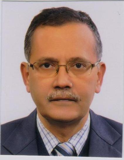 Arunaloke Chakrabarti合作论文数Department of Medical Microbiology, Postgraduate Institute of Medical Education & Research18
 Vishwajeet Singh合作论文数Department of Urology, King George’s Medical University17
 Rahul Janak Sinha合作论文数Department of Urology, King George’s Medical University17Amanjit Bal合作论文数16Indu Verma合作论文数Department of Biochemistry;graduate Institute of Medical Education and Research, Post;Department of Biochemistry|Postgraduate Institute of Medical Education and Research12Digamber Behera合作论文数11
 S C Sharma合作论文数11Maturu Venkata Nagarjuna合作论文数Department of Pulmonary Medicine, Postgraduate Institute of Medical Education and Research (PGIMER), Sector 12, Chandigarh, 160012, India.11
 Mandeep Kumar Garg合作论文数Department of Radiodiagnosis and Imaging;graduate Institute of Medical Education and Research, Post;Department of Radiodiagnosis and Imaging|Postgraduate Institute of Medical Education and Research11
 Nalini Gupta合作论文数Institute of Reproductive Medicine, Kolkata, India11Ajmal Khan合作论文数10G C Sood合作论文数10
 Srinivas Rajagopalan合作论文数9Bhagwant Rai Mittal合作论文数9
 Sethi Sunil Kumar合作论文数Yong Loo Lin School of Medicine, National University of Singapore8
 Arvind Rajwanshi合作论文数Departments of Gastroenterology and Cytology, Postgraduate Institute of Medical Education and Research, Chandigarh 160012, India8M Gulati合作论文数7K Joshi合作论文数7
 Abhishek Mewara合作论文数Department of Medical Parasitology, Postgraduate Institute of Medical Education and Research7
 Sampanna Jung Rayamajhi合作论文数Department of Nuclear Medicine and PET, Postgraduate Institute of Medical Education and Research, Chandigarh, India.7
 Biman Saikia合作论文数Pediatrics Intensive Care Unit, B L Kapur Super Speciality Hospital7
 Dhatwalia Sunil Kumar合作论文数Departments of Medical Microbiology, Postgraduate Institute of Medical Education and Research7
 Raj Kumar合作论文数6S Suri合作论文数6Sunil Chhabra合作论文数Vallabhbhai Patel Chest Institute, University of Delhi6
 Inderpaul Singh Sehgal合作论文数Department of Pulmonary Medicine, Postgraduate Institute of Medical Education and Research, Chandigarh6
 Ranjana Walker Minz合作论文数Department of Immunopathology, Post Graduate Institute of Medical Education and Research6S R Malik合作论文数6
 Ritu Aggarwal合作论文数Department of Immunopathology, Post Graduate Institute of Medical Education and Research (PGIMER), Chandigarh, 160012 India6G A D'Souza合作论文数5N K Panda合作论文数5
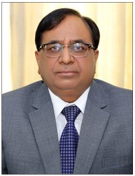 Surender Kashyap合作论文数Shri Lal Bahadur Med Coll & Hosp, Atal Med & Res Univ Campus5K Chaudhry合作论文数5B Shah合作论文数5
 S V Rana合作论文数Dept Biochem, All India Inst Med Sci5
 Janmeja Ashok K合作论文数Department of Pulmonary Medicine, Government Medical College and Hospital5Nath Bhola合作论文数5
 Satya N Sankhwar合作论文数5
 Rakesh Kumar Yadav合作论文数Department of Pharmacy, Banasthali University5Nidhi Mimani Gupta合作论文数4




