Re, 32,033 11/1985 Moreland et al. mor 430/31 3,959,105 6/1976 Feneberg et al. ................... 204/165 4,170,662 10/1979 Weiss et al. ........................... 427/38 4,178,097 12/1979 Sara ..................................... 355/110 4,179,312 12/1979 Keller et al. ......................... 148/15 4,260,649 4/1981 Dension et al. ... 427/53.1 4,283,482 8/1981 Hattori et al. ...................... 430/296 4,340,617 7/1982 Deutsch et al..... ... 427/53. 4,348,473 9/1982 Okumura et al. ................... 430/296 4,359,485 11/1982 Donnelly et al. ...... ... 427/53.1 4,444,456 4/1984 Jain et al. ............................. 350/3.7 4,458,994 7/1984 Jain et al. ................................ 354/4 4,490,211 12/1984 Chen et al. .......................... 156/643 4,516,832 5/1985 Jain et al. .............................. 350/96 4,521,087 6/1985 Hayes et al. ........................ 350/574 4,549,064 10/1985 Delfino ............................... 219/121 4,560,64l 12/1985 Kokaku et al. ..................... 430/312 4,568,632 2/1986 Blum et al. .......................... 430/322 4,608, 17 8/1986 Ehrlich et al. ...................... 156/610 4,615,904 10/1986 Ehrlich et al. ........................ 427/38 4,619,894 10/1986 Bozler et al........ 430/942 4,622,095 1 1/1986 Grobman et al. ................... 156/635 4,626,449 12/1986 Hirai et al. ......... ... 427/53.1 4,653,903 3/1987 Torigoe et al. ....................... 355/53 4,698,238 10/1987 Hayasaka et al. .................. 427/53.1 |||||||||||||| US005310624A
The parallel microfluidic cytometer (PMC) is an imaging flow cytometer that operates on statistical analysis of low-pixel-count, one-dimensional (1D) line scans. It is highly efficient in data collection and operates on suspension cells. In this article, we present a supervised automated pipeline for the PMC that minimizes operator intervention by incorporating multivariate logistic regression for data scoring. We test the self-tuning statistical algorithms in a human primary T-cell activation assay in flow using nuclear factor of activated T cells (NFAT) translocation as a readout and readily achieve an average Z′ of 0.55 and strictly standardized mean difference of 13 with standard phorbol myristate acetate/ionomycin induction. To implement the tests, we routinely load 4 µL samples and can readout 3000 to 9000 independent conditions from 15 mL of primary human blood (buffy coat fraction). We conclude that the new technology will support primary-cell protein-localization assays and “on-the-fly” data scoring at a sample throughput of more than 100,000 wells per day and that it is, in principle, consistent with a primary pharmaceutical screen.
Quantification of deletions in mtDNA is a long-standing problem in mutational analysis. We describe here an approach that combines the power of single-molecule PCR of the entire mitochondrial genome with the enrichment of the deletions by restriction digestion. This approach is indispensable if information about wide range of deletion types in a sample is critical, such as in studies concerning distribution of deletion breakpoints (as opposed to approaches where fraction of a single deletion or a limited set of deletions is used as a proxy for total deletion load). Because deletions in a sample are quantified almost exhaustively, the other important application of this approach involves studies where only small amounts of tissue, such as biopsies, are available.
A parallel microfluidic cytometer (PMC) is based on a one‐dimensional (1D) scanning detector, a parallel array of flow channels, and new multiparameter analysis algorithms that operate on low‐pixel‐count 1D images. In this article, we explore a series of image‐based live‐ and fixed‐cell screening assays, including two NF‐kB nuclear translocations and T‐cell capping. We then develop a new multiparametric linear weighted classifier that achieves a Z′ factor sufficient for scaled pharmaceutical discovery with Jurkat cells in suspension. We conclude that the PMC should have the throughput and statistical power to permit a new capability for image‐based high‐sample‐number pharmaceutical screening with suspension samples. © 2014 International Society for Advancement of Cytometry
We present measurements by deep-ultraviolet mass mapping of nucleic acid (NA) and protein for five commonly cultured and three primary cell types. The dry mass distribution at submicron resolution was determined on a single-cell basis for 250-500 cells from each of these types. Since the method carries a direct reference to a spectrophotometric standard (molar extinction coefficient), we are able to calibrate the absolute weight distributions both on a cell-to-cell basis within each type and across types. We also provide a calibration in absolute mass units for fluorescence-based measurements (flow cytometry and fluorescence microscopy). As might be expected the cultured cell lines show a high concentration of nucleic acids in the nuclear compartment, much larger than the genomic 2C number even in the G1 stage. The whole-cell nucleic-acid/protein ratio was found to be a characteristic of cell lines that persists independent of cell cycle and, as a result, this ratio has some value for phenotyping. Primary chicken red blood cells (cRBC), often used as a cytometry standard, were determined to have a nuclear-isolated nucleic acid content much closer to the genomic number than the cultured cell lines (cRBC: 3.00 pg total NA, 2.30 pg DNA, and 0.70 pg RNA). The individual blastomeres (n = 54) from mouse embryos at eight-cell stage were measured and found to vary by more than a factor or two in total protein and nucleic acid content (0.8-2.3 ng total protein, 70-150 pg total NA). The ratio of nucleic acid to protein was more nearly constant for each blastomere from a particular embryo and this ratio was found to be an identifying characteristic that varies from embryo to embryo obtained from a single flushing of a mouse.
Myanmar (Burma) exists in a timewarp and since recent political changes is becoming one of the most visited countries in the world. The country is eighty-seven per cent buddhist, studded with monastries, pagodas, dirt-track roads, oxcarts and elegant villages much as they were when the West intruded little more than 100 years ago. The country is still farmed by water buffalo, and its rituals remain true to their old-Asia form. Although tourism has increased significantly in the past 12 months there are many regions still off-limits. This book, in the form of a photo essay, captures an insider's view of a fragile and mystical aspect of Burmese culture. The curtain is drawn to reveal the backstage of the Burmese theatre; a world populated by animist spirit media (nakadaws), monsters from the Ramayana Buddhist texts, princesses (minthami) and princes (mintha). We go behind the scenes to see the preparations of these performers as they travel around the towns and countryside between temporary bamboo stages constructed for all-night festivals. With contributing essays from Professor Ward Keeler and U Ohn Maung, this book is both a visual and informative testament to Burmese performing arts.
Incorporation of one-dimensional imaging capability into a parallel microfluidic flow cytometer allows fast, low-resolution acquisition of images that permit classification of cells by automated analysis of preselected features. A parallel microfluidic cytometer (PMC) uses a high-speed scanning photomultiplier-based detector to combine low-pixel-count, one-dimensional imaging with flow cytometry. The 384 parallel flow channels of the PMC decouple count rate from signal-to-noise ratio. Using six-pixel one-dimensional images, we investigated protein localization in a yeast model for human protein misfolding diseases and demonstrated the feasibility of a nuclear-translocation assay in Chinese hamster ovary (CHO) cells expressing an NFκB-EGFP reporter.
Cobalt interconnects were deposited with a cw visiblewavelength laser from gaseous Co2 (CO)8. The deposited material was high-purity Co, and its electrical resistivity was as low as 13 μΩ-cm (twice that of bulk resistivity). The deposition is initiated by photochemical decomposition of Co2 (CO)8, and therefore process parameters (pressure, power, writing speed) are insensitive to the optical and thermal properties of the substrate.
By adding an additional degree of freedom from multichannel flow, the parallel microfluidic cytometer (PMC) combines some of the best features of fluorescence-activated flow cytometry (FCM) and microscope-based high-content screening (HCS). The PMC (i) lends itself to fast processing of large numbers of samples, (ii) adds a 1D imaging capability for intracellular localization assays (HCS), (iii) has a high rare-cell sensitivity, and (iv) has an unusual capability for time-synchronized sampling. An inability to practically handle large sample numbers has restricted applications of conventional flow cytometers and microscopes in combinatorial cell assays, network biology, and drug discovery. The PMC promises to relieve a bottleneck in these previously constrained applications. The PMC may also be a powerful tool for finding rare primary cells in the clinic. The multichannel architecture of current PMC prototypes allows 384 unique samples for a cell-based screen to be read out in ∼6-10 min, about 30 times the speed of most current FCM systems. In 1D intracellular imaging, the PMC can obtain protein localization using HCS marker strategies at many times for the sample throughput of charge-coupled device (CCD)-based microscopes or CCD-based single-channel flow cytometers. The PMC also permits the signal integration time to be varied over a larger range than is practical in conventional flow cytometers. The signal-to-noise advantages are useful, for example, in counting rare positive cells in the most difficult early stages of genome-wide screening. We review the status of parallel microfluidic cytometry and discuss some of the directions the new technology may take.
By using imaging spectrophotometry with paired images in the 200‐ to 280‐nm wavelength range, we have directly mapped intracellular nucleic acid and protein distributions across a population of Chinese hamster ovary (CHO‐K1) cells. A broadband 100× objective with a numerical aperture of 1.2 NA (glycerin immersion) and a novel laser‐induced‐plasma point source generated high‐contrast images with short (∼100 ms) exposures and a lateral resolution nearing 200 nm that easily resolves internal organelles. In a population of 420 CHO‐K1 cells and 477 nuclei, we found a G1 whole‐cell nucleic acid peak at 26.6 pg, a nuclear‐isolated total nucleic acid peak at 11.4 pg, and, as inferred by RNase treatment, a G1 total DNA mass of 7.4 pg. At the G1 peak, we found a whole‐cell protein mass of 95.6 pg, and a nuclear‐isolated protein mass of 39.3 pg. An algorithm for protein quantification that senses peptide‐bond (220‐nm) absorbance was found to have a higher signal‐to‐noise ratio and to provide more reliable nucleic acid and protein determinations when compared to more classical 280/260‐nm algorithms when used for intracellular mass mapping. Using simultaneous imaging with common nuclear stains (Hoechst 33342, Syto‐14, and Sytox Orange), we have compared staining patterns to deep‐UV images of condensed chromatin and have confirmed bias of these common nuclear stains related to nuclear packaging. The approach allows absolute mass measurements with no special sample preparation or staining. It can be used in conjunction with normal fluorescence microscopy and with relatively modest modification of the microscope. © 2011 International Society for Advancement of Cytometry
In this paper we describe progress in using the prodigious data-collecting ability of multilane microelectrophoresis instruments to bear on problems in scaled nucleic acid assays. We emphasize compound stacking and solid-support loading as means to concentrate <100 pg samples for direct injection. Reaction Mapping is applied to readout quantitative polymerase chain reaction gene-expression and as a way to practically overcome difficulty in interpreting amplification curves of multiplexed quantitative polymerase chain reaction at 20-50 gene/well complexity. We demonstrate multiplexed readout of gene expression over an abundancy range of 9 Log 2 units starting with reverse-transcribed samples as small as five molecules in each sample.
Vertical hydrodynamic focusing in microfluidic devices is investigated through simulation and through direct experimental verification using a confocal microscope and a novel form of stroboscopic imaging. Optimization for microfluidic cytometry of biological cells is examined. By combining multiple crossing junctions, it is possible to confine cells to a single analytic layer of interest. Subtractive flows are investigated as a means to move the analysis layer vertically in the channel and to correct the flatness of this layer. The simulation software (ADINA and Coventor) is shown to accurately capture the complex dependencies of the layer interfaces, which vary strongly with channel geometry and relative flow rates.
We have constructed a 384-channel parallel microfluidic cytometer (PMC). The multichannel architecture allows 384 unique samples for a cell-based screen to be read out in approximately 6-10 min, about 30-times the speed of a conventional fluorescence-activated cytometer system (FACS). This architecture also allows the signal integration time to be varied over a larger range than is practical in single-channel FACS and is suitable for detection of rare-cells in a high background of negatives. The signal-to-noise advantages have been confirmed by using the system to count rare clonal osteocytes in the most difficult early stages of an expression-cloning screen for the carboxy-terminal parathyroid hormone receptor (CPTHR). This problem requires finding several dozen positive cells in a background of one million negatives. The system is automated around a scanning laser confocal detector and a 96-tip robotic pipettor and can maintain in vitro cultures on-system in 384-well plates. It is therefore directly practical for biology applications using existing high-throughput culture facilities. The PMC system lends itself to high-sample-number cytometry with an unusual capability for time synchronization and rare-cell sensitivity. A limited ability to handle large sample numbers has restricted applications of single-channel FACS in combinatorial cell assays; therefore the PMC could have a significant application in high-throughput screening.
We have applied multiple-time-point reaction mapping to generate high-dynamic-range quantitative data from PCR multiplexes. The approach measures, then compensates, numerous PCR slope nonidealities across the multiplex without prejudice. A multilane microelectophoresis device with a novel scanning detector that reports redundantly over more than six decades in signal strength was used to collect data with multiple readings for each amplification point and with double internal calibration (lane standards and gene standards). We investigated scaling properties and sensitivity for readout of 12plex PCR reactions. The sensitive detection, stemming from confocal optics, allowed reduction of the PCR cycle number by approximately five cycles compared to commercial fluorometric readout. This increased sensitivity appears to allow quantitative PCR over a dynamic range of >9 log2 abundance ratio in multiplex reactions exceeding 20plexes. We argue that the combination of mapping, multiplexing, and an internal standard, improves the per-well efficiency of quantitative expression analysis by a factor of 50-100 relative to fluorometric qPCR readout. Therefore, the approach is attractive for analysis of large gene networks at reduced cost.
: The proposed research instrumentation will allow for greatly enhanced scope and efficiency of the ongoing programs, which have many important Air Force applications, particularly in monitoring hazardous materials, in health monitoring, and in fast forensic identification. The objective of this research instrumentation is to provide for the enhancement and augmentation of ongoing research in the area of laser photo chemistry. Research instrumentation will be procured to enhance and augment ongoing programs in biomolecular species identification. The funding will allow purchase of a ultraviolet laser system, which will be used for study of rapid direct detection and evaluation of genetic and molecular material in large parallel micro arrays.
We developed a deep-ultraviolet (UV) microscope capable of imaging cell mitosis and motility at 280 nm for 45 min with minimal UV-induced toxicity, and for 6 h before the onset of visible cell death in cultured human and mouse cells. Combined with computational methods that convert the intensity of each pixel into an estimate of mass, deep-UV microscopy images generate maps of nucleic acid mass, protein mass and fluorescence yield in unlabeled cells.
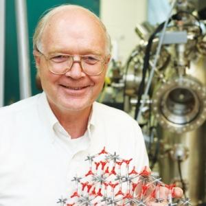 Richard M. Osgood合作论文数Department of Applied Physics and Applied Mathematics, Columbia University54
 Mordechai Rothschild合作论文数Lincoln Laboratory, Massachusetts Institute of Technology43
 Paul Thomas Matsudaira合作论文数Department of Biological Sciences, National University of Singapore31
 J. G. Black合作论文数20T F Deutsch合作论文数19
 Jeff Y Tsao合作论文数Material, Physical and Chemical Sciences Center 1800, Sandia National Laboratories19Brian Mckenna合作论文数15Thomas F. Deutsch合作论文数15Sameh El-Difrawy合作论文数Communications and Digital;Electrical and Computer Engineering Department, Center;Northeastern University11Mark A. Hollis合作论文数10Theodore M Bloomstein合作论文数10Jhc Sedlacek合作论文数9Sp Doran合作论文数9r a murphy合作论文数9DJ SILVERSMITH合作论文数9
 Roderick R. Kunz合作论文数Lincoln Laboratory, Massachusetts Institute of Technology9L Koutny合作论文数8
 Mitchell D. Eggers合作论文数8Michael E. Hogan合作论文数8
 Dennis D. Rathman合作论文数8Bernard B. Kosicki合作论文数8A Adourian合作论文数7D Schmalzing合作论文数7
 Dc Shaver合作论文数7Eggers, M.合作论文数7Samantha Desmarais合作论文数6
 Stella W. Pang (彭慧芝)合作论文数Department of Electronic Engineering, College of Engineering, City University of Hong Kong;Centre for Biosystems, Neuroscience, and Nanotechnology, Department of Electrical Engineering, City University of Hong Kong6Loucinda Carey合作论文数6
 Be Burke合作论文数5Kenneth Loren Beattie合作论文数5Cheung Man Ching合作论文数5Elizabeth A. Gismondi合作论文数5Aram Adourian合作论文数5Leo J Mahoney合作论文数5Chin-Long Chen合作论文数IBM eServer Group5
 Maribel Vazquez合作论文数The City College of The City, University of New York5S Silverman合作论文数4R Aucoin合作论文数4H. Zeiger合作论文数4Alok Srivastava合作论文数4
 George William Turner合作论文数4Chang-Lee Chen合作论文数4Goodhue, W.D.合作论文数4
 Gareth McKinley合作论文数School of Engineering, Massachusetts Institute of Technology;Department of Mechanical Engineering, Massachusetts Institute of Technology4Roger Lam合作论文数4Luba Mitnik合作论文数4Dieter Schmalzing合作论文数4R.W. Mountain合作论文数4V. Daneu合作论文数4




