Spinal cord injury (SCI) induces neuronal death, including apoptosis, which is completed within 24 hr at and around the impact site. We identified early proapoptotic transcriptional changes, including upregulation of proapoptotic Bax and downregulation of antiapoptotic Bcl‐xL, Bcl‐2, and Bcl‐w, using Affymetrix DNA microarrays. Because Bcl‐xL is the most robustly expressed antiapoptotic Bcl‐2 molecule in adult central nervous system, we decided to characterize better the effect of SCI on Bcl‐xL expression. We found Bcl‐xL expressed robustly throughout uninjured spinal cord in both neurons and glia cells. We also found Bcl‐xL localized in different cellular compartments: cytoplasmic, mitochondrial, and nuclear. Bcl‐xL protein levels decreased in the cytoplasm and mitochondria 2 hr after SCI and persisted for 24 hr. To test the contribution of proapoptotic decreases in Bcl‐xL to neuronal death, we augmented endogenous Bcl‐xL levels by administering Bcl‐xL fusion protein (Bcl‐xL FP) into injured spinal cords. Bcl‐xL FP significantly increased neuronal survival, suggesting that SCI‐induced changes in Bcl‐xL contribute considerably to neuronal death. Because Bcl‐xL FP increases survival of dorsal horn neurons and ventral horn motoneurons, it could become clinically relevant in preserving sensory and motor functions after SCI. © 2005 Wiley‐Liss, Inc.
Substance P (SP) is regarded as an important neurotransmitter in nociceptive transmission. It is abundantly expressed in the superficial layers of the spinal cord dorsal horn and its action is mediated through the activation of the neurokinin-1 (NK-1) receptor. Previous data have shown that intradermal capsaicin injection can evoke spinal cord central sensitization, which refers to an enhanced response of dorsal horn neurons resulting from innocuous and noxious stimuli of afferents that is unaffected by the capsaicin injection. This study investigated the role of SP in central sensitization induced by capsaicin injection. Using an Enzyme-Linked Immunosorbent Assay (ELISA) method, we investigated the change of SP synthesis and release in the dorsal spinal cord of rats after intradermal capsaicin injection. We found that both the synthesis and release of SP were significantly increased in response to peripheral inflammation. In electrophysiological experiments, local application of SP in the rat spinal cord enhanced the responses of wide-dynamic-range (WDR) neurons to mechanical stimuli. Such SP-induced responses were blocked by the administration of the NK-1 antagonist, L703,606. Furthermore, the NK-1 antagonist also inhibited the increased responses of WDR neurons evoked by capsaicin injection. These results suggest that: (1) SP synthesis and release in the spinal cord dorsal horn are induced by peripheral noxious stimuli; (2) The increased SP then activates the NK-1 receptors and contributes to capsaicin-induced central sensitization. Substance P (SP) is regarded as an important neurotransmitter in nociceptive transmission. It is abundantly expressed in the superficial layers of the spinal cord dorsal horn and its action is mediated through the activation of the neurokinin-1 (NK-1) receptor. Previous data have shown that intradermal capsaicin injection can evoke spinal cord central sensitization, which refers to an enhanced response of dorsal horn neurons resulting from innocuous and noxious stimuli of afferents that is unaffected by the capsaicin injection. This study investigated the role of SP in central sensitization induced by capsaicin injection. Using an Enzyme-Linked Immunosorbent Assay (ELISA) method, we investigated the change of SP synthesis and release in the dorsal spinal cord of rats after intradermal capsaicin injection. We found that both the synthesis and release of SP were significantly increased in response to peripheral inflammation. In electrophysiological experiments, local application of SP in the rat spinal cord enhanced the responses of wide-dynamic-range (WDR) neurons to mechanical stimuli. Such SP-induced responses were blocked by the administration of the NK-1 antagonist, L703,606. Furthermore, the NK-1 antagonist also inhibited the increased responses of WDR neurons evoked by capsaicin injection. These results suggest that: (1) SP synthesis and release in the spinal cord dorsal horn are induced by peripheral noxious stimuli; (2) The increased SP then activates the NK-1 receptors and contributes to capsaicin-induced central sensitization.
Amyotrophic lateral sclerosis (ALS) is a neurodegenerative disorder resulting in death of motor neurons from the motor cortex and spinal cord. Some familial cases of ALS are caused by missense mutations in the gene encoding Cu/Zn superoxide dismutase (SOD1). The deleterious effects of mutant SOD1 expression result from a toxic gain of function, however, the precise mechanisms of neurodegeneration are unknown. Gene therapy strategies for ALS have used viral vectors to deliver neurotrophic genes to vulnerable motor neurons. One alternative approach to treat SOD1-linked ALS would be to silence expression of mutant SOD1. Interfering RNA (RNAi) provides a powerful tool for investigating such therapeutic gene silencing approaches. Viral vectors generated to overexpress short hairpin RNA (shRNA) species provide an effective method for delivering RNAi. Lentiviral vectors based on the equine infectious anaemia virus (EIAV) mediate efficient and sustained transgene expression in the nervous system. Furthermore, these vectors have the capacity to be retrogradely transported along axons and this makes them of particular use for targeting motor neuron populations. We investigated the potential of EIAV vectors for delivering shRNAs targeted against mutant SOD1. Vectors carrying optimised RNAi target sequences were tested in primary neurons derived from SOD1 transgenic mice and mediated efficient ablation of mutant SOD1 expression. Further work will uncover the efficiency of such vectors in vivo and for alleviating disease phenotypes in transgenic mouse models of ALS.
The b-site amyloid precursor protein (APP)-cleaving enzyme (BACE1) is a prerequisite for the generation of b-amyloid peptides, which give rise to b-amyloid deposits in the brains of Alzheimer’s disease patients. It is believed that BACE1 is exclusively expressed by neurons. However, in the brains of transgenic Tg2576 mice that overexpress human APPsw, the BACE1 protein is also expressed by reactive astrocytes in proximity to b-amyloid plaques. Employing six different experimental strategies to activate brain glial cells we demonstrate the expression of BACE1 by reactive astrocytes but not by activated microglial cells in models of chronic rather than acute gliosis. To identify elements which drive tissueor cell type-specific BACE1 expression we cloned and sequenced a 1.5 kb fragment of the rat BACE1 promoter and generated BACE1 promoterluciferase reporter constructs. The basal activity of this promoter construct was highest in neuronal cell lines and in the pancreatic cell line AR42J, somewhat lower in rat primary neurons, astrocytic and microglial cultures, very low in hepatocytes and almost absent in fibroblasts and in the monocyte-macrophage cell line RAW264.7. The analysis of promoter activities of deletion mutants suggests the presence of activators of BACE1 transcription between bases )88 to )326 and the existence of inhibitors of BACE1 transcription between bases )327 and )1114. Together, our data indicate that brain glial cells, in particular astrocytes, may contribute to increased generation of b-amyloid peptides and to accelerated formation of b-amyloid plaques in the course of Alzheimer’s disease. The hypothesis is supported by our observation of BACE1-immunoreactive astrocytes in the brains of Alzheimer’s disease patients.
Spinal cord injury (SCI)‐induced neurodegeneration leads to irreversible and devastating motor and sensory dysfunction. Post‐traumatic outcomes are determined by events occurring during the first 24 hours after SCI. An increase in extracellular glutamate concentration to neurotoxic levels is one of the earliest events after SCI. We used Affymetrix DNA oligonucleotide microarrays (with 1,322 DNA probes) analysis to measure gene expression in order to test the hypothesis that SCI‐induced N‐methyl‐D‐aspartate (NMDA) receptor activation triggers significant postinjury transcriptional changes. Here we report that SCI, 1 hour after trauma, induced change in mRNA levels of 165 genes and expression sequence tags (ESTs). SCI affected mRNA levels of those genes that regulate predominantly transcription factors, inflammation, cell survival, and membrane excitability. We also report that NMDA receptor inhibition (with ‐(+)‐5‐methyl‐10,11‐dihydro‐5H‐dibenzo[a,d]‐cyclohepten‐5,10‐imine hydrogen maleate [MK‐801]) reversed the effect of SCI on about 50% of the SCI‐affected mRNAs. Especially interesting is the finding that NMDA receptor activation participates in the up‐regulation of inflammatory factors. Therefore, SCI‐induced NMDA receptor activation is one of the dominant, early signals after trauma that leads to changes in mRNA levels of a number of genes relevant to recovery processes. The majority of MK‐801 effects on the SCI‐induced mRNA changes reported here are novel. Additionally, we found that the MK‐801 treatment also changed the mRNA levels of 168 genes and ESTs that had not been affected by SCI alone, and that some of their gene products could have harmful effects on SCI outcome. © 2002 Wiley‐Liss, Inc.
We hypothesized that blocking receptors for pro‐inflammatory cytokine, interleukin‐1 (IL‐1) would ameliorate SCI‐dependent cell loss and resulting motor dysfunction. We found that chronic administration of recombinant IL‐1 receptor antagonist (rIL‐1ra) reduced SCI‐induced increases in cell death markers and caspase‐3 activity, suggesting that IL‐1ra prevents apoptosis in injured spinal cords. We also found that IL‐1ra significantly improved locomotor recovery. To answer the question how is the inhibition of IL‐1 receptors preventing cell death in injured spinal cord and improving motor recovery, we analyzed transcriptional changes in injured vs. injured spinal cord treated with IL‐1ra, via DNA microarrays. We found a large number of mRNAs whose expression levels were significantly changed after SCI and reversed in the presence of IL‐1ra. For example, we found that iNOS and COX‐2 mRNA are significantly up‐regulated after injury and down‐regulated in IL‐1ra‐treated spinal cords. If iNOS and COX‐2 proteins are synthesized, it is very likely that their activity will produce reactive oxygen species (ROS), which will cause cell death via different converging apoptotic pathways. We also found that SCI induces IL‐1 dependent up‐regulation of pro‐apoptotic cathepsins and cell cycle regulators, a novel finding. Therefore, we hypothesize that the anti‐inflammatory IL‐1ra could be therapeutic and alleviate secondary damage after SCI.Acknowledgements: Supported in part by grant NS39161 from NINDS and Mission Connect Foundation.
After contusion-derived spinal cord injury, (SCI) there is localized tissue disruption and energy failure that results in early necrosis and delayed apoptosis, events that contribute to chronic central pain in a majority of patients. We assessed the extent of contusion-induced apoptosis of neurons in a known central pain-signaling pathway, the spinothalamic tract (STT), which may be a contributor to SCI-induced pain. We observed the loss of STT cells and localized increase of DNA fragmentation and cytoplasmic histone-DNA complexes, which suggested potential apoptotic changes among STT neurons after SCI. We also showed SCI-associated changes in the expression of the antiapoptotic protein Bcl-xL, especially among STT cells, consistent with the hypothesis that Bcl-xL regulates the extent of apoptosis after SCI. Apoptosis in the injured spinal cord correlated well with prompt decreases in Bcl-xL protein levels and Bcl-xL/Bax protein ratios at the contusion site. We interpret these results as evidence that regulation of Bcl-xL may play a role in neural sparing after spinal injury and pain-signaling function.
Toxic products are formed when tryptophan is irradiated with light in the presence of photosensitizers such as riboflavin. In order to further investigate this phenomenon, solutions of tryptophan (48 mumol.mL-1) were irradiated with broad spectrum fluorescent light in the presence or absence of riboflavin (0.01 mg.mL-1). Solutions of riboflavin were similarly irradiated; control solutions were the respective solutions not exposed to light. Two-week-old suckling gerbils were then assigned to receiving 7 days of intraperitoneal injections of the light-exposed or non-light-exposed solutions. There were significant differences in the concentrations of tryptophan in serum, liver, and brain; activity of gamma-glutamyl transferase (GGT) as well as liver protein were also significantly different among the groups. Body and liver weights were also significantly different among the groups. In order to identify the photoproducts responsible for these changes, solutions of tryptophan that had been irradiated with light in the presence of riboflavin were then fractionated based on time of elution during high-pressure liquid chromatography and the fractions were then injected into the gerbils as before. GGT responses to one of the fractions was similar to that of the parent compound. Chromatographic studies indicated the presence of numerous photoadduct compounds of tryptophan and riboflavin after exposure to light. Both the presence of riboflavin and the exposure of the solutions to light alter brain concentrations of tryptophan in the developing gerbil indicating differing availability to the brain of this serotonin precursor. Clinical implications of the infusion of amino acids in the presence of photosensitizers and light must be considered.
Neurons R3-R14 of the marine mollusc Aplysia are model neuroendocrine cells thought to regulate cardiovascular activity in vivo. The cells express a gene encoding three peptides--peptides I, II and the histidine-rich basic peptide (HRBP)--each of which has been chemically characterized in Aplysia californica. In the studies presented here, HRBP and its prohormone (proHRBP) were purified from A. brasiliana abdominal ganglion extracts by reversed-phase high-performance liquid chromatography and characterized by amino acid compositional and sequence analyses. ProHRBP was an 85-residue peptide whose sequence was: NH2-Glu-Glu-Val-Phe-Asp-Asp-Thr-Asp-Val-Gly-Asp-Glu-Leu-Thr-Asn-Ala-Leu- Glu-Ser - Val-Leu-Thr-Asp-Leu-Lys-Asp-Lys-Arg-Asp-Ala-Glu-Glu-Pro-Ser-Ala-Phe-Met- Thr-Arg - Leu-Arg-Arg-Gln-Val-Ala-Gln-Met-His-Ile-Trp-Arg-Ala-Asn-His-Asp-Arg-His- His-Ser - Thr-Gly-Ser-Gly-Arg-His-Ser-Arg-Phe-Leu-Thr-Arg-Asn-Arg-Tyr-Gly-Gly-Gly- His-Leu - Ser-Asp-Ala-COOG. It differed from A. californica pro-HRBP at seven of the 85 positions. Compositional and sequence analyses demonstrated that A. brasiliana HRBP was a 43-residue peptide corresponding to residues 43 through 85 of proHRBP, and that a significant proportion of the isolated peptide possessed a blocked NH2 terminus. Although this sequence differed from that of A. californica HRBP at five of 43 residues, the two peptides were approximately equipotent in inducing contractions of A. californica crop muscle in vitro, suggesting that the substituted residues may not be critical for biological activity.
In the frog spinal cord primary afferent depolarization (PAD) constitutes a powerful inhibitory control mechanism. It has been suggested that γ-aminobutyric acid (GABA) is the transmitter substance involved in the genesis of PAD. In these studies we show that maximal glutamic acid decarboxylase activity is localized roughly 400–600 μm from the dorsal surface, and that correlates well with the intraspinal distribution of field potentials associated with PAD. Measurement of GABA in serial spinal cord sections cut in a dorsal-ventral direction shows that high levels of GABA are seen at 400–600 μm, with a peak at 800 μm from the dorsal surface. Stimulation at frequencies shown to produce PAD augments the release of endogenous GABA from a superfused frog hemicord preparation.
 J. Regino Perez-Polo合作论文数11Olivera Nesic-Taylor合作论文数Medical Branch, The University of Texas10GY Xu合作论文数8Claire E. Hulsebosch合作论文数8Karin N Westlund合作论文数8Z Ye合作论文数5NM Svrakic合作论文数4Jx Qiu合作论文数3G UNABIA合作论文数3ZM Ye合作论文数3Diana M. Cittelly合作论文数Medical Branch, University of Texas3JC Lee合作论文数2Claire E Hulsebosch合作论文数2O. Nesic-Taylor合作论文数2M. Paintlia合作论文数1L. Nie合作论文数1U Zeitschel合作论文数1Harriet Rea合作论文数1Shaffiat Karmally合作论文数1Ej Green合作论文数1Jr Perezpolo合作论文数1Valerie Bracchi-Ricard合作论文数1Tg Wood合作论文数1
 Sandra J Hewett合作论文数1KM Johnson合作论文数1Kw High合作论文数1AG Gilg合作论文数1J Bhatia合作论文数1B Haber合作论文数1J Yan合作论文数1
 Zhifeng Wu (吴志峰)合作论文数School of Geographical Sciences and Remote Sensing, Guangzhou University;Institute Aerospace Remote Sensing Innovation, Guangzhou University;Key Laboratory of Geo-Environment Monitoring of Greater Bay Area, MNR1
 Nicholas Mazarakis合作论文数Neuroscience, Department of Brain Sciences, Faculty of Medicine, Imperial College London1
 Ajaib Paintlia合作论文数1
 Richard J. Youle合作论文数Division of Intramural Research, National Institute of Neurological Disorders and Stroke, National Institutes of Health1
 Nenad Svrakic合作论文数1Singh Inderjit合作论文数1J. Lee合作论文数1
 Michael G. Hughes合作论文数School of Geosciences, University of Sydney1Ren Sun合作论文数1
 Wise Young合作论文数Department of Cell Biology & Neuroscience, Rutgers University1
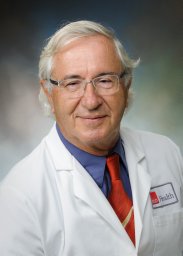 David K Rassin合作论文数Department of Pediatric Research Institute for Basic Research in Mental Retardation1Wenhui Hu合作论文数1J. Dunsmore合作论文数1Kent Johnson合作论文数1P Tolias合作论文数1Silvio Glusman合作论文数Department of Anesthesia and Pain Service, Cook County Hospital1Ralph S. Baric合作论文数1Christine Lange-Dohna合作论文数1Ernest Barbosa合作论文数1




