Background: Homozygous or compound heterozygous loss-of-function variants in GNPTAB cause mucolipidosis type II/III, a progressive multisystem disorder characterized by skeletal abnormalities, short stature, coarse facial features and cardiorespiratory disease. ML III is milder, with an older age of onset and a less severe phenotype. We report two siblings with arrhythmogenic cardiomyopathy and ventricular arrhythmias with compound heterozygous variants in GNPTAB. Methods and Results: The family presented due to the sudden cardiac death of a male in his early 30’s with arrhythmogenic cardiomyopathy identified at autopsy. His sister was found to have cardiomyopathy and experienced a ventricular tachycardia storm. Both siblings had history of skeletal dysplasia first investigated during adolescence. Clinical genetic testing did not identify a cause for the cardiomyopathy, and they were enrolled in the Elusive Hearts study. Following whole genome sequencing, we detected a heterozygous frameshift variant, NM_024312.5( GNPTAB ): c.3503_3504del, and a heterozygous missense variant, NM_024312.5( GNPTAB ): c.1400A>G, p.(Asp467Gly), occurring in trans . Enzymatic testing showed elevated plasma lysosomal enzyme activity, confirming the clinical diagnosis. Cardiomyopathy has rarely been reported in patients with ML III. Conclusion: We expand the cardiac phenotypic features of ML III and suggest that GNPTAB could be considered for testing in patients with genetically undiagnosed cardiomyopathy and/or sudden cardiac arrest.
Between 1975 and 1977, my collaborators and I conducted a whole-of-population study in Victoria, Australia, examining the various presentations and clinical manifestations of osteogenesis imperfecta (OI) and familial forms of bone fragility. In 1975, the prevailing view was that all presentations of OI reflected variable expression of pathogenic genomic variants at a single gene locus-possibly involving the recently identified protein, type I collagen. We concluded that OI was in fact genetically heterogeneous, setting the scene for future biochemical and genomic discoveries. Currently, OI is recognised to result from pathological variants in >20 genes, with variants in many further loci resulting in related forms of familial osteoporosis or special syndromes characterised by bone fragility. A dyadic nosology has been adopted to help clinicians, researchers and affected individuals in accessing OI diagnosis, treatment and research with a focus on precision medicine.
A paper published in Orphanet Journal of Rare Diseases proposes a new classification of osteogenesis imperfecta (OI) based upon underlying pathological mechanisms. The proposed numbering of OI types conflicts with the currently used numbering and is likely to lead to confusion. In addition, classification of OI according to underlying pathogenic mechanisms is not novel.
In 2023 following extensive consultation with key stakeholders, the expert Nosology Working Group of the International Skeletal Dysplasia Society (ISDS) published the new Dyadic Nosology for Genetic Disorders of the Skeleton. Some 770 entities were delineated associated with 552 genes. From these entities, over 40 genes resulting in distinct forms of Osteogenesis Imperfecta (OI) and Bone Fragility and/or Familial Osteoporosis were identified. To assist clinicians and lay stake holders and bring the considerable body of knowledge of the matrix biology and genomics to people with OI as well as to clinicians and scientists, a dyadic nosology has been recommended. This combines a genomic co-descriptor with a phenotypic naming based on the widely used Sillence nosology for the OI syndromes and the many other syndromes characterized in part by bone fragility. This review recapitulates and explains the evolution from the simple Congenita and Tarda subclassification of OI in the 1970 nosology, which was replaced by the Sillence types I–IV nosology which was again replaced in 2009 with 5 clinical groups, type 1 to 5. Qualitative and quantitative defects in type I collagen polypeptides were postulated to account for the genetic heterogeneity in OI for nearly 30 years, when OI type 5, a non-collagen disorder was recognized. Advances in matrix biology and genomics since that time have confirmed a surprising complexity both in transcriptional as well as post-translational mechanisms of collagens as well as in the many mechanisms of calcified tissue homeostasis and integrity.
The "Nosology of genetic skeletal disorders" has undergone its 11th revision and now contains 771 entries associated with 552 genes reflecting advances in molecular delineation of new disorders thanks to advances in DNA sequencing technology. The most significant change as compared to previous versions is the adoption of the dyadic naming system, systematically associating a phenotypic entity with the gene it arises from. We consider this a significant step forward as dyadic naming is more informative and less prone to errors than the traditional use of list numberings and eponyms. Despite the adoption of dyadic naming, efforts have been made to maintain strong ties to the MIM catalog and its historical data. As with the previous versions, the list of disorders and genes in the Nosology may be useful in considering the differential diagnosis in the clinic, directing bioinformatic analysis of next-generation sequencing results, and providing a basis for novel advances in biology and medicine.
Stefan Mundlos and colleagues report the identification of mutations in PYCR1 that cause autosomal recessive cutis laxa. PYCR1 encodes an enzyme involved in proline metabolism and localizes to mitochondria.
Microcephalic primordial dwarfism (MPD) is a group of rare single-gene disorders characterized by the extreme reduction in brain and body size from early development onwards. Proteins encoded by MPD-associated genes play important roles in fundamental cellular processes, notably genome replication and repair. Here we report the identification of four MPD individuals with biallelic variants in DNA2, which encodes an adenosine triphosphate (ATP)-dependent helicase/nuclease involved in DNA replication and repair. We demonstrate that the two intronic variants (c.1764-38_1764-37ins(53) and c.74+4A>C) found in these individuals substantially impair DNA2 transcript splicing. Additionally, we identify a missense variant (c.1963A>G), affecting a residue of the ATP-dependent helicase domain that is highly conserved between humans and yeast, with the resulting substitution (p.Thr655Ala) predicted to directly impact ATP/ADP (adenosine diphosphate) binding by DNA2. Our findings support the pathogenicity of these variants as biallelic hypomorphic mutations, establishing DNA2 as an MPD disease gene.
The application of massively parallel sequencing technology to the field of skeletal disorders has boosted the discovery of the underlying genetic defect for many of these diseases. It has also resulted in the delineation of new clinical entities and the identification of genes and pathways that had not previously been associated with skeletal disorders. These rapid advances have prompted the Nosology Committee of the International Skeletal Dysplasia Society to revise and update the last (2015) version of the Nosology and Classification of Genetic Skeletal Disorders. This newest and tenth version of the Nosology comprises 461 different diseases that are classified into 42 groups based on their clinical, radiographic, and/or molecular phenotypes. Remarkably, pathogenic variants affecting 437 different genes have been found in 425/461 (92%) of these disorders. By providing a reference list of recognized entities and their causal genes, the Nosology should help clinicians achieve accurate diagnoses for their patients and help scientists advance research in skeletal biology.
Background Pathogenic PLOD3 variants cause a connective tissue disorder (CTD) that has been described rarely. We further characterise this CTD and propose a clinical diagnostic label to improve recognition and diagnosis of PLOD3 -related disease. Methods Reported PLOD3 phenotypes were compared with known CTDs utilising data from three further individuals from a consanguineous family with a homozygous PLOD3 c.809C>T; p.(Pro270Leu) variant. PLOD3 mRNA expression in the developing embryo was analysed for tissue-specific localisation. Mouse microarray expression data were assessed for phylogenetic gene expression similarities across CTDs with overlapping clinical features. Results Key clinical features included ocular abnormalities with risk for retinal detachment, sensorineural hearing loss, reduced palmar creases, finger contractures, prominent knees, scoliosis, low bone mineral density, recognisable craniofacial dysmorphisms, developmental delay and risk for vascular dissection. Collated clinical features showed most overlap with Stickler syndrome with variable features of Ehlers-Danlos syndrome (EDS) and epidermolysis bullosa (EB). Human lysyl hydroxylase 3/ PLOD3 expression was localised to the developing cochlea, eyes, skin, forelimbs, heart and cartilage, mirroring the clinical phenotype of this disorder. Conclusion These data are consistent with pathogenic variants in PLOD3 resulting in a clinically distinct Stickler-like syndrome with vascular complications and variable features of EDS and EB. Early identification of PLOD3 variants would improve monitoring for comorbidities and may avoid serious adverse ocular and vascular outcomes.
Alpha-mannosidosis is a rare inherited metabolic disorder (OMIM #248500) caused by mutations in the enzyme α-mannosidase encoded by the gene MAN2B1. Patients have distinct physical and developmental features, but only limited information regarding standardized cognitive functioning of patients has been published. Here we contribute intellectual ability scores (IQ) on 12 patients with alpha-mannosidosis (ages 8-59 years, 10 males, 2 females). In addition, a pooled analysis was performed with data collected from this investigation and 31 cases obtained from the literature, allowing a comprehensive analysis of intellectual functioning in this rare disease. The initial and pooled analyses show that patients with alpha-mannosidosis have variable degrees of intellectual disability but show decline in IQ with age, particularly during the first decade of life. Patients treated with hematopoietic stem cell transplantation tend to show stabilized cognitive abilities.
Spondylocarpotarsal synostosis syndrome (SCTS) is characterized by intervertebral fusions and fusion of the carpal and tarsal bones. Biallelic mutations in FLNB cause this condition in some families, whereas monoallelic variants in MYH3, encoding embryonic heavy chain myosin 3, have been implicated in dominantly inherited forms of the disorder. Here, five individuals without FLNB mutations from three families were hypothesized to be affected by recessive SCTS on account of sibling recurrence of the phenotype. Initial whole-exome sequencing (WES) showed that all five were heterozygous for one of two independent splice-site variants in MYH3. Despite evidence indicating that three of the five individuals shared two allelic haplotypes encompassing MYH3, no second variant could be located in the WES datasets. Subsequent genome sequencing of these three individuals demonstrated a variant altering a 5' UTR splice donor site (rs557849165 in MYH3) not represented by exome-capture platforms. When the cohort was expanded to 16 SCTS-affected individuals without FLNB mutations, nine had truncating mutations transmitted by unaffected parents, and six inherited the rs557849165 variant in trans, an observation at odds with the population allele frequency for this variant. The rs557849165 variant disrupts splicing in the 5' UTR but is still permissive of MYH3 translational initiation, albeit with reduced efficiency. Although some MYH3 variants cause dominant SCTS, these data indicate that others (notably truncating variants) do not, except in the context of compound heterozygosity for a second hypomorphic allele. These observations make genetic diagnosis challenging in the context of simplex presentations of the disorder.
Frontometaphyseal dysplasia (FMD) is caused by gain‐of‐function mutations in the X‐linked gene FLNA in approximately 50% of patients. Recently we characterized an autosomal dominant form of FMD (AD‐FMD) caused by mutations in MAP3K7, which accounts for the condition in the majority of patients who lack a FLNA mutation. We previously also described a patient with a de novo variant in TAB2, which we hypothesized was causative of another form of AD‐FMD. In this study, a cohort of 20 individuals with AD‐FMD is clinically evaluated. This cohort consists of 15 individuals with the recently described, recurrent mutation (c.1454C>T) in MAP3K7, as well as three individuals with missense mutations that result in substitutions in the N‐terminal kinase domain of TGFβ‐activated kinase 1 (TAK1), encoded by MAP3K7. Additionally, two individuals have missense variants in the gene TAB2, which encodes a protein with a close functional relationship to TAK1, TAK1‐associated binding protein 2 (TAB2). Although the X‐linked and autosomal dominant forms of FMD are very similar, there are distinctions to be made between the two conditions. Individuals with AD‐FMD have characteristic facial features, and are more likely to be deaf, have scoliosis and cervical fusions, and have a cleft palate. Furthermore, there are features only found in AD‐FMD in our review of the literature including valgus deformity of the feet and predisposition to keloid scarring. Finally, intellectual disability is present in a small number of subjects with AD‐FMD but has not been described in association with X‐linked FMD.
BACKGROUNDCongenital malformations can be manifested as combinations of phenotypes that co-occur more often than expected by chance. In many such cases, it has proved difficult to identify a genetic cause. We sought the genetic cause of cardiac, vertebral, and renal defects, among others, in unrelated patients.METHODSWe used genomic sequencing to identify potentially pathogenic gene variants in families in which a person had multiple congenital malformations. We tested the function of the variant by using assays of in vitro enzyme activity and by quantifying metabolites in patient plasma. We engineered mouse models with similar variants using the CRISPR (clustered regularly interspaced short palindromic repeats)-Cas9 system.RESULTSVariants were identified in two genes that encode enzymes of the kynurenine pathway, 3-hydroxyanthranilic acid 3,4-dioxygenase (HAAO) and kynureninase (KYNU). Three patients carried homozygous variants predicting loss-of-function changes in the HAAO or KYNU proteins (HAAO p.D162*, HAAO p.W186*, or KYNU p.V57Efs*21). Another patient carried heterozygous KYNU variants (p.Y156* and p.F349Kfs*4). The mutant enzymes had greatly reduced activity in vitro. Nicotinamide adenine dinucleotide (NAD) is synthesized de novo from tryptophan through the kynurenine pathway. The patients had reduced levels of circulating NAD. Defects similar to those in the patients developed in the embryos of Haao-null or Kynu-null mice owing to NAD deficiency. In null mice, the prevention of NAD deficiency during gestation averted defects.CONCLUSIONSDisruption of NAD synthesis caused a deficiency of NAD and congenital malformations in humans and mice. Niacin supplementation during gestation prevented the malformations in mice. (Funded by the National Health and Medical Research Council of Australia and others.)
1968 -1969, with Dr Victor McKusick, the 4th edition of Heritable Disorders of Connective Tissue was in preparation. [1]PB had already concluded that Ehlers-Danlos syndrome (EDS) was a group of disorders with distinct but overlapping phenotypes and genotypes. [2]McKusick [1] extensively acknowledged the contribution which PB had made to delineating the heterogeneity in EDSs in Chapter 6.In Chapter 7 of the same volume, McKusick reviewed the knowledge about osteogenesis imperfecta (OI) and noted that 'the exceptionally wide range of
For 40 years the pathogenesis of the group of brittle bone disorders collectively named osteogenesis imperfect (OI) has been ascribed to mutations in type I collagen. Recent discoveries in matrix biology have transformed our perspectives on the role of mutations in the α1- and α2-chains of type I collagen (COLIA1, COLIA2), their post-translational modifications, trafficking and matrix interactions. Furthermore progress in gene discovery has identified 22 genes including the 2 COLI genes, in which mutations result in at least one OI phenotype. The International Bone Dysplasia Committee has grouped the syndromes arising from mutations in these genes into five OI phenotypes. All 3 modes of inheritance, Autosomal Dominant (4 genes) and Recessive (16 genes), X-linked (2 genes) have been discovered. The gene products of the recessive genes have a variety of functions. Mutations in LEPRE1, CRTAP and PIPB regulate prolyl-3-hydroxylation. A recent study in Crtap−/− mice showed upregulation of TGF-β target genes and reduced binding of type 1 collagen to the proteoglycan decorin. A similar pattern of TGFB dysregulation was observed in the tissues of heterozygous Col1a2tm1.1 Mcbr mice. Mutations in FKBP10, SERPINF1 (HSP10), SERPINH1 affect polypeptide trafficking but have other matrix functions. Mutations in PLOD2 and FKBP10 both have extra-skeletal effects on matrices resulting in joint contractures. Mineralisation and osteoclast function are affected by mutations in LRP5, SP7, TMEM38B, WNT1, IFITM5 and CREB3L1 (OASIS), SPARC as do hemizygous mutations in the X-linked gene PLS3. A role for the unfolded protein response (UPR) is observed in the pathogenesis of OI resulting from mutation in CREB3L1. There is some evidence that the frequency of the varying types of OI may vary in and between populations in Asia and the Pacific. OI with Congenital Joint contractures for example is of high frequency in Samoa and Tonga and may well be common in a source community in Asia. Similarly my colleagues have observed a number of families with OI type 5 in the Philippines. This heterogeneity is becoming relevant to management as there is evidence of resistance to bisphosphonate therapy in patients with homozygous mutations in SERPINF1 also known as OI type VI. Non-COL1 related OI is the most prevalent form of OI in some parts of Africa so that it would not be unusual if non-COLI related OI was more prevalent in some communities in the Asia Pacific region. Targeted exome Multiple Parallel sequencing panels are being developed and may be needed in the future to resolve the question of exact diagnosis to facilitate patient care.
Glycosylphosphatidylinositol (GPI)-anchored proteins are ubiquitously expressed in the human body and are important for various functions at the cell surface. Mutations in many GPI biosynthesis genes have been described to date in patients with multi-system disease and together these constitute a subtype of congenital disorders of glycosylation. We used whole exome sequencing in two families to investigate the genetic basis of disease and used RNA and cellular studies to investigate the functional consequences of sequence variants in the PIGY gene. Two families with different phenotypes had homozygous recessive sequence variants in the GPI biosynthesis gene PIGY. Two sisters with c.137T>C (p.Leu46Pro) PIGY variants had multi-system disease including dysmorphism, seizures, severe developmental delay, cataracts and early death. There were significantly reduced levels of GPI-anchored proteins (CD55 and CD59) on the surface of patient-derived skin fibroblasts (∼20–50% compared with controls). In a second, consanguineous family, two siblings had moderate development delay and microcephaly. A homozygous PIGY promoter variant (c.-540G>A) was detected within a 7.7 Mb region of autozygosity. This variant was predicted to disrupt a SP1 consensus binding site and was shown to be associated with reduced gene expression. Mutations in PIGY can occur in coding and non-coding regions of the gene and cause variable phenotypes. This article contributes to understanding of the range of disease phenotypes and disease genes associated with deficiencies of the GPI-anchor biosynthesis pathway and also serves to highlight the potential importance of analysing variants detected in 5′-UTR regions despite their typically low coverage in exome data.
The purpose of the nosology is to serve as a "master" list of the genetic disorders of the skeleton to facilitate diagnosis and to help delineate variant or newly recognized conditions. This is the 9th edition of the nosology and in comparison with its predecessor there are fewer conditions but many new genes. In previous editions, diagnoses that were phenotypically indistinguishable but genetically heterogenous were listed separately but we felt this was an unnecessary distinction. Thus the overall number of disorders has decreased from 456 to 436 but the number of groups has increased to 42 and the number of genes to 364. The nosology may become increasingly important today and tomorrow in the era of big data when the question for the geneticist is often whether a mutation identified by next generation sequencing technology in a particular gene can explain the clinical and radiological phenotype of their patient. This can be particularly difficult to answer conclusively in the prenatal setting. Personalized medicine emphasizes the importance of tailoring diagnosis and therapy to the individual but for our patients with rare skeletal disorders, the importance of tapping into a resource where genetic data can be centralized and made available should not be forgotten or underestimated. The nosology can also serve as a reference for the creation of locus-specific databases that are expected to help in delineating genotype-phenotype correlations and to harbor the information that will be gained by combining clinical observations and next generation sequencing results.
 Verity Pacey合作论文数The Connective Tissue Dysplasia Clinic, The Children's Hospital at Westmead;Rehabilitation Department, The Children's Hospital at Westmead;Discipline of Paediatrics and Child Health, University of Sydney;Physiotherapy Department, The Children's Hospital at Westmead12
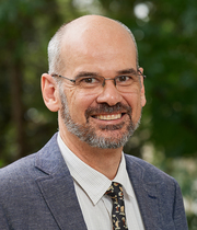 Craig Munns合作论文数School of Medicine, Faculty of Medicine and Health, The University of Sydney11Ravi Savarirayan合作论文数11Valérie Cormier-Daire合作论文数8
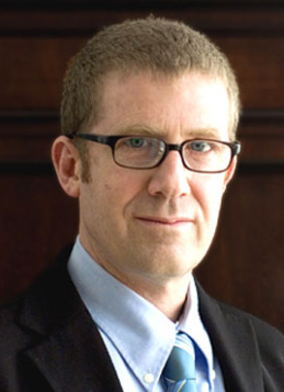 Andrea Superti Furga合作论文数Centre Hospitalier Universitaire Vaudois8Julie Briody合作论文数8
 Geert Mortier合作论文数7Andreas Zankl合作论文数School of Medicine, Faculty of Medicine and Health, The University of Sydney7
 Sheila Unger合作论文数Institute of Human Genetics, University of Freiburg Hospital7
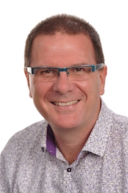 Stephen Robertson合作论文数Department of Women's and Children's Health, School of Biomedical Sciences, University of Otago7J. Ault合作论文数6
 Stefan Mundlos合作论文数Max-Planck-Institut fur Molekulare Genetik;Institute for Medical and Human Genetics, Charité Universitätsmedizin Berlin6Jenny Ault合作论文数6M.B. Alcausin合作论文数5
 Deborah Krakow合作论文数Department of Human Genetics, David Geffen School Of Medicine, University of California, Los Angeles5
 Michel Tchan合作论文数5Kazimierz Kozlowski合作论文数5M. McQuade合作论文数4Elizabeth Thompson合作论文数4Sharron Townshend合作论文数4Felicity Collins合作论文数4james mcgill合作论文数4
 Leanne Johnston合作论文数School of Health and Rehabilitation Sciences, Faculty of Health and Behavioural Sciences, The University of Queensland4Le Merrer Martine合作论文数4
 Matthew Warman合作论文数Department of Orthopaedic Surgery, Boston Children's Hospital;Department of Genetics, Blavatnik Institute Genetics, Harvard Medical School4Ireland Penelope Jane合作论文数4Arnold Munnich合作论文数Necker-Enfants malades de Paris4Gen Nishimura合作论文数Department of Molecular Medicine and Surgery, Karolinska Institutet4
 Chris Cowell合作论文数The Children's Hospital at Westmead;Discipline of Paediatrics & Child Health, University of Sydney;The Children's Hospital at Westmead, University of Sydney4
 Robert Ware合作论文数Centre for Applied Health Economics, Griffith University;Menzies Health Institute Queensland, Griffith University4Sara Cathey合作论文数4Christine M. Hall合作论文数4Jürgen Spranger合作论文数4Samantha Donaghey合作论文数3
 Karen Waters合作论文数School of Medicine, Faculty of Medicine and Health, The University of Sydney3
 Bernhard Zabel合作论文数Otto-von-Guericke-Universität Magdeburg3Alison Colley合作论文数3Sally L. Dunwoodie合作论文数3R. H. H. Engelbert合作论文数3Ralph S. Lachman合作论文数University of California, Los Angeles3
 Yasemin Alanay合作论文数Department of Pediatrics, Hacettepe University Faculty of Medicine3Céline Huber合作论文数3Tony Roscioli合作论文数Univeristy of New South Wales3
 Melanie Alcausin合作论文数Department of Genetic Medicine, University of Sydney3David Markie合作论文数3
 Outi Makitie合作论文数2Elizabeth M Thompson合作论文数2HE Ritchie合作论文数2Satz Mengensatzproduktion合作论文数2




