Abstract Purpose: Translation of the patient-derived xenograft (PDX) model into a method for practical personalized cancer treatment is prevented by the intense resources and time necessary to generate and test each tumorgraft. We aimed to develop a high-throughput ex vivo drug testing approach that can be used for personalized cancer treatment design. Experimental Design: We developed a unique ex vivo live tissue sensitivity assay (LTSA), in which precision-cut and uniform small tissue slices derived from pancreatic ductal adenocarcinoma PDX tumors were arrayed in a 96-well plate and screened against clinically relevant regimens within 3 to 5 days. The correlation between the sensitivities of tissue slices to the regimens and patients' clinical responses and outcome were statistically analyzed. The results of LTSA assay were further confirmed with biochemical methods in vitro and animal PDX model in vivo. Results: The ex vivo tissue slices remain viable for at least 5 days, and the tumor parenchyma, including stroma, vascular structures, and signaling pathways, are all retained. The sensitivities of the ex vivo tissue slices to gemcitabine and irinotecan was consistent with the clinical responses and outcomes of the patients from whom the tumorgrafts were derived (r = 0.77; P = 0.0002). Retrospective analysis showed that the patients who received LTSA-sensitive regimens had remarkably longer progression-free survival than patients who received LTSA-resistant regimens (16.33 vs. 3.8 months; n = 18, P = 0.011). Conclusions: The results from these PDX and LTSA methods reflect clinical patients' responses and could be used as a personalized strategy for improving systemic therapy effectiveness in patients with pancreatic cancer. Clin Cancer Res; 22(24); 6021–30. ©2016 AACR.
Overexpression and aberrant activation of signal transducer and activator of transcription 3 (STAT3) contribute to tumorigenesis, drug resistance, and tumor-immune evasion, making it a potential cancer therapeutic target. BP1003 is a neutral liposome incorporated with a nuclease-resistant P-ethoxy antisense oligodeoxynucleotide (ASO) targeting the STAT3 mRNA. Its unique design enhances BP1003 stability, cellular uptake, and target affinity. BP1003 efficiently reduces STAT3 expression and enhances the sensitivity of breast cancer cells (HER2+, triple negative) and ovarian cancer cells (late stage, invasive ovarian cancer) to paclitaxel and 5-fluorouracil (5-FU) in both 2D and 3D cell cultures. Similarly, ex vivo and in vivo patient-derived models of pancreatic ductal adenocarcinoma (PDAC) show reduced tissue viability and tumor volume with BP1003 and gemcitabine combination treatments. In addition to directly affecting tumor cells, BP1003 can modulate the tumor microenvironment. Unlike M1 differentiation, monocyte differentiation into anti-inflammatory M2 macrophages is suppressed by BP1003, indicating its potential contribution to immunotherapy. The broad anti-tumor effect of BP1003 in numerous preclinical solid tumor models, such as breast, ovarian, and pancreatic cancer models shown in this work, makes it a promising cancer therapeutic.
Supplemental Materials and Methods: Immunohistochemical staining of cleaved caspase 3, statistical analysis of the correlation of LTSA values with patients' progression free survival; Supplemental Figure legend
Supplementary Figures 6-7 from Critical Role and Regulation of Transcription Factor FoxM1 in Human Gastric Cancer Angiogenesis and Progression
Supplementary Figure 5 from Critical Role and Regulation of Transcription Factor FoxM1 in Human Gastric Cancer Angiogenesis and Progression
Supplementary Figures S1-S6.
Supplementary Figure 3 from Critical Role and Regulation of Transcription Factor FoxM1 in Human Gastric Cancer Angiogenesis and Progression
Supplementary Table 1. Wild type and mutant lumican SBE oligos for EMSA. Supplementary Table 2. Clinicopathologic and oncologic outcome correlation of stromal lumican expression in PDAC. Supplementary Table 3. Amount of Lumican Secretion in HPSC and PDACs Supernatant at 8 and 24 hours.
Tissue slices after treatment with Gemcitabine (100 µM) or Irinotecan (30 µM) for 48 hours were fixed and embedded for immunohistochemical staining of cleaved-Caspase 3 (40X magnification, Bars = 20 µm).
Mutant p53-associated genes
Supplementary Figure 1 from FoxM1B Transcriptionally Regulates Vascular Endothelial Growth Factor Expression and Promotes the Angiogenesis and Growth of Glioma Cells
HPaSteC and Media: Western blot and ELISA assay: Vectra Automated Quantitative Pathology Imaging System (VAQPIS, PerkinElmer): Double Immunofluorescence staining (IF), and Vectra multispectral analysis: Supplementary figure legends:
This table summarizes the dose (multiplicity of infection, MOI) of Delta-24-RGD that causes 50% of viability inhibition of infected pancreatic cancer cell lines (IC50).
PDAC cells matrix adhesions were affect by HPSC supernatant of 24 hour's collection.
Lumican expression and secretion in HPaSteC with or without TGF-β exposure at 8 and 24 hours.
Supplementary Tables 1- 3 from Critical Role and Regulation of Transcription Factor FoxM1 in Human Gastric Cancer Angiogenesis and Progression
 Jason B. Fleming合作论文数Department of Surgery, UT Southwestern Medical Center29
 Ya'an Kang合作论文数21
 Xinqun Li合作论文数20
 David Roife合作论文数16
 Mayrim Rios合作论文数14
 Jack A. Roth合作论文数Department of Thoracic and Cardiovascular Surgery, Division of Surgery, The University of Texas MD Anderson Cancer Center14Suyun Huang合作论文数Department of Neurosurgery, The University of Texas MD Anderson Cancer Center14
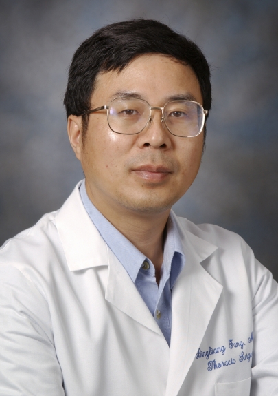 Bingliang Fang合作论文数Thoracic and Cardiovascular Surgery, MD Anderson Cancer Center14Michael P. Kim合作论文数13
 Michael Pratt合作论文数12
 Eugene J. Koay合作论文数Department of Radiation Oncology, Division of Radiation Oncology, The University of Texas MD Anderson Cancer Center;Department of Gastrointestinal Radiation Oncology, Division of Radiation Oncology, The University of Texas MD Anderson Cancer Center;Department of Nanomedicine, Houston Methodist Research Institute12
 Keping Xie (谢克平)合作论文数Center for Pancreatic Cancer Research, South China University of Technology12
 Jithesh J Augustine合作论文数10
 Rong Cai (蔡蓉)合作论文数Shanghai Jiao Tong University College of Basic Medical Sciences10
 Yeon-Ju Lee合作论文数Marine Natural Products Laboratory, Korea Ocean Research and Development Institute8Jenying Deng合作论文数7Jieru Meng合作论文数7
 John Minna合作论文数Hamon Center for Therapeutic Oncology Research, The University of Texas Southwestern Medical Center;Department of Internal Medicine, The University of Texas Southwestern Medical Center;Department of Pharmacology, The University of Texas Southwestern Medical Center7
 Matthew H.G. Katz合作论文数Department of Surgical Oncology, Division of Surgery, The University of Texas MD Anderson Cancer Center7
 Guanxiang Qian合作论文数Research Center for Human Gene Therapy, Shanghai Second Medical University;Fax: +86-21-63842916 e-mail: sbqian@shsmu.edu.cn CN CN;Research Center for Human Gene Therapy, Shanghai Second Medical University7Tara G. Hughes合作论文数6Christian Siangco合作论文数6Shishu Chen合作论文数6
 Nu Zhang (张弩)合作论文数The First Affiliated Hospital, Sun Yat-Sen University6
 Chatterjee Deyali合作论文数Dept Surg Oncol, Univ Texas MD Anderson Canc Ctr6Ling Jianhua合作论文数6
 Huamin Wang (王华民)合作论文数Department of Pathology, Division of Pathology/Lab Medicine, The University of Texas MD Anderson Cancer Center;MD Anderson UT Health Graduate School of Biomedical Sciences, The University of Texas6Majidi Mourad合作论文数6
 Lin Ji (吉琳)合作论文数Department of Chemistry, Capital Normal University6Daoyan Wei合作论文数6Xiangdong Le合作论文数6Shuhong Wu合作论文数6Rei Suzuki合作论文数Gastroenterology and Rheumatology, Fukushima Medical University6
 Paul J. Chiao合作论文数Department of Molecular and Cellular Oncology, Division of Basic Science Research, The University of Texas MD Anderson Cancer Center6Zhiliang Jia合作论文数6Ryan M. Thomas合作论文数5WANG Jia-min (王家敏)合作论文数5LU Jian (卢健)合作论文数5WenHan Mei (梅文瀚)合作论文数5
 Kenneth Aldape合作论文数Laboratory of Pathology, Center for Cancer Research, National Cancer Institute, National Institutes of Health5Christopher A. Bristow合作论文数University of Texas MD Anderson Cancer Center5Jian Lü合作论文数5
 li Wang合作论文数5
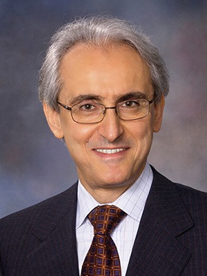 Raymond Sawaya合作论文数The University of Texas MD Anderson Cancer Center;American University of Beirut5
 Stephen G. Swisher合作论文数Department of Thoracic and Cardiovascular Surgery, Division of Surgery, The University of Texas MD Anderson Cancer Center;Lung Cancer5Timothy P. Heffernan合作论文数5
 Mark J Truty合作论文数Department of Surgical Oncology, The University of Texas MD Anderson Cancer Center5Shaoyu Yan合作论文数4Wang Yu合作论文数4




