Supplementary Figure 1. Coverage of pericyte and the basal membrane by LPA. Expression of LPA4 in several tumors. Supplementary Figure 2. Survival of wild-type and LPA4-KO mice harboring LLC brain tumors treated with or without LPA in the presence or absence of 5-FU. Supplementary Figure 3. Junctional protein expressioon in vascular endothelial cells by LPA. Supplementary Figure 4. LPA induces ICAM-1 expression but doesn't alter the expression of an HEV marker protein on endothelial cells Supplementary Figure 5. Investigation of LPA mediated VCAM-1 expression in MS-1 cells by using several inhibitors. Supplementary Figure 6. PD-L1 expression in GL261 brain tumors and LLC subcutaneous tumors. HE staining of the brain after anti-PD-1 antibody and LPA treatment.
EC-SP and -MP cells are in the active phase of the cell cycle in the tumor microenvironment.
Background/Aim: This study aimed to evaluate the association between the change in peripheral eosinophil count during postoperative pelvic radiotherapy and gastrointestinal (GI) toxicities in patients with cervical cancer. Patients and Methods: The medical records of 163 patients with cervical cancer who underwent postoperative concurrent chemoradiotherapy between 2000 and 2016 were analyzed. Results: Among the peripheral blood cell counts, transient elevation of the eosinophil count was observed during radiotherapy. Of the 163 patients, 117 developed grade ≥2 diarrhea during radiotherapy, and 25 patients developed grade ≥2 late GI toxicities. In multivariate analysis, the maximum eosinophil count and age emerged as independent predictors of grade ≥2 acute diarrhea during radiotherapy, while bowel bag V40 Gy and age were predictive of grade ≥2 late GI toxicities. Conclusion: Early detection of transient elevation of eosinophil may facilitate early treatment of acute diarrhea during radiotherapy.
Background: The present study aimed to estimate geometric changes in applicators and prostate over 3 days in patients with high-dose-rate brachytherapy (HDR-BT) and to assess the need for daily replanning. Patients and Methods: This study retrospectively investigated 18 patients who underwent HDR-BT as monotherapy from February 2016 to October 2018. Results: Without replanning, the planning target volume coverage significantly worsened on day 2 (p<0.001) and day 3 (p=0.003). The minimum dose distributed to the highest irradiated rectal volume of 5 cc became significantly higher on day 2 (p=0.02), and the maximum dose distributed to the urethra became significantly higher on day 2 (p=0.01). Conclusion: Conformal, high-dose delivery of HDR-BT is impaired without replanning not only on the second day but also on the third day. Daily replanning is required for achieving accuracy of HDR-BT.
TNF-α is a pleiotropic cytokine that has the potential to induce apoptosis under inflammation. How endothelial cells (ECs) are spared from this fate in inflammatory environments where TNF-α is present is not known. Here, we show that TGF-β-activated kinase 1 (TAK1) ensures EC survival and maintains vascular integrity upon TNF-α stimulation. Endothelial-specific TAK1 knockout mice exhibit intestinal and liver hemorrhage due to EC apoptosis, leading to vascular destruction and rapid death. This EC apoptosis was induced by TNF-α from myeloid cells responding to intestinal microbiota. TNF-α secretion associated with inflammation also induced vascular defects in inflamed organs. Additionally, we determined that TAK1 deletion in tumor ECs resulted in blood vessel and hence tumor regression. Our results illuminate mechanisms ensuring survival of intestinal and liver ECs under physiological conditions and ECs of other organs under inflammatory conditions that could be exploited for anti-angiogenic therapy to treat cancer.
Spontaneous regression in intracranial germ cell tumors has been reported in some literatures, but the mechanism has not been well known. We retrospectively measured the tumor volume before chemoradiotherapy and analyzed factors that influence shrinkage of tumor volume. PATIENTS AND Thirty-nine cases with primary intracranial germinomas and HCG-producing germinomas were treated in our hospital from 1994 to 2017. In twenty-one of them, plural MRI scans were done before the first course of chemotherapy regimen. Their age ranged from 8 to 31 years. Three cases were bifocal type, one case had multiple lesions. Biopsies were performed in all regions. Tumor volume of twenty-four lesions was analyzed by volumetric assessment based on MRI. Ratio of volumetric change between the first MRI and the scan immediately before chemotherapy was defined as shrinking rate (%). Period between disease onset and the first chemotherapy was 20 to 47 days. Diagnostic radiation dose was calculated in each case. Initial tumor volume ranged from 0.962 to 72.356 cubic centimeters (mean: 9.67). Diagnostic radiation dose: 52.2 to 910.1 mGy. Shrinking rate ranged from -57.8 to 85.4% (mean: 28.7). In only 7 regions (29.1%), shrinking rate was within 30%. Shrinking rate was significant positively influenced by diagnostic radiation dose (p<0.05) and negatively influenced by initial volume (p<0.05). But, it had no correlation with age, sex, histopathological parameters. This study shows that the volume of intracranial germ cell tumors is changing dynamically in many cases. Diagnostic exposure to low-dose radiation influences spontaneous regression in intracranial germinomas.
Abstract The structure and function of tumor blood vessels profoundly affects the tumor microenvironment. Signals mediated through the lysophosphatidic acid receptor 4 (LPA4) promote vascular network formation to restore normal vascular barrier function in subcutaneous tumors and thus improve drug delivery. However, the characteristics of the vasculature vary by organ and tumor types, and how drug delivery and leukocyte trafficking are affected by modification of vascular function by LPA in different cancers is unclear. Here, we show that LPA4 activation promotes the formation of fine vascular structures in brain tumors. RhoA/ROCK signaling contributed to LPA-induced endothelial cell–cell adhesion, and RhoA/ROCK activity following LPA4 stimulation regulated expression of VCAM-1. This resulted in increased lymphocyte infiltration into the tumor. LPA improved delivery of exogenous IgG into brain tumors and enhanced the anticancer effect of anti–programmed cell death-1 antibody therapy. These results indicate the effects of LPA on vascular structure and function apply not only to chemotherapy but also to immunotherapy. Significance: These findings demonstrate that lysophosphatidic acid, a lipid mediator, promotes development of a fine capillary network in brain tumors by inducing tightening of endothelial cell-to-cell adhesion, facilitating improved drug delivery, and lymphocyte penetration.
Vascular normalization in tumors may improve drug delivery and anti-tumor immunity. Angiogenesis inhibitors induce hypoxia, which may facilitate malignant progression; therefore, we investigated other methods to promote vascular maturation. Here, we show that lysophosphatidic acid (LPA) enhances blood flow by promoting fine vascular networks, thereby improving vascular permeability and suppressing tumor growth when combined with anti-cancer drug treatment. Six different G protein-coupled receptors have been identified as LPA receptors (LPA1-6). In studies using mutant mice, we found that LPA4 is involved in vascular network formation. LPA4 activation induces circumferential actin bundling beneath the cell membrane and enhances linear adherens junction formation by VE-cadherin in endothelial cells. Therefore, we conclude that activation of LPA4 is a promising approach for vascular regulation.
Recent studies have reported strong associations between tumor locations, clinical features and genetic alterations in meningiomas. Although several predilection sites including cerebral convex, parasagittal areas or several skull base regions have been well-documented, those evaluations were traditionally based on descriptive methods. This study aimed to re-evaluate preferential areas of meningiomas more accurately by means of voxel-based lesion mapping (VBLM) and three-dimensional (3D) image rendering techniques. Magnetic resonance images of a consecutive series of 248 cases with treatment naïve 260 meningiomas were retrospectively analyzed. All images were registered to a T1-weighted brain atlas provided by the Montreal Neurological Institute (MNI152), and a lesion frequency map was created followed by 3D volume rendering to visualize the predilection sites of the tumors. In order to evaluate the validity of the cohort, the frequencies of tumor locations were compared with the results of previous publication including the Japan Brain Tumor Registry (JBTR). The 3D lesion frequency map clearly showed the preferential areas of meningiomas including the cerebral convexity and skull base regions. In parasagittal areas, the mid one-third of the superior sagittal sinus was most commonly affected. Substantial lesion accumulation was observed around the leptomeninges covering the central sulcus and the Sylvian fissure while very few lesions were observed at the frontal, parietal and occipital convexities. In skull base areas, parasellar, sphenoid wing and petroclival regions were commonly affected by the tumor. In the descriptive evaluation, frequencies for the tumor incidences in each area including the cerebral convex or falx, parasellar region were comparable with previous reports. The VBLM technique successfully visualized the meningioma predilection areas on the 3D lesion frequency map, and particularly indicated the preference of the mid-third area among the parasagittal regions. These observations warrant further studies for meningioma biology using this technique.
Alterations to the tumor stromal microenvironment induced by chemotherapy could influence the behavior of cancer cells. In the tumor stromal microenvironment, cancer-associated fibroblasts (CAFs) play an important role. Because the receptor tyrosine kinase Axl and its ligand Gas6 could be involved in promoting non-small cell lung cancer (NSCLC), we investigated the role of Gas6 secreted by CAFs during chemotherapy in NSCLC. In a murine model, we found that Gas6 expression by CAFs was upregulated following cisplatin treatment. Gas6 expression might be influenced by intratumoral hypoperfusion during chemotherapy, and it increased after serum starvation in a human lung CAF line, LCAF(hTERT). Gas6 is associated with LCAF(hTERT) cell growth. Recombinant Gas6 promoted H1299 migration, and conditioned medium (CM) from LCAF(hTERT) cells activated Axl in H1299 cells and promoted migration. Silencing Gas6 in LCAF(hTERT) reduced the Axl activation and H1299 cell migration induced by CM from LCAF(hTERT). In clinical samples, stromal Gas6 expression increased after chemotherapy. Five-year disease-free survival rates for patients with tumor Axl-and stromal Gas6-positive tumors (n = 37) was significantly worse than for the double negative group (n = 12) (21.9% vs 51.3%, p = 0.04). Based on these findings, it is presumed that Gas6 derived from CAFs promotes migration of Axl-expressing lung cancer cells during chemotherapy and is involved in poor clinical outcome.
Abstract Angiogenesis plays a crucial role in tumor growth, with an undisputed contribution of resident endothelial cells (EC) to new blood vessels in the tumor. Here, we report the definition of a small population of vascular-resident stem/progenitor–like EC that contributes predominantly to new blood vessel formation in the tumor. Although the surface markers of this population are similar to other ECs, those from the lung vasculature possess colony-forming ability in vitro and contribute to angiogenesis in vivo. These specific ECs actively proliferate in lung tumors, and the percentage of this population significantly increases in the tumor vasculature relative to normal lung tissue. Using genetic recombination and bone marrow transplant models, we show that these cells are phenotypically true ECs and do not originate from hematopoietic cells. After treatment of tumors with antiangiogenic drugs, these specific ECs selectively survived and remained in the tumor. Together, our results established that ECs in the peripheral vasculature are heterogeneous and that stem/progenitor–like ECs play an indispensable role in tumor angiogenesis as EC-supplying cells. The lack of susceptibility of these ECs to antiangiogenic drugs may account for resistance of the tumor to this drug type. Thus, inhibiting these ECs might provide a promising strategy to overcome antiangiogenic drug resistance. Cancer Res; 76(11); 3200–10. ©2016 AACR.
PSF1 (Partner of SLD Five 1) is an evolutionarily conserved DNA replication factor that is part of the GINS (Go, Ichi, Nii, and San) complex. The objective of this study was to evaluate the relationship between PSF1 expression and prognosis in patients with non-small cell lung cancer (NSCLC) treated with surgery following preoperative chemotherapy or chemoradiotherapy.
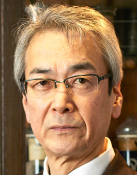 Nobuyuki Takakura (髙倉伸幸)合作论文数Department of Signal Transduction Research Institute for Microbial Diseases, Osaka University10
 Naito Hisamichi合作论文数Research Institute for Microbial Diseases, Osaka University10Takara Kazuhiro合作论文数9Tomohiro Iba合作论文数8
 Hiroyasu Kidoya合作论文数Dept Signal Transduct, Osaka Univ8Muramatsu Fumitaka合作论文数8
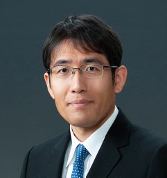 Taku Wakabayashi合作论文数Department of Ophthalmology, Osaka University Medical School8
 Naoki Kagawa合作论文数8Yasunori Fujimoto合作论文数7
 Manabu Kinoshita合作论文数6
 Ryuichi Hirayama合作论文数6
 Kishima Haruhiko合作论文数Osaka University6Yasuyoshi Chiba合作论文数5Shogo Fukuya合作论文数4Yamane Keitaro合作论文数4Chisato Yokota合作论文数3Toshiki Yoshimine合作论文数3Koji Takano合作论文数3Hashimoto Naoya合作论文数3Hideyuki Arita合作论文数3Yohei Tsukada合作论文数Graduate School of Pharmaceutical Sciences, Osaka University3Jyunichi Hara合作论文数2Kazuyoshi Kise合作论文数2Takamune Achiha合作论文数2Fukuko Yamamoto合作论文数2Takanori Fukunaga合作论文数2Katsuhiko Nakanishi合作论文数2Umehara Toru合作论文数2Kazuhiko Ogawa合作论文数2Toru Kimura合作论文数2
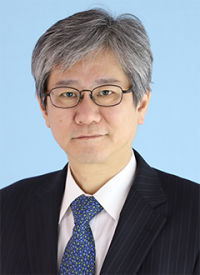 Meinoshin Okumura (奥村明之進)合作论文数Department of General Thoracic Surgery, Graduate School of Medicine, Osaka University2
 Shintani Yasushi (新谷康)合作论文数Osaka University2Soichiro Funaki合作论文数2Tomohiro Kawamura合作论文数Yodogawa Christian Hospital2Masato Minami (南正人)合作论文数Surgical Center, Osaka University Hospital2Yoshiko Hashii合作论文数2Ryu Kanzaki合作论文数2Yonehiro Kanemura合作论文数2Michio Oda合作论文数1Youhei Banba合作论文数1Syogo Fukuya合作论文数1
 Satoshi Ishii合作论文数1Hiroyuki Sakurai合作论文数1Isohashi Fumiaki合作论文数1Sakimoto Susumu合作论文数1Shusuke Moriuchi合作论文数1
 Michael Taylor合作论文数Department of Medical Biophysics, Temerty Faculty of Medicine, University of Toronto;Departments of Pediatrics, Baylor College of Medicine;Texas Children’s Hospital1Kijima Noriyuki合作论文数1Jia Weizhen合作论文数1




