Psilocybin has profound therapeutic potential for various mental health disorders, but its mechanisms of action are unknown. Functional MRI studies have reported the effects of psilocybin on brain activity and connectivity; however, these measurements rely on neurovascular coupling to infer neural activity changes and assume that blood flow responses to neural activity are not altered by psilocybin. Using two-photon excited fluorescence imaging in the visual cortex of awake mice to simultaneously measure neural activity and capillary blood flow dynamics, we found that psilocybin administration prolonged the increase in visual stimulus-evoked capillary blood flow - an effect which was reduced by pretreatment with a 5-HT2AR antagonist - despite not causing changes in the stimulus-evoked neural response. Multimodal widefield imaging also showed that psilocybin extends the stimulus-evoked vascular responses in surface vessels with no observed effect on the population neural response. Computational simulation with a whole-brain neural mass model showed that prolonged neurovascular coupling responses can lead to spurious increases in BOLD-based measures of functional connectivity. Together, these findings demonstrate that psilocybin broadens neurovascular responses in the brain and highlights the importance of accounting for these effects when interpreting human neuroimaging data of psychedelic drug action.
Apolipoprotein E4 (ApoE4) genotype, hypertension, and biological sex are critical risk factors for Alzheimer's disease and related dementias. Yet, their combined impact on early cerebrovascular dysfunction, brain inflammation, and memory impairment remains poorly understood. We developed a translational mouse model incorporating human ApoE4, hypertension via angiotensin II infusion, and induced accelerated ovarian failure (AOF) to mimic perimenopause in females to investigate these interactions. Hypertensive ApoE4 mice of both sexes exhibited impaired spatial working memory, decreased cerebral blood flow, increased neuroinflammation, and decreased blood brain barrier integrity, recapitulating key early clinical features observed in human populations with these risk factors. Brain blood flow reduction was associated with an increased incidence of capillary stalling, with notable sex differences in the extent and cellular composition of stalls: in males, stalling was strongly elevated and mostly due to red blood cell arrest, while stalling was modestly elevated in peri-AOF females with most stalls including leukocytes. Treatment with prasugrel, a P2Y12 receptor inhibitor, improved memory performance in both sexes but was correlated with different physiological effects - restored cerebral blood flow in males and reduced microglia motility and inflammation in peri-AOF females. Platelet depletion mimicked prasugrel's blood flow and cognitive benefits in males, while microglia depletion selectively rescued memory in females. Our work emphasizes the necessity of including translationally relevant female mouse models in neurodegenerative disease studies, and our findings highlight the importance of risk profile-specific interventions and demonstrate that early vascular dysfunction may be a key, sex-dependent driver of cognitive decline.
Several genetic and cardiovascular risk factors increase incidence of Alzheimer’s disease and related dementias (ADRD). Hypertension and the ε4 allele of apolipoprotein E (ApoE) are powerful drivers of cognitive impairment in ADRD. These risk factors are also associated with decreased cerebral blood flow (CBF). Experimental data suggest that the CBF reduction may be due to temporary capillary occlusions (capillary stalls) caused by circulating vascular and immune cells. To gain insight into how these risk factors may contribute to cognitive decline, we induced hypertension in mice expressing human ApoE3 or E4 (ApoE3- or E4-TR mice), and focused on the resulting CBF and cognitive alterations, and their potential reversal by preventing capillary stalling. Male ApoE3 and 4-TR mice (n = 9-15/group) were assessed at three timepoints: Baseline, following two weeks of angiotensin II-induced hypertension (AngII, 500ng/kg/min), and after an additional week of anti-platelet treatment (prasugrel, 10mg/kg, daily). We tested spatial memory function in a Y-maze, and imaged cortical microvascular flow using two-photon microscopy. At baseline, ApoE3 and 4-TR mice showed similar memory performance, but with hypertension (∼25 mmHg increase in blood pressure) ApoE4-TR mice exhibited a spatial working memory deficit, which coincided with a 26% decrease in capillary flow speed (4.11±0.3 vs. 3.05±0.29 mm/s, p <0.01), and a 1.8X increase in the incidence of non-flowing capillaries. Most stalled capillaries contained only red blood cells in the stalled segment (57%), with some also containing leukocytes (30%), or platelets (12%) (Fig. 1). Treatment with prasugrel led to a 55% reduction in stalling (65±9 vs. 144±24 stalls/mm 3 ), and a concomitant 23% increase in capillary blood flow (3.75±0.36 vs. 3.05±0.29 mm/s, p = 0.11), and significantly improved blood flow in penetrating arterioles that correlated with restored performance on the Y-maze task (Fig. 2). Immunohistochemistry further revealed improved BBB integrity and reduced gliosis following prasugrel treatment. Paralleling human findings, we show that mice with the e4 allele of ApoE experience more deleterious consequences from hypertension, as compared to their e3 counterparts. Prasugrel treatment led to an almost complete recovery of these deficits, suggesting that anti-platelet agents may be of therapeutic value by improving CBF and cognitive function in the presence of these risk factors.
Small animal studies in biomedical research often require anesthesia to reduce pain or stress experienced by research animals and to minimize motion artifact during imaging or other measurements. Anesthetized animals must be closely monitored for the safety of the animals and to prevent unintended effects of altered physiology on experimental outcomes. Many currently available monitoring devices are expensive, invasive, or interfere with experimental design. Here, we present MousePZT, a low-cost device based on a simple piezoelectric sensor, with a custom circuit and computer software that allows for measurements of both respiratory rate and heart rate in a non-invasive, minimal contact manner. We find the accuracy of the MousePZT device in measuring respiratory and heart rate matches those of commercial systems. Using the widely-used gas isoflurane and injectable ketamine/xylazine combination, we also demonstrate that changes in respiratory rate are more easily detected and can precede changes in heart rate associated with variations in anesthetic depth. Additional circuitry on the device outputs a respiration-locked trigger signal for respiratory-gating of imaging or other data acquisition and has high sensitivity and specificity for detecting respiratory cycles. We provide detailed instruction documents and all necessary microcontroller and computer software, enabling straightforward construction and utilization of this device.
In partial onset epilepsy, seizures arise focally in the brain and often propagate, causing acute behavior changes, chronic cognitive decline, and increased mortality. Patients frequently become refractory to medical management, leaving neurosurgical resection of the seizure focus as a primary treatment, which can cause neurologic deficits. In the cortex, focal seizures are thought to spread through horizontal connections in layers II/III, suggesting that selectively severing these connections could block seizure propagation while preserving normal columnar circuitry and function. We induced focal neocortical epilepsy in mice and used tightly-focused femtosecond-duration laser pulses to create a sub-surface, opencylinder cut surrounding the seizure focus and severing cortical layers II-IV. We monitored seizure propagation using electrophysiological recordings at the seizure focus and at distant electrodes for 3-8 months. With laser cuts, only 5% of seizures propagated to the distant electrodes, compared to 85% in control animals. Laser cuts also decreased the number of seizures that were initiated, so that the average number of propagated seizures per day decreased from 42 in control mice to 1.5 with laser cuts. Physiologically, these cuts produced a modest decrease in cortical blood flow that recovered within days and, at one month, left a ~20-μm wide scar with increased gliosis and localized inflammatory cell infiltration but minimal collateral damage. When placed over motor cortex, cuts did not cause notable deficits in a skilled reaching task. Femtosecond laser produced sub-surface cuts hold promise as a novel neurosurgical approach for intractable focal cortical epilepsy, as might develop following traumatic brain injury. Once sentence summary In a mouse model of focal epilepsy, sub-surface laser-produced cuts encircling the seizure focus attenuate propagation without behavioral impairment.
RNA macromolecules, like proteins, fold to assume shapes that are intimately connected to their broadly recognized biological functions; however, because of their high charge and dynamic nature, RNA structures are far more challenging to determine. We introduce an approach that exploits the high brilliance of x-ray free-electron laser sources to reveal the formation and ready identification of angstrom-scale features in structured and unstructured RNAs. Previously unrecognized structural signatures of RNA secondary and tertiary structures are identified through wide-angle solution scattering experiments. With millisecond time resolution, we observe an RNA fold from a dynamically varying single strand through a base-paired intermediate to assume a triple-helix conformation. While the backbone orchestrates the folding, the final structure is locked in by base stacking. This method may help to rapidly characterize and identify structural elements in nucleic acids in both equilibrium and time-resolved experiments.
Background: Chronic inflammation and myocardial hypoperfusion are implicated in the pathogenesis of heart failure with preserved ejection fraction (HFpEF), but the cellular and molecular mechanisms are unknown. Intravital cardiac multiphoton microscopy (MPM) enables direct visualization of blood flow and inflammatory cells within the capillary bed of the actively beating heart. We hypothesize that transient arrest of neutrophils in myocardial capillaries leads to perfusion deficits, resulting in hypoxia and driving disease progression. Methods: Male and female 8-12-week-old C57BL/6 mice received a high fat diet and L-NAME in drinking water (HFpEF) or standard diet and water (Chow) for 15 weeks. Intravital imaging of the actively beating heart was performed in mechanically ventilated mice to visualize neutrophils and capillary perfusion. An antibody against the neutrophil surface marker Ly6G (αLy6G) was administered intraperitoneally to test the effect of neutrophil depletion. Results: In vivo MPM revealed increases in arrested neutrophils in myocardial capillaries in HFpEF mice compared to Chows. Myocardial hypoxia, assessed by pimonidazole staining, and diastolic function, assessed as mitral e/e’ using echocardiography, were rescued with both extended neutrophil depletion (2mg/kg every 3d for 4 wk) and acutely (24 hrs after a single dose of 4mg/kg). To further test the immediate effects of αLy6G we implemented multi-exposure laser speckle imaging for perfusion and spectral imaging for tissue and blood oxygenation with electrocardiograph-driven reconstruction. Conclusion: Arrested neutrophils within capillaries appear to compromise myocardial perfusion in HFpEF. Neutrophil depletion to restore blood flow improved myocardial oxygenation and diastolic function rapidly. Effects were sustained with prolonged treatment. Capillary stall perfusion deficits may be a future therapeutic target to slow disease progression and offer rapid symptomatic relief.
Laser speckle contrast imaging (LSCI) is a widefield imaging technique that enables high spatiotemporal resolution measurement of blood flow. Laser coherence, optical aberrations, and static scattering effects restrict LSCI to relative and qualitative measurements. Multi-exposure speckle imaging (MESI) is a quantitative extension of LSCI that accounts for these factors but has been limited to post-acquisition analysis due to long data processing times. Here we propose and test a real-time quasi-analytic solution to fitting MESI data, using both simulated and real-world data from a mouse model of photothrombotic stroke. This rapid estimation of multi-exposure imaging (REMI) enables processing of full-frame MESI images at up to 8 Hz with negligible errors relative to time-intensive least-squares methods. REMI opens the door to real-time, quantitative measures of perfusion change using simple optical systems.
Optical imaging of wholemount tissue samples provides greater understanding of structure-function relationships as the architecture of these specimens is generally well preserved. However, difficulties arise when attempting to stitch together images of multiple regions of larger, oddly shaped specimens. These difficulties include (1) maintaining consistent signal-to-noise ratios when the overlying sample surface is uneven, (2) ensuring sample viability when live samples are required, and (3) stabilizing the specimen in a fixed position in a flowing medium without distorting the tissue sample. To address these problems, we designed a simple and cost-efficient device that can be 3D-printed and machined. The design for the device, named the Platform for Planar Imaging of Curved Surfaces (PICS), consists of a sample holder, or cap with gaps for fluid flow and a depression for securing the sample in a fixed position without glue or pins, a basket with two arms that move along an external radius to rotate the sample around a central axis, and a customizable platform designed to fit on a commercially available temperature control system for slice electrophysiology. We tested the system using wholemounts of the murine subventricular zone (SVZ), which has a high degree of curvature, to assess sample viability and image quality through cell movement forover an hour for each sample. Using the PICS system, tissues remained viable throughout the imaging sessions, there were no noticeable decreases in the image SNR across an imaging plane, and there was no noticeable displacement of the specimen due to fluid flow.
Cerebral blood flow (CBF) reductions in Alzheimer's disease patients and related mouse models have been recognized for decades, but the underlying mechanisms and resulting consequences for Alzheimer's disease pathogenesis remain poorly understood. In APP/PS1 and 5xFAD mice we found that an increased number of cortical capillaries had stalled blood flow as compared to in wild-type animals, largely due to neutrophils that had adhered in capillary segments and blocked blood flow. Administration of antibodies against the neutrophil marker Ly6G reduced the number of stalled capillaries, leading to both an immediate increase in CBF and rapidly improved performance in spatial and working memory tasks. This study identified a previously uncharacterized cellular mechanism that explains the majority of the CBF reduction seen in two mouse models of Alzheimer's disease and demonstrated that improving CBF rapidly enhanced short-term memory function. Restoring cerebral perfusion by preventing neutrophil adhesion may provide a strategy for improving cognition in Alzheimer's disease patients.
 Chris Schaffer合作论文数Meinig School of Biomedical Engineering, College of Engineering, Cornell University8
 Nozomi Nishimura合作论文数Schaffer-Nishimura Lab, Meinig School of Biomedical Engineering, College of Engineering, Cornell University5
 Anne E. Buglione合作论文数2
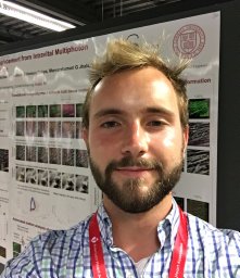 Nathaniel H. Allan-Rahill合作论文数Nancy E. and Peter C. Meinig School of Biomedical Engineering, Cornell University2
 Costantino Iadecola合作论文数Graduate School of Medical Sciences, Weill Cornell Medical College;Feil Family Brain and Mind Research Institute, Weill Cornell Medical College2
 Bracko Oliver合作论文数Institute of Molecular Health Sciences, Swiss Federal Institute of Technology Zurich2Benjamin S Kemp合作论文数1Gabriel Otte合作论文数1Jeffrey D. Beverly合作论文数1Myriam Peyrounette合作论文数1Lindsay K. Vinarcsik合作论文数1Ryan Radwanski合作论文数1
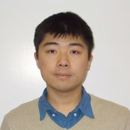 Shuo Sui合作论文数Dept Chem Engn, Univ Massachusetts1
 Kara A Zielinski合作论文数Sch Appl & Engn Phys, Cornell Univ1
 Maithri M Kashipathy合作论文数Protein Structure and X-ray Crystallography Laboratory, The University of Kansas1
 Clara Liao合作论文数Yale University1Seth Lieberman合作论文数1Iryna Ivasyk合作论文数1
 Jean Cruz合作论文数1Yiming Kang合作论文数Ctr Genome Sci & Syst Biol, Washington Univ1Maxime Berg合作论文数Institut de Mecanique des Fluides de Toulouse1Stella Lisova合作论文数Arizona State Univ, Dept Phys, Tempe, AZ 85287 USA1
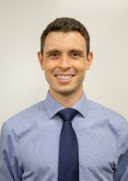 Calvin J Kersbergen合作论文数The Solomon H. Snyder Department of Neuroscience, Johns Hopkins University1
 Mohammad Haft-Javaherian合作论文数institution Meinig School of Biomedical Engineering, Cornell University1
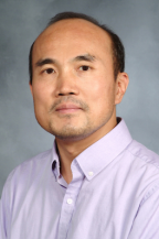 Laibaik Park合作论文数Feil Family Brain and Mind Research Institute, Weill Cornell Medicine, Cornell University1Mingrui Zhao合作论文数Feil Family Brain and Mind Research Institute, Weill Cornell Medicine, Cornell University1
 Lois Pollack合作论文数School of Applied and Engineering Physics, College of Engineering, Cornell University1
 Alex Kwan合作论文数Neuroscience, Biological & Biomedical Sciences, Yale University1
 David Small合作论文数Menzies Institute for Medical Research, University of Tasmania1Joan Zhou合作论文数1
 Sidney Strickland合作论文数Laboratory of Neurobiology and Genetics, The Rockefeller University1
 Cortes-Canteli Marta合作论文数Laboratory of Neurobiology and Genetics, The Rockefeller University1
 Teresa A. Murray合作论文数Barrow Neurological Institute1
 Elizabeth M. Davenport合作论文数1
 Teresa Southard合作论文数Biomedical Sciences, College of Veterinary Medicine, Cornell University1
 Sylvie Lorthois合作论文数1
 Moss Frank合作论文数Dept Chem, Stanford Univ1
 Thomas Grant合作论文数Hauptman-Woodward Medical Research Institute1
 Tong Wang合作论文数Shandong University, Shandong University1Mark S. Hunter合作论文数Department of Chemistry and Biochemistry, Arizona State University1
 Christopher Kupitz合作论文数Department of Chemistry and Biochemistry, Arizona State University1Smith Amy合作论文数1
 Theodore H. Schwartz合作论文数Minimally Invasive Surgery, Weill Cornell Medicine, Cornell University;Neurological Surgery, Weill Cornell Medicine, Cornell University1
 Amy Kuceyeski合作论文数Department of Radiology, Weill Cornell Medicine;Department of Computational Biology, Cornell University1Yohan Davit合作论文数IMFT (Institut de Mécanique des Fluides de Toulouse),, Université de Toulouse1Lynn Schooley Johnson合作论文数1
 Suzette A. Pabit合作论文数Cornell University1Valerio Mariani合作论文数Institute for Structural Biology1
 Michael Lamont合作论文数Meinig School of Biomedical Engineering, Cornell Engineering1




