Inflammatory cues affect hematopoietic stem cell (HSC) homeostasis and drive proliferation and myeloid skewing of HSCs. The HSC niche in the bone marrow (BM) is populated by a variety of stromal and immune cells that sense and respond to cellular stress. We investigated how BM-resident type 2 innate lymphoid cells (ILC2s) regulate HSC homeostasis and differentiation in steady state, during aging, and after genotoxic stress. We documented that PDGFR-α+sca-1+ mesenchymal stromal cells in the BM produced interleukin (IL)-33 with elevated levels after irradiation and during aging. IL-33/ST2 signaling in BM-resident ILC2s activated MAPK/NF-κB/JAK-STAT signaling and induced cytokine secretion. IL-6 and granulocyte-macrophage colony-stimulating factor (GM-CSF), secreted by ILC2s, promoted HSCs to proliferate and differentiate into the myeloid lineage. Taken together, we identified that IL-33 produced by MSCs induced ILC2s to secrete myeloid differentiation factors leading to myeloid-skewed HSCs with reduced self-renewal during aging.
Background Omalizumab, a therapeutic mAb targeting IgE, is approved for the treatment of multiple allergic indications. However, its moderate affinity for IgE necessitates frequent high-dose administrations, limiting its therapeutic use and efficacy. Attempts to develop next-generation anti-IgE antibodies with improved affinity, such as ligelizumab or HAE1, have yielded alternatives that are either less safe or not demonstrably superior. Objective We sought to generate optimized omalizumab variants featuring 2 specific molecular enhancements: increased IgE binding affinity while preserving epitope specificity to enhance target neutralization and improved potency to actively dissociate prebound IgE from its high-affinity receptor FcεRI. Methods Using a targeted yeast display selection strategy applied to mutated omalizumab libraries, we identified the anti-IgE clone C03 and engineered 2 flexible variants, C03-H1L2 and C03-H2L2. Results The C03 antibodies demonstrated approximately 10-fold higher IgE binding affinity compared with omalizumab, resulting in superior inhibition of IgE binding to FcεRI. Furthermore, C03-H1L2 and C03-H2L2 exhibited enhanced potency in displacing FcεRI-bound IgE from humanized mouse mast cells and human basophils without triggering spontaneous cell activation. In a systemic anaphylaxis mouse model, single-dose administration of the flexible C03 variants, in contrast to omalizumab, desensitized allergic effector cells within 36 hours, fully preventing antigen-induced anaphylaxis. Conclusions These findings underscore the importance of engineering next-generation anti-IgE therapies with higher affinity and disruptive potency to optimize current treatment approaches.
Due to its key roles in the development and manifestation of allergic reactions, immunoglobulin E (IgE) has become an interesting molecular target. The monoclonal anti-IgE antibody omalizumab, which primarily inhibits IgE binding to its high-affinity receptor, FcεRI⍺, on allergic effector cells has shown remarkable therapeutic success in various allergic indications. Recently, we have described an alternative anti-IgE molecule, termed KIH_E07_79, which in addition to the neutralization of free IgE rapidly disrupts pre-formed IgE-FcεRI complexes and thereby stops pre-initiated systemic anaphylaxis in mice.
To the Editor, Early in the coronavirus disease 2019 (COVID19) pandemic, age has been recognized as one of the major risk factors for poor clinical outcome.1 Based on hospitalization rates, it has also rapidly become evident that fewer women than men were affected by severe disease manifestation.2 With the primary goal to protect the most vulnerable populations, those older than 65, scientists around the world have successfully developed different vaccines with unprecedented speed.3 Although it is well established that immune responses against infections decline with age,4 it is less clear how vaccineelicited immunity varies between different sex and age groups.5 Given the importance of understanding these biological parameters, which may directly affect translatability of research findings into the clinic, we sought to investigate the immune response against severe acute respiratory syndrome coronavirus 2 (SARSCoV2) in a proteinbased and vesicular stomatitis virus (VSV)vectored vaccination approach in young and aged mice of both sexes. First, we used the recombinant receptorbinding domain (RBD) of the SARSCoV2 spike protein from the original reference strain emulsified in an aluminum hydroxide containing wet gel suspension (i.e. Alum) to subcutaneously immunize C57BL/6 mice (Figure S1). Seven days later, they received a booster injection and the vaccine response was assessed on day 28 (Figure 1A,B). To test the induction of humoral immunity as a function of age, we measured antigenspecific IgG in young (2 months old) and aged (18– 19 months old) mice by ELISA. Consistent with other studies, the systemic RBDspecific IgG response was significantly diminished in aged mice (Figure 1C,D). This agerelated decline in total RBDspecific IgG is primarily due to a loss of IgG1 production since the other subclasses remained barely detectable (Figure S2). To further characterize humoral immunity, we measured the total number of plasma Bcells in spleen by flow cytometry and quantified RBDspecific plasma Bcells in the spleen of immunized mice by ELISpot. While the total number of splenic plasma Bcells was increased in aged mice the RBDspecific IgG positive Bcells were significantly diminished (Figure 1E,F) and correlated with serum IgG levels (Figure 1G), suggesting that the agerelated reduction of RBDspecific plasma cell formation might contribute to the concomitant decrease in antibody titers. Previous studies have reported significant alterations in T follicular helper (Tfh) and regulatory (Tfr) cell numbers in lymphoid organs in aged mice contributing to impaired plasma Bcell generation and defective antibody production.6 Indeed, we measured an agerelated increase in both Tfr and Tfh populations in the spleen as quantified by flow cytometry, while the number of classical T regulatory cells (Tregs) remained unchanged (Figure S3A). Most importantly, the live SARSCoV2 neutralization potency of serum from aged mice was significantly reduced for the original reference strain and different other variants of concern (i.e. alpha, gamma and delta), which is in line with the agerelated decrease in RBDspecific serum antibody titers and plasma Bcells (Figure 1H). Additionally, we evaluated sexspecific differences in vaccination response across age in the same cohorts of immunized C57BL/6 mice (Figure 2A). While RBDspecific IgG and IgG1 responses in serum were higher in young females as compared to young male controls (Figure 2B,C), these differences were no longer apparent in the aged mice, and there were no detectable sexspecific differences in the number of splenic plasma Bcells (Figure 2D). In line with higher RBDspecific antibody titers, young female C57BL/6 mice also showed more potent virus neutralization of the SARSCoV2 reference strain when subcutaneously immunized with a proteinbased vaccine or intramuscularly injected with two VSVvectored COVID19 vaccine candidates (i.e., VSVSD21 and VSVMqSD21) as compared to male controls (Figure 2E and Figure S4A,B). However, neutralization of the other tested variants of concern was diminished and equally weak in both sexes (Figure 2E), indicating that mutations in the RBD domain of these variants were sufficient to escape the vaccineinduced antibody response. To test whether these findings were conserved across different mouse strains, we repeated the same immunization regimen in young BALB/c mice. The observed outcome was essentially the same with females showing a better vaccination response than male mice (Figure S5A– E). The sexspecific differences in humoral immune response of young C57BL/6 mice persisted even after an additional injection with a proteinbased vaccine 21 days after the first boost as assessed on day 42 (Figure 2F– J). Strikingly, we found increased numbers of RBDspecific plasma Bcells in the bone marrow of young female mice in this context. In summary, our data demonstrate significant ageand sexrelated differences in the humoral immune response to different
Background: Clinical management of allergic diseases has been hampered by the lack of safe and convenient tests to reliably identify culprit allergens and to closely follow changes in disease activity over time. Because allergy diagnosis is a complex and laborious multistep procedure, there is an urgent need for simpler but still functionally accurate ex vivo assays allowing objective diagnosis, substantiating treatment choices, and quantifying therapeutic responses. Objective: In this study, we sought to develop a novel functional cell-based assay that relies on passive sensitization of allergic effector cells with patient serum, circumventing current limitations in allergy diagnosis. Methods: We genetically engineered a conditional homeobox B8 (Hoxb8)-immortalized progenitor line from the bone marrow of mice that are transgenic for the human high-affinity IgE receptor (Fc epsilon RIa). These cells can be reproducibly differentiated into mature Hoxb8 mast cells within 5 days of culture in virtually unlimited numbers. Results: We demonstrate that the established Hoxb8 mast cell assay can be used to accurately measure total IgE levels, identify culprit allergens, longitudinally monitor allergen-specific immunotherapy, and potentially determine the time point of tolerance induction upon allergen-specific immunotherapy in patients with allergy. To facilitate the analysis of large testing volumes, we demonstrate a proof-of-concept for a high-throughput screening application based on fluorescent cell barcoding using the engineered Hoxb8 mast cells. Conclusions: Our results indicate that this novel mast cell assay could represent a valuable tool to support clinicians in the identification of IgE-mediated allergies and in the quantification of treatment efficacy as well as duration of therapeutic response.
Background: Serological tests are a powerful tool in the monitoring of infectious diseases and the detection of host immunity. However, manufacturers often provide diagnostic accuracy data generated through biased studies and the performance in clinical practice is essentially unclear. Objectives: We aimed to determine the diagnostic accuracy of various serological testing strategies for (a) identification of patients with previous coronavirus disease-2019 (COVID-19) and (b) prediction of neutralizing antibodies against SARS-CoV-2 in real-life clinical settings. Methods: We prospectively included 2’573 consecutive health-care workers and 1’085 inpatients with suspected or possible previous COVID-19 at a Swiss University Hospital. Various serological immunoassays based on different analytical techniques (enzyme-linked immunosorbent assays, ELISA; chemiluminescence immunoassay, CLIA; electrochemiluminescence immunoassay, ECLIA; lateral-flow immunoassay, LFI), epitopes of SARS-CoV-2 (nucleocapsid, N; receptor-binding domain, RBD; extended RBD, RBD+; S1 or S2 domain of the spike [S] protein, S1/S2), and antibody subtypes (IgG, pan-Ig) were conducted. A positive real-time PCR test from a nasopharyngeal swab was defined as previous COVID-19. Neutralization assays with live SARS-CoV-2 were performed in a subgroup of patients to assess neutralization activity (n=201). Results: The sensitivity to detect patients with previous COVID-19 was ≥85% in anti-N ECLIA (86.8%) and anti-S1 ELISA (86.2%). Sensitivity was 84.7% in anti-S1/S2 CLIA, 84.0% in anti-RBD+ LFI, 81.0% in anti-N CLIA, 79.2% in anti-RBD ELISA, and 65.6% in anti-N ELISA. The specificity was 98.4% in anti-N ECLIA, 98.3% in anti-N CLIA, 98.2% in anti-S1 ELISA, 97.7% in anti-N ELISA, 97.6% in anti-S1/S2 CLIA, 97.2% in anti-RBD ELISA, and 96.1% in anti-RBD+ LFI. The sensitivity to detect neutralizing antibodies was ≥85% in anti-S1 ELISA (92.7%), anti-N ECLIA (91.7%), anti-S1/S2 CLIA (90.3%), anti-RBD+ LFI (87.9%), and anti-RBD ELISA (85.8%). Sensitivity was 84.1% in anti-N CLIA, and 66.2% in anti-N ELISA. The specificity was ≥97% in anti-N CLIA (100%), anti-S1/S2 CLIA (97.7%), and anti-RBD+ LFI (97.9%). Specificity was 95.9% in anti-RBD ELISA, 93.0% in anti-N ECLIA, 92% in anti-S1 ELISA, and 65.3% in anti-N ELISA. Diagnostic accuracy measures were consistent among subgroups. Conclusions: The diagnostic accuracy of serological tests for SARS-CoV-2 antibodies varied remarkably in clinical practice, and the sensitivity to identify patients with previous COVID-19 deviated substantially from the manufacturer’s specifications. The data presented here should be considered when using such tests to estimate the infection burden within a specific population and determine the likelihood of protection against re-infection.
Background: Anaphylaxis represents one of the most severe and fatal forms of allergic reactions. Like most other allergies, it is caused by activation of basophils and mast cells by allergen-mediated cross-linking of IgE bound to its high-affinity receptor, Fc epsilon RI, on the cell surface. The systemic release of soluble mediators induces an inflammatory cascade, rapidly causing symptoms with peak severity in minutes to hours after allergen exposure. Primary treatment for anaphylaxis consists of immediate intramuscular administration of adrenaline. Objective: While adrenaline alleviates life-threatening symptoms of an anaphylactic reaction, there are currently no disease-modifying interventions available. We sought to develop potent and fast-acting IgE inhibitors with the potential to rapidly terminate acute allergic reactions. Methods: Using affinity maturation by yeast display and structure-guided molecular engineering, we generated 3 optimized disruptive IgE inhibitors based on designed ankyrin repeat proteins and assessed their ability to actively remove IgE from allergic effector cells in vitro as well as in vivo in mice. Results: The engineered IgE inhibitors rapidly dissociate preformed IgE:Fc epsilon RI complexes, terminate IgE-mediated signaling in preactivated human blood basophils in vitro, and shut down preinitiated allergic reactions and anaphylaxis in mice in vivo. Conclusions: Fast-acting disruptive IgE inhibitors demonstrate the feasibility of developing kinetically optimized inhibitors for the treatment of anaphylaxis and the rapid desensitization of allergic individuals.
Targeting of immunoglobulin E (IgE) represents an interesting approach for the treatment of allergic disorders. A high-affinity monoclonal anti-IgE antibody, ligelizumab, has recently been developed to overcome some of the limitations associated with the clinical use of the therapeutic anti-IgE antibody, omalizumab. Here, we determine the molecular binding profile and functional modes-of-action of ligelizumab. We solve the crystal structure of ligelizumab bound to IgE, and report epitope differences between ligelizumab and omalizumab that contribute to their qualitatively distinct IgE-receptor inhibition profiles. While ligelizumab shows superior inhibition of IgE binding to FcεRI, basophil activation, IgE production by B cells and passive systemic anaphylaxis in an in vivo mouse model, ligelizumab is less potent in inhibiting IgE:CD23 interactions than omalizumab. Our data thus provide a structural and mechanistic foundation for understanding the efficient suppression of FcεRI-dependent allergic reactions by ligelizumab in vitro as well as in vivo.
Background: Serological immunoassays that are able to identify protective immunity against SARS-CoV-2 are urgently required to adapt quarantine measures, assess vaccination responses and evaluate donor plasma. To date, however the utility of such immunoassays remains unclear. In a mixed-design evaluation study, we compared the performance of serological immunoassays that are based on various SARS-CoV-2 proteins and assessed the neutralizing activity of COVID-19 patient sera. Methods: Patients admitted with confirmed SARS-CoV-2 infection were prospectively followed alongside medical staff and biobank samples from winter 2018/2019. An in-house enzyme-linked immunosorbent assay utilizing recombinant receptor binding domain (RBD) of the SARS-CoV-2 spike protein was developed and compared to three commercially available enzyme-linked immunosorbent assays (ELISAs) targeting the nucleocapsid (N), the S1 domain of the spike protein (S1) and a lateral flow immunoassay (LFI) based on full-length spike protein. Neutralisation assays with live SARS-CoV-2 were performed to determine immunity. Findings: With a prevalence of 19.9%, we find 106 COVID-19 positive results among the 534 tested individuals. Time to IgG seroconversion occurred between day 0 and day 21 after the start of symptoms or positive RT-PCR. While the ELISAs showed sensitivities of 84.9% for RBD, 85.9% for S1 and 68.9% for Nprotein, the measured specificity amounted to 94.6% for the RBD, 97.7% for the S1 and 96.3% for the N protein. The LFI performed inferior with 71.4% sensitivity and 95.8% specificity. Out of 54 COVID-19 positive individuals 96.3% showed full neutralisation of live SARS-CoV-2 at serum dilutions ≥1:16, while none of the 6 COVID-19 negative sera revealed neutralizing activity. Interpretation: Our data suggest that ELISAs based on RBD and S1 protein of SARS-CoV-2 are adequate diagnostic predictors of protective immunity. Funding Statement: Development of SARS-CoV-2 reagents was partially supported by the NIAID Centers of Excellence for Influenza Research and Surveillance (CEIRS) contract HHSN272201400008C. MN is supported by a research grant of the Swiss National Science Foundation (#179334). AE received grant support from the Research Fund of the Swiss Lung Association, Bern and the Uniscientia foundation. Declaration of Interests: All authors declare that there is no conflict of interests. Ethics Approval Statement: The study protocol was approved by the appropriate ethics committee and the authorities of the University Hospital and conducted in accordance with the Declaration of Helsinki.
BACKGROUND:Serological immunoassays that can identify protective immunity against SARS-CoV-2 are needed to adapt quarantine measures, assess vaccination responses, and evaluate donor plasma. To date, however, the utility of such immunoassays remains unclear. In a mixed-design evaluation study, we compared the diagnostic accuracy of serological immunoassays that are based on various SARS-CoV-2 proteins and assessed the neutralizing activity of antibodies in patient sera.METHODS:Consecutive patients admitted with confirmed SARS-CoV-2 infection were prospectively followed alongside medical staff and biobank samples from winter 2018/2019. An in-house enzyme-linked immunosorbent assay utilizing recombinant receptor-binding domain (RBD) of the SARS-CoV-2 spike protein was developed and compared to three commercially available enzyme-linked immunosorbent assays (ELISAs) targeting the nucleoprotein (N), the S1 domain of the spike protein (S1), and a lateral flow immunoassay (LFI) based on full-length spike protein. Neutralization assays with live SARS-CoV-2 were performed.RESULTS:One thousand four hundred and seventy-seven individuals were included comprising 112 SARS-CoV-2 positives (defined as a positive real-time PCR result; prevalence 7.6%). IgG seroconversion occurred between day 0 and day 21. While the ELISAs showed sensitivities of 88.4% for RBD, 89.3% for S1, and 72.9% for N protein, the specificity was above 94% for all tests. Out of 54 SARS-CoV-2 positive individuals, 96.3% showed full neutralization of live SARS-CoV-2 at serum dilutions ≥ 1:16, while none of the 6 SARS-CoV-2-negative sera revealed neutralizing activity.CONCLUSIONS:ELISAs targeting RBD and S1 protein of SARS-CoV-2 are promising immunoassays which shall be further evaluated in studies verifying diagnostic accuracy and protective immunity against SARS-CoV-2.
Adipose tissue eosinophils (ATEs) are important in the control of obesity-associated inflammation and metabolic disease. However, the way in which ageing impacts the regulatory role of ATEs remains unknown. Here, we show that ATEs undergo major age-related changes in distribution and function associated with impaired adipose tissue homeostasis and systemic low-grade inflammation in both humans and mice. We find that exposure to a young systemic environment partially restores ATE distribution in aged parabionts and reduces adipose tissue inflammation. Approaches to restore ATE distribution using adoptive transfer of eosinophils from young mice into aged recipients proved sufficient to dampen age-related local and systemic low-grade inflammation. Importantly, restoration of a youthful systemic milieu by means of eosinophil transfers resulted in systemic rejuvenation of the aged host, manifesting in improved physical and immune fitness that was partially mediated by eosinophil-derived IL-4. Together, these findings support a critical function of adipose tissue as a source of pro-ageing factors and uncover a new role of eosinophils in promoting healthy ageing by sustaining adipose tissue homeostasis.
Background: Significant hope and expectation has been placed on serological immunoassays to test for previous COVID-19 infection, to determine immunity against SARS-CoV2 and to assess the response to vaccination. To date, however the diagnostic utility of such immunoassays remains unclear. In a mixed-design evaluation study, we compared the performance of four serological immunoassays targeting various proteins of the SARS-CoV-2 virus: (a) the receptor binding domain (RBD; ELISA, IgG, IgM), (b) the nucleocapsid (N; ELISA, IgG, IgM), (c) the S1 domain of the spike protein (S1; ELISA, IgG) and (d) the spike protein (S; lateral flow immunoassay, IgG, IgM). Methods: Patients admitted between March and April 2020 to Inselspital University Hospital with confirmed COVID-19 infection were prospectively followed alongside medical staff in a cross-sectional design. Biobank samples from the previous winter period 2018/2019 were additionally tested. An in-house enzyme-linked immunosorbent assay utilising recombinant RBD from the SARS-CoV-2 spike protein was developed and compared to three commercially available serological assays. The primary reference standard was defined as a positive RT-PCR (nasopharyngeal swab). Findings: One-hundred and fifty-nine individuals were included, comprising 25 patients (with 187 data points), 102 medical staff, and 32 biobank samples. Prevalence was 30.8% (n=49). Time to IgG seroconversion occurred between day 2 and day 21 after the start of symptoms and between day 0 and 21 after a positive RT-PCR. Regarding IgG, the area under the receiver operating characteristic curve was 0.97 for RBD (95% confidence interval 0.94 to 1.00), 0.97 for S1 (0.95, 1.00), and 0.94 for N ELISA (0.88, 0.99). Corresponding sensitivity was 79.6% (RBD; 66.4, 88.5), 87.8% (S1; 75.8, 94.3), 67.4% (95% CI 52.5, 80.1), and 71.4% (S LFI; 58.7, 82.1). Specificity was 98.2% (RBD; 93.6, 99.7), 99.1 (S1; 95.0, 100.0), 95.5% (N ELISA 89.7, 98.5), and 95.8% (S LFI). Interpretation: Diagnostic performance of serological immunoassays targeting RBD, S1, N and S appears be adequate for identification of patients with previous COVID-19 infection. This observation should be confirmed by full diagnostic accuracy studies to also derive the precise time-point of seroconversion and presence and duration of immunity. Funding Statement: No specific funding was obtained for the purpose of this study.Declaration of Interests: All authors declare that there is no conflict of interests.Ethics Approval Statement: The study protocol was approved by the appropriate ethics committee and the authorities of the University Hospital and conducted in accordance with the Declaration of Helsinki.
 Alexander Eggel合作论文数Institute of Immunology, University of Bern26
 P. Gasser合作论文数11
 Ted Jardetzky合作论文数Department of Structural Biology, School of Medicine, Stanford University11Pennington Luke F合作论文数Department of Urology, Stanford University School of Medicine10
 Mario P. Tschan合作论文数Department of Hematology;Department of Clinical Research;University and Inselspital Berne;Department of Clinical Research, University and Inselspital Berne10
 Michael P Horn合作论文数5N. Zbaren合作论文数5P. Guntern合作论文数5Pascal Guntern合作论文数5Schläfli Anna M合作论文数5
 Martin F. Fey合作论文数Institut für Medizinische Onkologie, Universität, Inselspital Bern;University of Bern5Lauro Damonti合作论文数4Noemi Zbaren合作论文数4
 Nagler Michael合作论文数Division of Hematology and Central Hematology Laboratory, Luzerner Kantonsspital4
 Tanja K Froehlich合作论文数4
 Weber Benjamin合作论文数Institute of Pathology, University of Bern4Peter Villiger合作论文数Medical Faculty, University of Bern4
 Jonas Marschall合作论文数4
 Stephen L. Leib合作论文数Institute for Infectious Diseases, University of Bern4
 Carlo R. Largiader合作论文数Zoological Institute;University of Bern;Zoological Institute, University of Bern4Vanja Piezzi合作论文数3Patricia Iseli合作论文数3Pascal Bittel合作论文数3Abigail E Powell合作论文数Stanford Univ, Stanford ChEM H, Stanford, CA 94305 USA3
 Christoph Hauser合作论文数3Proikas-Cezanne Tassula合作论文数3
 Patrick Buschor合作论文数3
 Aladin Haimovici合作论文数Institute of Pathology, University of Bern;Graduate School for Cellular and Biomedical Sciences, University of Bern;University of Bern, University of Bern3
 Fabian Zellweger合作论文数3
 Peter S. Kim合作论文数Department of Biochemistry, Stanford University;School of Medicine, Stanford University3
 Hanspeter Stalder合作论文数3
 Martin Fiedler合作论文数Inselspital, University Hospital of Bern3Svetlana S. Tarchevskaya合作论文数3M. Noti合作论文数2Denise Siegrist合作论文数2
 Daniela Bakula合作论文数Department of Cellular and Molecular Medicine, Faculty of Health and Medical Sciences, University of Copenhagen2T. Jardetzky合作论文数2Lindsay Pennington合作论文数Populat Hlth Sci Inst, Newcastle Univ2
 Bruce Edward Torbett合作论文数2
 Carsten Riether合作论文数2Engler Olivier合作论文数2
 Johannes Schmid合作论文数2
 Hulda R. Jonsdottir合作论文数2
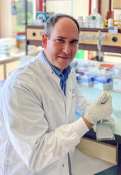 Noti Mario合作论文数Perelman School of Medicine, University of Pennsylvania2Horst Robenek合作论文数2Deborah Shan合作论文数2Tancred Frickey合作论文数2Thomas Kaufmann合作论文数2
 Monique Vogel合作论文数Institute of Immunology and Allergology, Sahli Haus 2, Inselspital, 3010 Bern, Switzerland2




