This chapter discusses two flow-imaging techniques that are useful for measuring flow on a microscopic scale: time of flight (ToF) and phase contrast (PC). It explores the physical limitations to the resolution and applicable parameter ranges of the flow. The chapter presents some specific examples, including the characterization of liquid exchange in different aneurysm models, the measurements of velocity fields, and the determination of wall shear stress (WSS) from the measured velocity field. ToF magnetic resonance imaging (MRI) is a possible method for observing flow on a microscopic scale. The PC method is well established for non-microscopic applications and is also suitable for flow imaging on microscopic scales. The ToF technique is used to measure the liquid exchange in different aneurysm models with a resolution of < 150 µm and validated these results with computer simulations.
Diffusion MRI is an efficient and widely used technique for the investigation of tissue structure and organisation in vivo. Multiple phenomenological and biophysical diffusion models are intensively exploited for the analysis of the diffusion experiments. However, the verification of the applied diffusion models remains challenging. In order to provide a "gold standard" and to assess the accuracy of the derived parameters and the limitations of the diffusion models, anisotropic diffusion phantoms with well known architecture are demanded. In the present work we built four anisotropic diffusion phantoms consisting of hollow microcapillaries with very small inner diameters of 5, 10 and 20μm and outer diameters of 90 and 150μm. For testing the suitability of these phantoms, we performed diffusion measurements on all of them and evaluated the resulting data with a set of popular diffusion models, such as diffusion tensor and diffusion kurtosis imaging, a two compartment model with intra- and extra-capillary water spaces using bi-exponential fitting, and time-dependent diffusion coefficients in Mitra's limit. The perspectives and limitations of these diffusion phantoms are presented and discussed.
Visualisation of living tissue structure and function is a challenging problem of modern imaging techniques. Diffusion MRI allows one to probe in vivo structures on a micrometer scale. However, conventional diffusion measurements are time-consuming procedures, because they require several measurements with different gradient directions. Considerable time savings are therefore possible by measurement schemes that generate an isotropic diffusion weighting in a single shot. Multiple approaches for generating isotropic diffusion weighting are known and have become very popular as useful tools in clinical research. Thus, there is a strong need for a comprehensive comparison of different isotropic weighting approaches. In the present work we introduce two new sequences based on simple (co)sine modulations and compare their performance to established q-space magic-angle spinning sequences and conventional DTI, using a diffusion phantom assembled from microcapillaries and in vivo experiments at 7T. The advantages and disadvantages of all compared schemes are demonstrated and discussed.
Using microcapsules for medical and technical purposes relies on capsule properties such as stability, shape, and permeability, which depend on the production process and the capsule material itself. The methods presented here, which are based on nuclear magnetic resonance microscopy, allow one to quantitatively determine these parameters for individual microcapsules with or without coatings. The experiments are noninvasive and can access such parameters as geometry, diffusion constants, or molecular correlation times in different chemical environments with a spatial resolution of ~10 μm. The influence of production parameters as well as the dynamic behavior of capsules in various test environments are characterized for potential applications in targeted drug delivery.
PURPOSE:The impact and the development of aneurysms depend to a significant degree on the exchange of liquid between the regular vessel and the pathological extension. A better understanding of this process will lead to improved prediction capabilities. The aim of the current study was to investigate fluid-exchange in aneurysm models of different complexities by combining microscopic magnetic resonance measurements with numerical simulations. In order to evaluate the accuracy and applicability of these methods, the fluid-exchange process between the unaltered vessel lumen and the aneurysm phantoms was analyzed quantitatively using high spatial resolution.METHODS:Magnetic resonance flow imaging was used to visualize fluid-exchange in two different models produced with a 3D printer. One model of an aneurysm was based on histological findings. The flow distribution in the different models was measured on a microscopic scale using time of flight magnetic resonance imaging. The whole experiment was simulated using fast graphics processing unit-based numerical simulations. The obtained simulation results were compared qualitatively and quantitatively with the magnetic resonance imaging measurements, taking into account flow and spin-lattice relaxation.RESULTS:The results of both presented methods compared well for the used aneurysm models and the chosen flow distributions. The results from the fluid-exchange analysis showed comparable characteristics concerning measurement and simulation. Similar symmetry behavior was observed. Based on these results, the amount of fluid-exchange was calculated. Depending on the geometry of the models, 7% to 45% of the liquid was exchanged per second.CONCLUSIONS:The result of the numerical simulations coincides well with the experimentally determined velocity field. The rate of fluid-exchange between vessel and aneurysm was well-predicted. Hence, the results obtained by simulation could be validated by the experiment. The observed deviations can be caused by the noise in the measurement and by the limited resolution of the simulation. The resulting differences are small enough to allow reliable predictions of the flow distribution in vessels with stents and for pulsed blood flow.
Microcapsules consisting of natural polysaccharide hydrogels such as pectinates have a large potential for the transportation of liquid drugs with food. If a controlled and site specific release of the encapsulated drugs is desired, they must be stored sufficiently long under different environmental conditions. Here, we optimize the capsules with respect to the retention time of the encapsulated drugs and the chemical stability under gastrointestinal conditions. To achieve this, we coat the pure hydrogel capsules with the natural resin shellac. NMR microscopy is used for the characterization of the structure and suitability of these microcapsules. We measure the permeability of differently prepared capsules by using paramagnetic probe molecules. By an acquisition of T1-weighted images as a function of time we can monitor the diffusion of the molecules into the capsules. Diffusion coefficients for the probe molecules in the capsule membranes can be extracted from these measurements for quantizing the permeabilities. The obtained permeabilities show that shellac can seal pure pectinate capsules under acidic conditions. In addition, we also monitor the behavior of the capsules under simulated gastrointestinal conditions. These measurements show that the capsules do not undergo changes under gastric and intestinal conditions, but dissolve under colonic conditions on timescales between 1 and 28 hours, depending on the preparation process.
Alterations of the blood flow are associated with various cardiovascular diseases. Precise knowledge of the velocity distribution is therefore important for understanding these diseases and predicting the effect of different medical intervention schemes. The goal of this work is to estimate the precision with which the velocity field can be measured and predicted by studying two simple model geometries with NMR micro imaging and computational fluid dynamics. For these initial experiments, we use water as an ideal test medium. The phantoms consist of tubes simulating a straight blood vessel and a step between two tubes of different diameters, which can be seen as a minimal model of the situation behind a stenosis. For both models, we compare the experimental data with the numerical prediction, using the experimental boundary conditions. For the simpler model, we also compare the data to the analytical solution. As an additional validation, we determine the divergence of the velocity field and verify that it vanishes within the experimental uncertainties. We discuss the resulting precision of the simulation and the outlook for extending this approach to the analysis of specific cases of arteriovascular problems.
Microscopic capsules made from polysaccharides are used as carriers for drugs and food additives. Here, we use NMR microscopy to assess the permeability of capsule membranes and their stability under different environmental conditions. The results allow us to determine the suitability of different capsules for controlled drug delivery. As a measure of the membrane permeability, we monitor the diffusion of paramagnetic molecules into the microcapsules by dynamic NMR microimaging. We obtained the diffusion coefficients of the probe molecules in the membranes and in the capsule core by comparing the measured time dependent concentration maps with numerical solutions of the diffusion equation. The results reveal that external coatings strongly decrease the permeability of the capsules. In addition, we also visualized that the capsules are stable under gastric conditions but dissolve under simulated colonic conditions, as required for targeted drug delivery. Depending on the capsule, the timescales for these processes range from 1 to 28h.
 Dieter Suter合作论文数Department of Physics, Technische Universität Dortmund8
 Henning Stefan合作论文数3Sebastian Vellmer合作论文数Dept Neurosurg, Charite Univ Med Berlin2
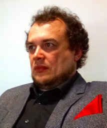 Maximov Ivan I合作论文数Interdisciplinary Nanoscience Centre (iNANO),, University of Aarhus2
 Heinz Rehage合作论文数University of Essen2
 Sabine Leick合作论文数2Lars Walczak合作论文数2Frank Weichert合作论文数Universitat Dortmund2
 Frauke Frank合作论文数1
 Rudiger Stirnberg合作论文数1Ernst Benedikt合作论文数1
 Tony Stoecker合作论文数Institute of Neuroscience and Medicine, Research Center Jülich1Inge Schmitz合作论文数Institute of Pathology, University of Bochum1
 Patrick Degen合作论文数1




