Current options for aortic valve replacements are non-viable and thus lack the ability to grow and remodel, which can be problematic for paediatric applications. Toward the development of living valve substitutes that can grow and remodel, porcine aortic valve interstitial cells (VICs) were isolated and encapsulated within proteolytically degradable and cell-adhesive poly(ethylene glycol) (PEG) hydrogels, in an effort to study their phenotypes and functions. The results showed that encapsulated VICs maintained high viability and proliferated within the hydrogels. The VICs actively remodelled the hydrogels via secretion of matrix metalloproteinase-2 (MMP-2) and deposition of new extracellular matrix (ECM) components, including collagens I and III. The soft hydrogels with compressive moduli of ~4.3 kPa quickly reverted VICs from an activated myofibroblastic phenotype to a quiescent, unactivated phenotype, evidenced by the loss of α-smooth muscle actin expression upon encapsulation. In an effort to promote VIC-mediated ECM production, ascorbic acid (AA) was supplemented in the medium to investigate its effects on VIC function and phenotype. AA treatment enhanced VIC spreading and proliferation, and inhibited apoptosis. AA treatment also promoted VIC-mediated ECM remodelling by increasing MMP-2 activity and depositing collagens I and III. AA treatment did not significantly influence the expression of α-smooth muscle actin (myofibroblast activation marker) and alkaline phosphatase (osteogenic differentiation marker). No calcification or nodule formation was observed within the cell-laden hydrogels, with or without AA treatment. These results suggest the potential of this system and the beneficial effect of AA in heart valve tissue engineering. Copyright © 2015 John Wiley & Sons, Ltd.
As catheter-based structural heart interventions become increasingly complex, the ability to effectively model patient-specific valve geometry as well as the potential interaction of an implanted device within that geometry will become increasingly important. Our aim with this investigation was to combine the technologies of high-spatial resolution cardiac imaging, image processing software, and fused multi-material 3D printing, to demonstrate that patient-specific models of the mitral valve apparatus could be created to facilitate functional evaluation of novel trans-catheter mitral valve repair strategies. Clinical 3D transesophageal echocardiography and computed tomography images were acquired for three patients being evaluated for a catheter-based mitral valve repair. Target anatomies were identified, segmented and reconstructed into 3D patient-specific digital models. For each patient, the mitral valve apparatus was digitally reconstructed from a single or fused imaging data set. Using multi-material 3D printing methods, patient-specific anatomic replicas of the mitral valve were created. 3D print materials were selected based on the mechanical testing of elastomeric TangoPlus materials (Stratasys, Eden Prairie, Minnesota, USA) and were compared to freshly harvested porcine leaflet tissue. The effective bending modulus of healthy porcine MV tissue was significantly less than the bending modulus of TangoPlus (p < 0.01). All TangoPlus varieties were less stiff than the maximum tensile elastic modulus of mitral valve tissue (3697.2 ± 385.8 kPa anterior leaflet; 2582.1 ± 374.2 kPa posterior leaflet) (p < 0.01). However, the slopes of the stress-strain toe regions of the mitral valve tissues (532.8 ± 281.9 kPa anterior leaflet; 389.0 ± 156.9 kPa posterior leaflet) were not different than those of the Shore 27, Shore 35, and Shore 27 with Shore 35 blend TangoPlus material (p > 0.95). We have demonstrated that patient-specific mitral valve models can be reconstructed from multi-modality imaging datasets and fabricated using the multi-material 3D printing technology and we provide two examples to show how catheter-based repair devices could be evaluated within specific patient 3D printed valve geometry. However, we recognize that the use of 3D printed models for the development of new therapies, or for specific procedural training has yet to be defined.
The aortic valve is a deceptively complex tissue. Valves experience rigorous and dynamic mechanical activity due to the movement of the heart and blood. Consequently, the cells found within and lining the heart valves are strongly influenced by mechanical stimulation, which regulates their phenotypic characteristics, functional behavior, production of extracellular matrix (ECM), and susceptibility to disease. Scientists have developed a number of strategies for studying the mechanobiology of aortic valves, including approaches that stretch cells isolated from heart valves, grow valve cells within synthetic or native scaffold materials, culture cells or intact valve tissues within bioreactor systems, and analyze in vivo models in which the valve environment has been mechanically disrupted. These investigations are revealing new information about the mechanical regulation of the biology and function of heart valves, which can be applied to the design of novel treatments and diagnostic tools for aortic valve disease.
Advanced tissue engineered heart valves must be constructed from multiple materials to better mimic the heterogeneity found in the native valve. The trilayered structure of aortic valves provides the ability to open and close consistently over a full human lifetime, with each layer performing specific mechanical functions. The middle spongiosa layer consists primarily of proteoglycans and glycosaminoglycans, providing lubrication and dampening functions as the valve leaflet flexes open and closed. In this study, hyaluronan hydrogels were tuned to perform the mechanical functions of the spongiosa layer, provide a biomimetic scaffold in which valve cells were encapsulated in 3D for tissue engineering applications, and gain insight into how valve cells maintain hyaluronan homeostasis within heart valves. Expression of the HAS1 isoform of hyaluronan synthase was significantly higher in hyaluronan hydrogels compared to blank-slate poly(ethylene glycol) diacrylate (PEGDA) hydrogels. Hyaluronidase and matrix metalloproteinase enzyme activity was similar between hyaluronan and PEGDA hydrogels, even though these scaffold materials were each specifically susceptible to degradation by different enzyme types. KIAA1199 was expressed by valve cells and may play a role in the regulation of hyaluronan in heart valves. Cross-linked hyaluronan hydrogels maintained healthy phenotype of valve cells in 3D culture and were tuned to approximate the mechanical properties of the valve spongiosa layer. Therefore, hyaluronan can be used as an appropriate material for the spongiosa layer of a proposed laminate tissue engineered heart valve scaffold.
In this study, a composite scaffold consisting of an electrospun polyurethane and poly(ethylene glycol) hydrogel was investigated for aortic valve tissue engineering. This multilayered approach permitted the fabrication of a scaffold that met the desired mechanical requirements while enabling the 3D culture of cells. The scaffold was tuned to mimic the tensile strength, anisotropy, and extensibility of the natural aortic valve through design of the electrospun polyurethane mesh layer. Valve interstitial cells were encapsulated inside the hydrogel portion of the scaffold around the electrospun mesh, creating a composite scaffold approximately 200 μm thick. The stiffness of the electrospun fibers caused the encapsulated cells to exhibit an activated phenotype that resulted in fibrotic remodeling of the scaffold in a heterogeneous manner. Remodeling was further explored by culturing the scaffolds in both a mechanically constrained state and in a bent state. The constrained scaffolds demonstrated strong fibrotic remodeling with cells aligning in the direction of the mechanical constraint. Bent scaffolds demonstrated that applied mechanical forces could influence cell behavior. Cells seeded on the outside curve of the bend exhibited an activated, fibrotic response, while cells seeded on the inside curve of the bend were a quiescent phenotype, demonstrating potential control over the fibrotic behavior of cells. Overall, these results indicate that this polyurethane/hydrogel scaffold mimics the structural and functional heterogeneity of native valves and warrants further investigation to be used as a model for understanding fibrotic valve disease.
Tissue oxygenation often plays a significant role in disease and is an essential design consideration for tissue engineering. Here, oxygen diffusion profiles of porcine aortic and mitral valve leaflets were determined using an oxygen diffusion chamber in conjunction with computational models. Results from these studies revealed the differences between aortic and mitral valve leaflet diffusion profiles and suggested that diffusion alone was insufficient for normal oxygen delivery in mitral valves. During fibrotic valve disease, leaflet thickening due to abnormal ECM likely reduces regional oxygen availability. To assess the impact of low oxygen levels on valve behaviour, whole leaflet organ cultures were created to induce leaflet hypoxia. These studies revealed a loss of layer stratification and elevated levels of hypoxia inducible factor 1-alpha in both aortic and mitral valve hypoxic groups. Mitral valves also exhibited altered expression of angiogenic factors in response to low oxygen environments when compared with normoxic groups. Hypoxia affected aortic and mitral valves differently, and mitral valves appeared to show a stenotic, rheumatic phenotype accompanied by significant cell death. These results indicate that hypoxia could be a factor in mid to late valve disease progression, especially with the reduction in chondromodulin-1 expression shown by hypoxic mitral valves.
Tissue oxygenation often plays a significant role in disease and is an essential design consideration for tissue engineering. Here, oxygen diffusion profiles of porcine aortic and mitral valve leaflets were determined using an oxygen diffusion chamber in conjunction with computational models. Results from these studies revealed the differences between aortic and mitral valve leaflet diffusion profiles and suggested that diffusion alone was insufficient for normal oxygen delivery in mitral valves. During fibrotic valve disease, leaflet thickening due to abnormal extracellular matrix is likely to reduce regional oxygen availability. To assess the impact of low oxygen levels on valve behaviour, whole leaflet organ cultures were created to induce leaflet hypoxia. These studies revealed a loss of layer stratification and elevated levels of hypoxia inducible factor 1-alpha in both aortic and mitral valve hypoxic groups. Mitral valves also exhibited altered expression of angiogenic factors in response to low oxygen environments when compared with normoxic groups. Hypoxia affected aortic and mitral valves differently, and mitral valves appeared to show a stenotic, rheumatic phenotype accompanied by significant cell death. These results indicate that hypoxia could be a factor in mid to late valve disease progression, especially with the reduction in chondromodulin-1 expression shown by hypoxic mitral valves.
With an increasing number of patients requiring valve replacements, there is heightened interest in advancing heart valve tissue engineering (HVTE) to provide solutions to the many limitations of current surgical treatments. A variety of materials have been developed as scaffolds for HVTE including natural polymers, synthetic polymers, and decellularized valvular matrices. Among them, biocompatible hydrogels are generating growing interest. Natural hydrogels, such as collagen and fibrin, generally show good bioactivity but poor mechanical durability. Synthetic hydrogels, on the other hand, have tunable mechanical properties; however, appropriate cell-matrix interactions are difficult to obtain. Moreover, hydrogels can be used as cell carriers when the cellular component is seeded into the polymer meshes or decellularized valve scaffolds. In this review, we discuss current research strategies for HVTE with an emphasis on hydrogel applications. The physicochemical properties and fabrication methods of these hydrogels, as well as their mechanical properties and bioactivities are described. Performance of some hydrogels including in vitro evaluation using bioreactors and in vivo tests in different animal models are also discussed. For future HVTE, it will be compelling to examine how hydrogels can be constructed from composite materials to replicate mechanical properties and mimic biological functions of the native heart valve.
Physiologically relevant in vitro models are needed to study disease progression and to develop and screen potential therapeutic interventions for disease. Heart valve disease, in particular, has no early intervention or non-invasive treatment because there is a lack of understanding the cellular mechanisms which lead to disease. Here, we establish a novel, customizable synthetic hydrogel platform that can be used to study cell cell interactions and the factors which contribute to valve disease. Spatially localized cell adhesive ligands bound in the scaffold promote cell growth and organization of valve interstitial cells and valve endothelial cells in 3D co-culture. Both cell types maintained phenotypes, homeostatic functions, and produced zonally localized extracellular matrix. This model extends the capabilities of in vitro research by providing a platform to perform direct contact co-culture with cells in their physiologically relevant spatial arrangement. (C) 2015 Elsevier Ltd. All rights reserved.
The development of advanced scaffolds that recapitulate the anisotropic mechanical behavior and biological functions of the extracellular matrix in leaflets would be transformative for heart valve tissue engineering. In this study, anisotropic mechanical properties were established in poly(ethylene glycol) (PEG) hydrogels by crosslinking stripes of 3.4 kDa PEG diacrylate (PEGDA) within 20 kDa PEGDA base hydrogels using a photolithographic patterning method. Varying the stripe width and spacing resulted in a tensile elastic modulus parallel to the stripes that was 4.1-6.8 times greater than that in the perpendicular direction, comparable to the degree of anisotropy between the circumferential and radial orientations in native valve leaflets. Biomimetic PEG-peptide hydrogels were prepared by tethering the cell-adhesive peptide RGDS and incorporating the collagenase-degradable peptide PQ (GGGPQG↓IWGQGK) into the polymer network. The specific amounts of RGDS and PEG-PQ within the resulting hydrogels influenced the elongation, de novo extracellular matrix deposition and hydrogel degradation behavior of encapsulated valvular interstitial cells (VICs). In addition, the morphology and activation of VICs grown atop PEG hydrogels could be modulated by controlling the concentration or micro-patterning profile of PEG-RGDS. These results are promising for the fabrication of PEG-based hydrogels using anatomically and biologically inspired scaffold design features for heart valve tissue engineering.
1 1 4 7 jci.org Volume 125 Number 3 March 2015 Introduction Collagen is the most abundant intrinsic matrix scaffolding protein and contributes to tissue tensile strength (1). Various aspects of collagen metabolism, including its expression, deposition, organization, and turnover, are aberrant in epithelial cancer and have been implicated in tumor progression (2). Evidence from multiple types of human epithelial cancers and experimental tumor models suggests that tumor extracellular matrix (ECM) stiffness is enhanced by an accumulation of collagen stabilized by covalent intraand inter-molecular cross-links (3). Collagen cross-linking is initiated in the extracellular space by the action of lysyl oxidase (LOX), a secreted copper-dependent amine oxidase that converts telopeptidyl lysine (Lys) and hydroxylysine (Hyl) residues to reactive aldehydes (Lysald and Hylald, respectively) that condense with juxtaposed Lys, Hyl, and histidine (His) residues to form various types of cross-links (4). The discovery that tumor cells have high expression of LOX and LOX-like family members led to the development of pharmacological inhibitors of these enzymes that, when administered to tumor-bearing animals, decrease the amount of collagen cross-links in tumors and suppress metastasis, suggesting that the amount of collagen cross-links in tumors controls disease progression (5). The types of collagen cross-links that form in normal connective tissues are determined prior to cross-link formation by the hydroxylation of telopeptidyl and helical Lys residues on collagen (4). Mediating these changes are lysyl hydroxylases (LH1, LH2, and LH3) encoded by distinct procollagen-lysine, 2-oxoglutarate 5-dioxygenase (PLOD) genes; these intracellular enzymes induce hydroxylation of Lys residues on collagen prior to the formation of triple helical pro-collagen molecules (6–11). Following secretion of pro-collagen molecules into the extracellular space, the telopeptidyl Hyl residues undergo LOX-induced oxidative deamination into Hylald, which then forms Hylald-derived aldimine cross-links Epithelial tumor metastasis is preceded by an accumulation of collagen cross-links that heighten stromal stiffness and stimulate the invasive properties of tumor cells. However, the biochemical nature of collagen cross-links in cancer is still unclear. Here, we postulated that epithelial tumorigenesis is accompanied by changes in the biochemical type of collagen crosslinks. Utilizing resected human lung cancer tissues and a p21CIP1/WAF1-deficient, K-rasG12D-expressing murine metastatic lung cancer model, we showed that, relative to normal lung tissues, tumor stroma contains higher levels of hydroxylysine aldehyde– derived collagen cross-links (HLCCs) and lower levels of lysine aldehyde–derived cross-links (LCCs), which are the predominant types of collagen cross-links in skeletal tissues and soft tissues, respectively. Gainand loss-of-function studies in tumor cells showed that lysyl hydroxylase 2 (LH2), which hydroxylates telopeptidyl lysine residues on collagen, shifted the tumor stroma toward a high-HLCC, low-LCC state, increased tumor stiffness, and enhanced tumor cell invasion and metastasis. Together, our data indicate that LH2 enhances the metastatic properties of tumor cells and functions as a regulatory switch that controls the relative abundance of biochemically distinct types of collagen cross-links in the tumor stroma. Lysyl hydroxylase 2 induces a collagen cross-link switch in tumor stroma
Epithelial tumor metastasis is preceded by an accumulation of collagen cross-links that heighten stromal stiffness and stimulate the invasive properties of tumor cells. However, the biochemical nature of collagen cross-links in cancer is still unclear. Here, we postulated that epithelial tumorigenesis is accompanied by changes in the biochemical type of collagen cross-links. Utilizing resected human lung cancer tissues and a p21(CIP1/WAF1)-deficient, K-ras(G12D)-expressing murine metastatic lung cancer model, we showed that, relative to normal lung tissues, tumor stroma contains higher levels of hydroxylysine aldehyde-derived collagen cross-links (HLCCs) and lower levels of lysine aldehyde-derived cross-links (LCCs), which are the predominant types of collagen cross-links in skeletal tissues and soft tissues, respectively. Gain- and loss-of-function studies in tumor cells showed that lysyl hydroxylase 2 (LH2), which hydroxylates telopeptidyl lysine residues on collagen, shifted the tumor stroma toward a high-HLCC, low-LCC state, increased tumor stiffness, and enhanced tumor cell invasion and metastasis. Together, our data indicate that LH2 enhances the metastatic properties of tumor cells and functions as a regulatory switch that controls the relative abundance of biochemically distinct types of collagen cross-links in the tumor stroma.
The recapitulation of the material properties and structure of the native aortic valve leaflet, specifically its anisotropy and laminate structure, is a major design goal for scaffolds for heart valve tissue engineering. Poly(ethylene glycol) (PEG) hydrogels are attractive scaffolds for this purpose as they are biocompatible, can be modified for their mechanical and biofunctional properties, and can be laminated. This study investigated augmenting PEG hydrogels with polycaprolactone (PCL) as an analog to the fibrosa to improve strength and introduce anisotropic mechanical behavior. However, due to its hydrophobicity, PCL must be modified prior to embedding within PEG hydrogels. In this study, PCL was electrospun (ePCL) and modified in three different ways, by protein adsorption (pPCL), alkali digestion (hPCL), and acrylation (aPCL). Modified PCL of all types maintained the anisotropic elastic moduli and yield strain of unmodified anisotropic ePCL. Composites of PEG and PCL (PPCs) maintained anisotropic elastic moduli, but aPCL and pPCL had isotropic yield strains. Overall, PPCs of all modifications had elastic moduli of 3.79 +/- 0.90 MPa and 0.46 +/- 0.21 MPa in the parallel and perpendicular directions, respectively. Valvular interstitial cells seeded atop anisotropic aPCL displayed an actin distribution aligned in the direction of the underlying fibers. The resulting scaffold combines the biocompatibility and tunable fabrication of PEG with the strength and anisotropy of ePCL to form a foundation for future engineered valve scaffolds.
A Day of Launch flight loads assessment technique that determines running loads calculated from flexible body coupled loads was developed for the Ares I-X Flight Test Vehicle. The technique was developed to quantify DOL flight loads in terms of structural load components rather than the typically used q-alpha metric to provide more insight into the DOL loads. In this technique, running loads in the primary structure are determined from the combination of quasi-static aerodynamic loads and dynamic loads. The aerodynamic loads are calculated as a function of time using trajectory parameters passed from the DOL trajectory simulation and are combined with precalculated dynamic loads using a load combination equation. The potential change in aerodynamic load due to wind variability during the countdown is included in the load combination. In the event of a load limit exceedance, the technique allows the identification of what load component is exceeded, a quantification of how much the load limit is exceeded, and where on the vehicle the exceedance occurs. This technique was used to clear the Ares I-X FTV for launch on October 28, 2009. This paper describes the use of coupled loads in the Ares I-X flight loads assessment and summarizes the Ares I-X load assessment results.
Since many of the larger launch vehicles are operated near their design limits during the ascent phase of flight to optimize payload to orbit, it often becomes necessary to verify that the vehicle will remain within certification limits during the ascent phase as part of the go/no-go review made prior to launch. This paper describes the approach used to predict Ares I-X launch vehicle structural air loads and controllability prior to launch which represents a distinct departure from the methodology of the Space Shuttle and Evolved Expendable Launch Vehicle (EELV) programs. Protection for uncertainty of key environment and trajectory parameters is added to the nominal assessment of launch capability to ensure that critical launch trajectory variables would be within the integrated vehicle certification envelopes. This process was applied by the launch team as a key element of the launch day go/no-go recommendation. Pre-launch assessments of vehicle launch capability for NASA's Space Shuttle and the EELV heavy lift versions require the use of a high-resolution wind profile measurements, which have relatively small sample size compared with low-resolution profile databases (which include low-resolution balloons and radar wind profilers). The approach described in this paper has the potential to allow the pre-launch assessment team to use larger samples of wind measurements from low-resolution wind profile databases that will improve the accuracy of pre-launch assessments of launch availability with no degradation of mission assurance or launch safety.
Day of launch I-load uplink (DOLILU) and a corresponding day of launch trajectory and loads evaluation are being considered for the Constellation Program. Abort effectiveness, controllability, and ascent structural loads all improve with some of the flight software I-loads designed using current measured atmospheric conditions. While a generalized DOLILU framework has been assumed, specific implementation details have not yet been discussed in depth nor documented. Operational improvements can be leveraged from Space Shuttle experience to create a more cost-effective and streamlined process while maintaining the integrity of the results. A preliminary discussion of DOLILU design methodology is presented followed by some suggestions and implementation details for Constellation Program DOLILU operations
The National Aeronautics and Space Administration's (NASA) Space Shuttle utilizes atmospheric winds on day of launch to develop throttle and steering commands to best optimize vehicle performance while keeping structural loading on the vehicle within limits. The steering commands and resultant trajectory are influenced by both the high and low frequency component of the wind. However, the low frequency component has a greater effect on the ascent design. Change in the low frequency wind content from the time of trajectory design until launch can induce excessive loading on the vehicle. Wind change limits have been derived to protect from launching in an environment where these temporal changes occur. Process of developing wind change limits are discussed followed by an observational study of temporal wind change in low frequency wind profiles at the NASA's Kennedy Space Center area are presented.
 K. Jane Grande-Allen合作论文数Department of Bioengineering, School of Engineering and Computing, Rice University14
 Jennifer L. West合作论文数Department of Engineering and Applied Sciences, Pratt School of Engineering, University of Virginia8
 Yan Wu合作论文数6
 Takeshi Watanabe合作论文数2Bin Xu合作论文数2
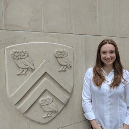 Zoe E. Punske合作论文数Dept Bioengn, Rice Univ2Matthew C Sapp合作论文数Department of Bioengineering, Rice University2Ronan O’Connell合作论文数Rice Univ2
 Daniela Pankova合作论文数2
 Xing Zhang (张兴)合作论文数Institute of Metal Research, Chinese Academy of Sciences2
 Li Sun (孙立)合作论文数Institute of Pharmaceutical Sciences, China Pharmaceutical University2
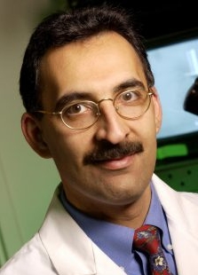 Farshid Guilak合作论文数Center of Regenerative Medicine, School of Medicine, Washington University in St. Louis;Department of Orthopaedic Surgery, Washington University in St. Louis2Sanchez-Adams Johannah合作论文数2
 Krishnamurthy Varun K合作论文数Cincinnati Children's Hospital Medical Center, the Heart Institute2Rodriguez Jaime合作论文数2Masahiko Terajima合作论文数2
 Young-Ho Ahn合作论文数2Thilaganathan Nishan合作论文数2
 Cuchiara Maude L合作论文数Department of Biomedical Engineering, Duke University2Alan R. Burns合作论文数University of Houston2Carmen Behrens合作论文数Department of Thoracic Head & Neck Medical Oncology, The University of Texas MD Anderson Cancer Center;Center for Cancer Epigenetics, The University of Texas MD Anderson Cancer Center2
 Chad Creighton合作论文数Dan L Duncan Comprehensive Cancer Center, Baylor College of Medicine;Human Genome Sequencing Center, Baylor College of Medicine2
 Mitsuo Yamauchi合作论文数Department of Oral and Craniofacial Health Sciences, University of North Carolina2
 Jonathan M. Kurie合作论文数Department of Thoracic-Head & Neck Med Onc, Division of Cancer Medicine, University of Texas MD Anderson Cancer Center2Yulong Chen (陈玉龙)合作论文数Laboratory of Molecular Biology, Henan University of Traditional Chinese Medicine2Rosalba Minelli合作论文数2
 Shanda Blackmon合作论文数The Methodist Hospital2Kenneth L. Scott合作论文数2Hubert Tseng合作论文数Department of Bioengineering, Rice University2
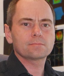 Tomasz Zal合作论文数Department of Immunology, The Scripps Research Institute2Debananda Pati合作论文数Department of Biological Sciences, University of Calgary2Elisabeth Martinez合作论文数2brett r starr合作论文数2frank leahy合作论文数1barry c roberts合作论文数1Jay V. Shah合作论文数1Ignacio I. Wistuba合作论文数1richard leach合作论文数1Alysha P. Kishan合作论文数Alysha Kishan and Taneidra Buie contributed equally to this work.1Hui Liu合作论文数1
 Prashanth Ravishankar合作论文数Department of Biomedical Engineering, University of Arkansas1
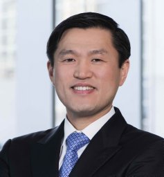 Kim Min P合作论文数1
 Elizabeth Cosgriff-Hernandez合作论文数Department of Biomedical Engineering, Cockrell School of Engineering, The University of Texas at Austin1Yonezawa Aline L合作论文数1Liu Hui合作论文数1
 Yanan Yang合作论文数Department of Medicine,Mayo Clinic College of Medicine1
 Min-Pyo Kim合作论文数1
 Marija Vukicevic合作论文数From the *Mechanical and Bioengineering Engineering, Clemson University;†Laboratory of Biological Structure Mechanics, Politecnico di Milano;and ‡Cardiothoracic Surgery, Great Ormond Street Hospital for Children;Great Ormond Street Hospital for Children, Clemson University1




