Purpose The purpose of this paper is to present a simulation study using a model of a new optically pumped magnetometer sensor for application in the field of magnetoencephalography. The effects of sensor distance and orientation on the measurement information and the sensitivity to neuronal sources are investigated. Further, this paper uses a combinatorial optimization approach for sensor placement to measure spontaneous activity in the region of the occipital cortex. Design/methodology/approach This paper studies the effects of sensor distance and orientation on sensitivity to cortical sources and measurement information. A three-compartment model of the head, using the boundary element method, is applied. For sensor setup optimization, a combinatorial optimization scheme is developed. Findings The sensor distance to sources considerably affects the sensitivity and the retrieved information. A specific arrangement of four sensors for measuring spontaneous activity over the occipital part of the head is optimized by effectively avoiding position conflicts. Research limitations/implications Individual head models, as well as more detailed noise and signal models, will increase the significance for specific-use cases in future studies. Originality/value Effects of sensor distance and orientation are specifically evaluated for a new optically pumped magnetometer. A discrete optimization scheme for sensor optimization is introduced. The presented methodology is applicable for other sensor characterization and optimization problems. The findings contribute significantly to the development of new sensors.
Lorentz force evaluation is a nondestructive evaluation method applied for the characterization of sub-surface defects in specimen consisting of layers of conducting material. The movement of a specimen under investigation relative to a permanent magnet leads to Lorentz forces that are perturbed in the presence of a defect. A minimum norm estimation (MNE) with Elastic Net Regularization (ENR) is compared to MNE with Tikhonov-Phillips-Regularization (TPR) regarding the ability to determine defect properties from force perturbation signals. A cylindrically shaped defect with diameter of 5 mm and height of 2 mm is examined at depths of 2, 4 and 8 mm. Simulated data obtained by the finite element method and measured data are considered. ENR and TPR estimated the defect depth correctly for simulation and measurement data at depths of 2 and 4 mm, while at 8 mm, only ENR estimated the depth correctly. The defect sizes were slightly overestimated for both ENR and TPR. TPR produced additional spurious solutions at the boundary of the reconstruction grid. We conclude that ENR is more suitable than TPR for MNE in Lorentz force evaluation.
Physical head phantoms allow the assessment of source reconstruction procedures in electroencephalography and electrical stimulation profiles during transcranial electric stimulation. Volume conduction in the head is strongly influenced by the skull, which represents the main conductivity barrier. Realistic modeling of its characteristics is thus important for phantom development. In the present study, we proposed plastic clay as a material for modeling the skull in phantoms. We analyzed five clay types varying in granularity and fractions of fire clay, each with firing temperatures from 550°C to 950°C. We investigated the conductivity of standardized clay samples when immersed in a 0.9% sodium chloride solution with time-resolved four-point impedance measurements. To test the reusability of the clay model, these measurements were repeated after cleaning the samples by rinsing in deionized water for 5 h. We found time-dependent impedance changes for approximately 5 min after immersion in the solution. Thereafter, the conductivities stabilized between 0.0716 S/m and 0.0224 S/m depending on clay type and firing temperatures. The reproducibility of the measurement results proved the effectiveness of the rinsing procedure. Clay provides formability, is permeable to ions, can be adjusted in conductivity value and is thus suitable for the skull modeling in phantoms.
Source imaging based on magnetoencephalography (MEG) and electroencephalography (EEG) allows for the non-invasive analysis of brain activity with high temporal and good spatial resolution. As the bioelectromagnetic inverse problem is ill-posed, constraints are required. For the analysis of evoked brain activity, spatial sparsity of the neuronal activation is a common assumption. It is often taken into account using convex constraints based on the l1-norm. The resulting source estimates are however biased in amplitude and often suboptimal in terms of source selection due to high correlations in the forward model. In this work, we demonstrate that an inverse solver based on a block-separable penalty with a Frobenius norm per block and a l0.5-quasinorm over blocks addresses both of these issues. For solving the resulting non-convex optimization problem, we propose the iterative reweighted Mixed Norm Estimate (irMxNE), an optimization scheme based on iterative reweighted convex surrogate optimization problems, which are solved efficiently using a block coordinate descent scheme and an active set strategy. We compare the proposed sparse imaging method to the dSPM and the RAP-MUSIC approach based on two MEG data sets. We provide empirical evidence based on simulations and analysis of MEG data that the proposed method improves on the standard Mixed Norm Estimate (MxNE) in terms of amplitude bias, support recovery, and stability.
Magnetoencephalography (MEG) and electroencephalography (EEG) source localization is a challenging illposed problem. To identify an appropriate solution out of an infinite set of possible candidates, the problem requires setting certain constraints depending on the assumptions or a priori knowledge about the source distribution. Different constraints have been proposed so far, including those that impose sparsity on the source reconstruction in both standard and time-frequency domains. Source localization in the time-frequency domain has already been investigated using Gabor dictionary in both a convex (TF-MxNE) and non-convex way (Iterative Reweighted TF-MxNE). The iterative reweighted (ir)TF-MxNE solver has been shown to outperform TF-MxNE in both source recovery and amplitude bias. However, the choice of an optimal dictionary remains unsolved. Due to a mixture of signals, i.e. short transient signals (right after the stimulus onset) and slower brain waves, the choice of a single dictionary explaining simultaneously both signals types in a sparse way is difficult. In this work, we introduce a method to improve the source estimation relying on a multi-scale dictionary, i.e. multiple dictionaries with different scales concatenated to fit short transients and slow waves at the same time. We compare our results with irTF-MxNE on realistic simulation, then we use somatosensory data to demonstrate the benefits of the approach in terms of reduced leakage (time courses mixture), temporal smoothness and detection of both signals types.
A controversy exists on photic driving in the human visual cortex evoked by intermittent photic stimulation. Frequency entrainment and resonance phenomena are reported for frequencies higher than 12 Hz in some studies while missing in others. We hypothesized that this might be due to different experimental conditions, since both high and low intensity light stimulation were used. However, most studies do not report radiometric measurements, which makes it impossible to categorize the stimulation according to photopic, mesopic, and scotopic vision. Low intensity light stimulation might lead to scotopic vision, where rod perception dominates. In this study, we investigated photic driving for rod-dominated visual input under scotopic conditions. Twelve healthy volunteers were stimulated with low intensity light flashes at 20 stimulation frequencies, leading to rod activation only. The frequencies were multiples of the individual alpha frequency (α) of each volunteer in the range from 0.40 to 2.30(∗)α. Three hundred and six-channel whole head magnetoencephalography recordings were analyzed in time, frequency, and spatiotemporal domains with the Topographic Matching Pursuit algorithm. We found resonance phenomena and frequency entrainment for stimulations at or close to the individual alpha frequency (0.90-1.10(∗)α) and half of the alpha frequency (0.40-0.55(∗)α). No signs of resonance and frequency entrainment phenomena were revealed around 2.00(∗)α. Instead, on-responses at the beginning and off-responses at the end of each stimulation train were observed for the first time in a photic driving experiment at frequencies of 1.30-2.30(∗)α, indicating that the flicker fusion threshold was reached. All results, the resonance and entrainment as well as the fusion effects, provide evidence for rod-dominated photic driving in the visual cortex.
Electrode and cap concepts for continuous and ubiquitous monitoring of brain activity will open up new fields of application and contribute to increased use of electroencephalography (EEG) in clinical routine, neurosciences, brain-computer-interfacing and out-of-the-lab monitoring. However, mobile and unobtrusive applications are currently hindered by the lack of applicable convenient and reliable electrode and cap systems. We propose a novel modular electrode concept based on a flexible polymer substrate, coated with electrically conductive metallic films. The overall concept enables design adaptation to different head regions and cap designs. We describe the single modules of the system and investigate the influence of electrode pin number, coating material and adduction force on electrode-skin impedance and perceived wearing comfort. Our results contribute to rapid and comfortable multichannel dry EEG.
Due to the excellent temporal resolution, MEG/EEG source imaging is an important measurement modality to study dynamic processes in the brain. As the bio electromagnetic inverse problem is ill-posed, constraints have to be imposed on the source estimates to find a unique solution. These constraints can be applied either in the standard or a transformed domain. The Time-Frequency Mixed Norm Estimate applies a composite convex regularization functional promoting structured sparsity in the time-frequency domain by combining an l2,1-mixed-norm and an l1-norm penalty on the coefficients of the Gabor TF decomposition of the source signals, to improve the reconstruction of spatially sparse neural activations with non-stationary and transient signals. Due to the l1-norm based constraints, the resulting source estimates are however biased in amplitude and often suboptimal in terms of source selection. In this work, we present the iterative reweighted Time-Frequency Mixed Norm Estimate, which employs a composite non-convex penalty formed by the sum of an l2,0.5-quasinorm and an l0.5-quasinorm penalty. The resulting non-convex problem is solved with a reweighted convex optimization scheme, in which each iteration is equivalent to a weighted Time-Frequency Mixed-Norm Estimate solved efficiently using a block coordinate descent scheme and an active set strategy. We compare our approach to alternative solvers using simulations and analysis of MEG data and demonstrate the benefit of the iterative reweighted Time-Frequency Mixed Norm Estimate with regard to active source identification, amplitude bias correction, and temporal unmixing of activations.
Due to the excellent temporal resolution, MEG/EEG source imaging is an important measurement modality to study dynamic processes in the brain. As the bio electromagnetic inverse problem is ill-posed, constraints have to be imposed on the source estimates to find a unique solution. These constraints can be applied either in the standard or a transformed domain. The Time-Frequency Mixed Norm Estimate applies a composite convex regularization functional promoting structured sparsity in the time-frequency domain by combining an l2,1-mixed-norm and an l1-norm penalty on the coefficients of the Gabor TF decomposition of the source signals, to improve the reconstruction of spatially sparse neural activations with non-stationary and transient signals. Due to the l1-norm based constraints, the resulting source estimates are however biased in amplitude and often suboptimal in terms of source selection. In this work, we present the iterative reweighted Time-Frequency Mixed Norm Estimate, which employs a composite non-convex penalty formed by the sum of an l2,0.5-quasinorm and an l0.5-quasinorm penalty. The resulting non-convex problem is solved with a reweighted convex optimization scheme, in which each iteration is equivalent to a weighted Time-Frequency Mixed-Norm Estimate solved efficiently using a block coordinate descent scheme and an active set strategy. We compare our approach to alternative solvers using simulations and analysis of MEG data and demonstrate the benefit of the iterative reweighted Time-Frequency Mixed Norm Estimate with regard to active source identification, amplitude bias correction, and temporal unmixing of activations.
Magneto encephalography (MEG) and electroen-cephalography (EEG) are imaging methods that measure neuronal dynamics non invasively with high temporal precision. It is often desired in MEG and EEG analysis to estimate the neural sources of the signals. Strategies used for this purpose often take into account the covariance between sensors to yield more precise estimates of the sources. Here we investigate in greater detail how the quality of such covariance estimates conditions the estimation of MEG and EEG sources. We investigated three distinct source localization methods: dynamic Statistical Parametric Maps (dSPM), the linearly constrained minimum variance (LCMV) beam former and Mixed-Norm Estimates (MxNE). We implemented and evaluated automated strategies for improving the quality of covariance estimates at different stages of data processing. Our results show that irrespective of the source localization method, accuracy can suffer from improper covariance estimation but can be improved by relying on automated regularization of covariance estimates.
With its millisecond temporal resolution, Magnetoencephalography (MEG) is well suited for real-time monitoring of brain activity. Real-time feedback allows the adaption of the experiment to the subject’s reaction and increases time efficiency by shortening acquisition and off-line analysis. Two formidable challenges exist in real-time analysis: the low signal-to-noise ratio (SNR) and the limited time available for computations. Since the low SNR reduces the number of distinguishable sources, we propose an approach which downsizes the source space based on a cortical atlas and allows to discern the sources in the presence of noise. Each cortical region is represented by a small set of dipoles, which is obtained by a clustering algorithm. Using this approach, we adapted dynamic statistical parametric mapping for real-time source localization. In terms of point spread and crosstalk between regions the proposed clustering technique performs better than selecting spatially evenly distributed dipoles. We conducted real-time source localization on MEG data from an auditory experiment. The results demonstrate that the proposed real-time method localizes sources reliably in the superior temporal gyrus. We conclude that real-time source estimation based on MEG is a feasible, useful addition to the standard on-line processing methods, and enables feedback based on neural activity during the measurements.
Important requirements for the analysis of multichannel EEG data are efficient techniques for signal enhancement, signal decomposition, feature extraction, and dimensionality reduction. We propose a new approach for spatial harmonic analysis (SPHARA) that extends the classical spatial Fourier analysis to EEG sensors positioned non-uniformly on the surface of the head. The proposed method is based on the eigenanalysis of the discrete Laplace-Beltrami operator defined on a triangular mesh. We present several ways to discretize the continuous Laplace-Beltrami operator and compare the properties of the resulting basis functions computed using these discretization methods. We apply SPHARA to somatosensory evoked potential data from eleven volunteers and demonstrate the ability of the method for spatial data decomposition, dimensionality reduction and noise suppression. When employing SPHARA for dimensionality reduction, a significantly more compact representation can be achieved using the FEM approach, compared to the other discretization methods. Using FEM, to recover 95% and 99% of the total energy of the EEG data, on average only 35% and 58% of the coefficients are necessary. The capability of SPHARA for noise suppression is shown using artificial data. We conclude that SPHARA can be used for spatial harmonic analysis of multi-sensor data at arbitrary positions and can be utilized in a variety of other applications.
 Jens Haueisen合作论文数Institute of Biomedical Engineering and Informatics, Technical University Ilmenau29
 Alexandre Gramfort合作论文数Meta Reality Labs;French Ministry of Industry17
 Patrique Fiedler合作论文数Technische Universität Ilmenau9
 Denis A. Engemann合作论文数F. Hoffmann-La Roche Ltd7
 Martin Luessi合作论文数Department of EECS, Northwestern University7
 Christian Brodbeck合作论文数6
 Christoph Dinh合作论文数6
 Uwe Graichen合作论文数6roman goj合作论文数5
 Matti Hämäläinen合作论文数Department of Neuroscience and Biomedical Engineering, School of Science, Aalto University5
 Brooks Teon L合作论文数Department of Psychology, New York University New York5
 Matthieu Kowalski合作论文数Laboratoire d'Analyse5emily ruzich合作论文数4alan leggitt合作论文数4
 Chris Holdgraf合作论文数International Computer Science Institute4tal linzen合作论文数4
 Dykstra Andrew R合作论文数Division of Health Sciences and Technology Cambridge, Harvard-MIT4
 Alexander Hunold合作论文数Institute of Biomedical Engineering and Informatics, Department of Computer Science and Automation, Technische Universität Ilmenau;Faculty of Computer Science and Automation, Technische Universität Ilmenau;Neuroconn Gmbh4
 Roland Eichardt合作论文数4
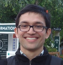 Mainak Jas合作论文数LTCI, Univ Paris Saclay4
 Marijn van Vliet合作论文数Laboratory for Neuro- and Psychofysiology, KU Leuven4Romain Trachel合作论文数4
 Eric C. Larson合作论文数University of Washington4
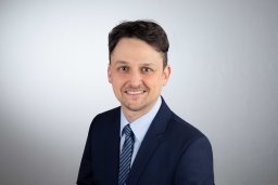 Stefan Griebel合作论文数3
 Eric R Larson合作论文数Department of Entomology, School of Integrative Biology, College of Liberal Arts and Sciences, University of Illinois at Urbana-Champaign3
 Daniel Baumgarten合作论文数Institute of Biomedical Engineering and Informatics, Technische Universität Ilmenau3
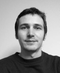 Alexandre Barachant合作论文数GIPSA-Lab, Grenoble Universities3
 Frank Zanow合作论文数3
 Satu Jumisko-Pyykkö合作论文数Tampere University of Technology3D. Guellmar合作论文数2ralph huonker合作论文数2roan laplante合作论文数2
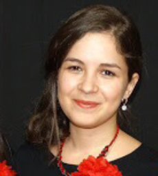 Yousra Bekhti合作论文数2
 Bharadwaj Hari M合作论文数Center for Computational Neuroscience and Neural Technology, Boston University Boston;Department of Biomedical Engineering, Boston University Boston;Boston University Boston, Boston University Boston2
 Klee Sascha合作论文数Institute of Biomedical Engineering and Informatics, Ilmenau University of Technology2
 Daniel Güllmar合作论文数Medical Physics Group, Jena University Hospital2Mario Liehr合作论文数2
 Lauri Parkkonen合作论文数Low Temperature Laboratory, Helsinki University of Technology2
 Otto W. Witte合作论文数Department of Neurology Jena University Hospital2
 Clemens Brunner合作论文数Institute for Knowledge Discovery, Graz University of Technology2
 Lorenz Esch合作论文数2Gregor Oelsner合作论文数1M Hämäläinen合作论文数1M S Hämäläinen合作论文数1S-S Poil合作论文数1R. Eichardt合作论文数1Anja Tränkner合作论文数1Matti S. Haemaelaeinen合作论文数1
 Michael M Plichta合作论文数1




