Massively multidimensional diffusion-relaxation correlation MRI provides detailed information on tissue microstructure by analyzing water populations at a sub-voxel level. This method correlates frequency-dependent tensor-valued diffusion MRI with longitudinal and transverse relaxation rates, generating non-parametric D(ω)-R 1-R 2-distributions. Traditionally, D(ω)-R 1-R 2-distributions are separated using manual binning of the diffusivity and anisotropy space to differentiate white matter (WM), gray matter (GM), and free water (FW) in brain tissue. However, while effective, this approach oversimplifies complex tissue fractions and does not fully utilize all available diffusion-relaxation parameters. In this study, we implemented an unsupervised clustering approach to automatically classify WM, GM, and FW and explore additional water populations using all components in the D(ω)-R 1-R 2-distributions on ex vivo and in vivo rat brain, and in vivo human brain. Results showed that a basic separation of WM, GM, and FW is possible using unsupervised clustering even under different multidimensional diffusion-relaxation protocols of rat brain and human brain. Additionally, when there is high frequency-dependent diffusion range, it is possible to obtain a cluster characterized by restriction localized in specific high cell density regions such as the dentate gyrus and cerebellum of rat brain. These findings were compared with rat histological sections of myelin and Nissl stainings. We demonstrated that unsupervised clustering of diffusion-relaxation MRI data can reveal tissue complexity beyond traditional WM, GM, and FW segmentation in rat and human brain without parameter assumptions. The unsupervised cluster approach could be used in other body parts (e.g., prostate and breast cancer) without requiring pre-defined bin limits. Furthermore, the characterization of the clusters by diffusivities, anisotropy, and relaxation rates can provide a better understanding of the subtle changes in different cellular fractions in tissue-specific pathologies.
Abstract Alzheimer’s disease (AD) pathology involves amyloid deposition, reactive gliosis, and localized tissue alterations that coexist within the same brain regions, creating heterogeneous microstructural environments within individual imaging voxels. Conventional diffusion MRI averages these environments into aggregate measures, potentially obscuring their distinct contributions. Frequency-dependent multidimensional MRI (ωMD-MRI) resolves distributions of water components with different diffusion length scales, anisotropies, and relaxation properties, providing sensitivity to microstructural restriction, heterogeneity, and shape-size correlations within a voxel. Whether these measurements reveal microstructural complexity associated with AD pathology remains unclear. Here, we performed ωMD-MRI on ex vivo brain specimens from approximately 8-month-old 5xFAD and wild-type mice and interpreted the imaging findings alongside complementary histology. ωMD-MRI revealed widespread but spatially nonuniform differences between 5xFAD and wild-type brains. Measurements sensitive to microstructural restriction, heterogeneity, and shape-size correlations consistently indicated greater microstructural heterogeneity in 5xFAD brains, with the most prominent differences in the hippocampal formation and major cerebral white matter tracts. Complementary qualitative histology demonstrated extensive amyloid deposition and glial activation in affected regions, while overall cytoarchitecture and myelin organization remained largely preserved. Thus, the ωMD-MRI abnormalities occurred in tissue characterized by multiple coexisting pathological and relatively preserved microstructural environments rather than widespread structural degeneration. These findings demonstrate that ωMD-MRI can reveal the spatial and microstructural heterogeneity associated with amyloid pathology and provide a more comprehensive characterization of AD-related tissue alterations.
Frequency-dependent diffusion-relaxation distribution MRI provides information beyond the traditional voxel-averaged metric that may better characterize the microstructural features of biological tissue. Frequency-dependent multidimensional ( ω $$ \omega $$ MD) MRI reproducibility has been established in clinical settings, but has yet to be thoroughly evaluated under preclinical conditions, where superior hardware and modulated gradient waveforms enhance its performance. In this study, we investigate the reproducibility of ω $$ \omega $$ MD-MRI using a micro-imaging system to investigate ex vivo mouse brains. Notably, the estimated signal fractions of intra-voxel spectral components in the ω $$ \omega $$ MD-MRI distribution, corresponding to white and gray matter, along with the frequency-dependent parameters, demonstrated high reproducibility. We identified bias between scan and rescan in some of the metrics, which we attribute to the time gap between repeated scans pointing to a long-time progressive fixation effect. We compare our results with in vivo results from clinical scanners and show the reproducibility of diffusion frequency-dependent metrics to benefit from the improved gradient hardware on our preclinical setup. Our results inform future micro-imaging ex vivo studies of the reproducibility of ω $$ \omega $$ MD-MRI metrics and their dependence on fixation time.
Non-invasive MRI is widely used to assess and monitor ischemic stroke, yet conventional approaches often lack sensitivity to subtle microstructural changes and struggle to evaluate tissue viability across lesion, penumbra, and distal regions. In this study, frequency dependent diffusion tensor distribution imaging (ωDTD) was combined with clustering of diffusion tensor distributions D(ω) and multivariate regression modeling to characterize ischemic tissue alterations in a whole brain section. Ex vivo ωDTD and histology were performed in rats subjected to middle cerebral artery occlusion (MCAO) or sham surgery (P = 17) 24 hours after reperfusion. Lesions showed cell loss and an increased presence of smaller, likely glial, cells. A random forest (RF) model was used to explain and predict histological parameters from diffusion tensor imaging (DTI), manually bin resolved ωDTD features, and cluster resolved ωDTD parameters. Model performance was evaluated using leave one animal out cross validation (LOO CV). ωDTD features better represented cell number than DTI metrics (ωDTD R2 = 0.73 vs. DTI R2 = 0.49), with similar advantages for nuclear area and circularity (ωDTD R2 = 0.64 and 0.61 vs. DTI R2 = 0.40 and 0.35). The RF model further proved beneficial in capturing complex, nonlinear relationships between MRI parameters and tissue characteristics. Overall, these results indicate that ωDTD provides richer microstructural information than standard DTI, and that combining ωDTD with advanced machine learning methods enhances interpretation of ischemic tissue damage.
The increasing demand for accessible phosphorus sources, essential for plant growth, is placing growing pressure on both industry and academia. Here, we investigate nanoplatelets as carriers for phosphate species, using Laponite® as a model system for sprayable phosphorus formulations. We examine how phosphate species chain length (1, 2, 3, 14, 60, and 130 monomers) governs dispersion stability and interparticle assembly of nanoplatelet dispersions at 1 wt%. A combination of cryogenic transmission electron microscopy (cryo-TEM), 31P magic-angle spinning NMR, small-angle X-ray scattering (SAXS), light scattering, and coarse-grained molecular dynamics simulations reveals that electrostatic interactions between anionic phosphate species and the anisotropically charged platelet surfaces dictate structural evolution. Short-chain phosphate species (1-3 monomers) enhance charge screening, increasing compressibility and promoting clustering. In contrast, longer-chain polyphosphates ( ≥ 14 monomers) preferentially associate with the positively charged platelet rims, suppressing dense rim-face aggregation while still permitting open, weakly connected structures, thereby stabilizing the dispersions against compact flocculation. Cryo-TEM directly visualizes this transition in assembly behavior as chain length increases. These results establish a molecular-level understanding of how multivalent polyelectrolytes regulate anisotropic colloidal interactions and demonstrate that chain length provides a direct handle for tuning nanoplatelet dispersion stability. The findings offer general design principles for stable nanoplatelet-polyphosphate formulations with controllable aggregation and phosphate release characteristics.
The compatibility and safety of contrast media injectors (CMIs) at ultra-high magnetic field strengths remains a critical challenge. This study aimed to investigate a custom-designed CMI powered by a ceramic motor in a newly developed 5T MRI environment, comparing it with a commercial CMI commonly used in a clinic. Three key performance aspects of the CMI were assessed in the 5T environment: translational attraction force, injection flow rates, and total injected volume. Potential imaging artifacts were checked. The custom-designed CMI demonstrated robust performance in the 5T environment, maintaining injection accuracy across all test locations and ensuring translational attraction forces remained within safe thresholds, even in the most challenging positions. Importantly, the custom-designed CMI exhibited no significant radiofrequency (RF) interference, and no imaging artifacts were observed across routine clinical sequences. In contrast, the commercial 3T CMI showed RF interference in several sensitive tests, such as the gradient echo (GRE) sequence with a 0° flip angle and frequency-based detection methods, underscoring the need for field-specific CMI designs tailored to ultra-high field environments. Further tests were performed in monkey livers and a human brain in vivo. The custom-designed CMI proved to be safe, accurate, and fully compatible with the 5T environment.
The Gaussian phase distribution approximation enables analysis of restricted diffusion encoded by general gradient waveforms but fails to account for the diffraction-like features that may occur for simple pore geometries. We investigate the range of validity of the approximation by random walk simulations of restricted diffusion in a cylinder using isotropic diffusion encoding sequences as well as conventional single gradient pulse pairs and oscillating gradient waveforms. The results show that clear deviations from the approximation may be observed at relative signal attenuations below 0.1 for one-dimensional sequences with few oscillation periods. Increasing the encoding dimensionality and/or number of oscillations while extending the total duration of the waveform diminishes the non-Gaussian effects while preserving the low apparent diffusivities characteristic of restriction.
Accurate grading and genotyping of gliomas are critical for tailoring personalized therapeutic strategies and predicting patient outcomes. This study investigates the potential of 5T glutamate chemical exchange saturation transfer (GluCEST) imaging in differentiating glioma grades and predicting pivotal molecular biomarker status. We first validated the specificity of GluCEST signals to glutamate via phantom studies, then quantified potential interference from other metabolites. Thirty-four newly diagnosed glioma patients underwent preoperative 5T GluCEST imaging. The correlation between GluCEST values and the Ki-67 labeling index (LI) was analyzed using Spearman’s rank correlation. Furthermore, the capability of GluCEST values to predict glioma grade and genotype was assessed using receiver operating characteristic (ROC) curves and the area under the curve (AUC) metrics. These results were compared to advanced 3T MRI techniques, including relative cerebral blood volume (rCBV), apparent diffusion coefficient (ADC), and fractional anisotropy (FA). GluCEST signals were predominantly driven by glutamate and exhibited a significant positive correlation with phantom glutamate concentration. A strong correlation was also observed between GluCEST values and the Ki-67 LI (r = 0.565, P < 0.001). Notably, GluCEST imaging demonstrated high diagnostic performance in distinguishing low-grade gliomas (LGG) from high-grade gliomas (HGG) (AUC = 0.90, P < 0.001), as well as in predicting IDH mutation status (AUC = 0.88, P < 0.001) and 1p/19q co-deletion status (AUC = 0.87, P < 0.001). By contrast, rCBV and ADC only showed moderate potential in identifying MGMT methylation (AUC = 0.73, P = 0.018) and EGFR amplification (AUC = 0.69, P = 0.049), respectively. In conclusion, 5T GluCEST imaging provides complementary metabolic information to 3T MRI, showing strong potential as a reliable non-invasive tool for differentiating LGG from HGG and for predicting IDH mutation and 1p/19q co-deletion status.
Massively multidimensional diffusion-relaxation correlation MRI (MMD-MRI) provides information beyond the traditional voxel-averaged metric that may better characterize the microstructural characteristics of biological tissues. MMD-MRI reproducibility has been established in clinical settings, but has yet to be thoroughly evaluated under preclinical conditions, where superior hardware and modulated gradient waveforms enhance its performance. In this study, we investigate the reproducibility of MMD-MRI on a micro-imaging system using ex vivo mouse brains. Notably, the estimated signal fractions of intra-voxel spectral components in the MD-MRI distribution, corresponding to white and gray matter, along with the frequency-dependent parameters, demonstrated high reproducibility. We identified bias between scan and rescan in some of the metrics, which we attribute to the time gap between repeated scans pointing to a long-time progressive fixation effect. We compare our results with in vivo results from clinical scanners and show the reproducibility of diffusion frequency-dependent metrics to benefit from the improved gradient hardware on our preclinical setup. Our results inform future micro-imaging ex vivo MMD-MRI studies of the reproducibility of MMD-MRI metrics and their dependence on fixation time.
Lipid-based formulations are widely utilized in various applications including food, cosmetics, and pharmaceuticals. The properties of these formulations can be influenced by changes in the external environment. As one example, dehydration can induce phase changes and alter the structural organization and molecular dynamic in the formulation, which potentially compromises the reversibility to a dispersed liquid crystalline state upon rehydration. A common strategy to prevent phase transitions and mitigate these effects involves the addition of small molecules with low vapor pressure. The protective effects of such additives will depend on their distribution within the lipid self-assembly structure. In this study, we investigate the effects of an intermediate polarity compound on phospholipid self-assembly in varying hydration conditions. As a model intermediate polarity compound, we use 1,2,3-trimethoxy propane (TMP), and we compare its effects with hydrophobic and hydrophilic compounds of similar molecular weight on the same lipid system. Lipid self-assembly structure and molecular dynamics were characterized using a multi-technique approach, including solid-state NMR, differential scanning calorimetry, and small- and wide-angle X-ray scattering. It is demonstrated that TMP influences lipid self-assembly at low water content while its effect becomes negligible at high water content. The observations can be rationalised based on the partitioning of TMP within the lamellar structure, where it behaves as a hydrophobic additive in dry conditions and as a hydrophilic additive in more hydrated conditions. The underlying principles of TMP's dual behavior highlight the potential of other intermediate polarity molecules in tailoring the properties of lipid-based formulations.
The skin acts as an effective barrier against the uptake of hazardous chemicals and microorganisms as well as prevention of extensive water loss. These barrier functions are mainly assured by the outermost layer of the skin, the stratum corneum (SC). In conditions such as atopic dermatitis (AD)-a chronic inflammatory skin disease-these barrier functions can become impaired. Although AD is driven by environmental triggers and inflammation in the deeper skin layers, its effects are also evident in the SC, which is a lipid-corneocyte composite membrane. This study characterizes molecular dynamics of lipids and protein components in SC samples from the plantar heel region of AD patients aged 60-80 years, as well as healthy volunteers in the same age range and in the 20-30 age group. Using solid-state NMR, we show that, compared with age-matched healthy controls, lipids in the AD SC exhibit reduced mobility under dry conditions. With increasing hydration, mobility of both lipids and the protein keratin increases, with a stronger response observed in the AD SC. These molecular-level insights could provide further insight in the development of therapeutic strategies aimed at restoring properties of healthy skin.
Early studies on water - n-alkane - ionic surfactant microemulsions provide first hints for the possible existence of a foam-like nanostructure, i.e. a dense packing of polyhedral nanometer-sized water droplets separated by a thin layer of a continuous oil phase. Indeed, we found a foam-like structure in the system water/NaCl - hexyl methacrylate (C6MA) - dioctyl sulfosuccinate sodium salt (AOT). We were able to locate an isotropic one-phase channel, the L3 phase, emanating from the pseudo-binary system water/NaCl - AOT at ambient temperature and extending towards lower NaCl content with increasing oil content. We showed in our previous work that already upon addition of small amounts of oil to the L3 phase the conductivities become very low and the viscosities very high. Freeze fracture electron microscopy allowed us to visualize the anticipated foam-like nanostructure. To complement our previous work, we investigated the structural transition in the L3 channel by NMR self-diffusion measurements. The new data unambiguously confirm the existence of a foam-like structure. Based on this confirmation we offer an explanation for the topological transition to a foam-like structure, which one can also consider as a "super-swollen reverse micellar phase" - the first of its kind reported so far.
Time- or frequency-dependent ("restricted") diffusion potentially provides useful information about cellular-scale structures in the brain but is challenging to interpret because of intravoxel tissue heterogeneity. Multidimensional diffusion-relaxation correlation MRI with tensor-valued diffusion encoding enables characterization of intravoxel heterogeneity in terms of nonparametric distributions of diffusion tensors and nuclear relaxation rates, and was recently augmented with explicit consideration of frequency-dependence to resolve the effects of restricted diffusion for distinct populations of tissue water. The simplest acquisition protocols for tensor-valued encoding unintentionally cover a frequency range of a factor 2-3, which can be extended in a more controlled way with oscillating gradient waveforms. While microimaging equipment with high-amplitude magnetic field gradients allows exploration of frequencies from tens to hundreds of Hz, clinical scanners with more moderate gradient capabilities are limited to narrower ranges that may be insufficient to observe restricted diffusion for brain tissues. We here investigate the effects of including or omitting frequency-dependence in the data inversion from isotropic and anisotropic liquids, excised tumor tissue, ex vivo mouse brain, and in vivo human brain. For microimaging measurements covering a wide frequency range, from 35 to 320 Hz at b-values over 4·109 sm-2, the inclusion of frequency-dependence drastically reduces fit residuals and avoids bias in the diffusion metrics for tumor and brain voxels with micrometer-scale structures. Conversely, for the case of in vivo human brain investigated in the narrow frequency range from 5 to 11 Hz at b = 3·109 sm-2, analyses with and without inclusion of frequency-dependence yield similar fit residuals and diffusion metrics for all voxels. These results indicate that frequency-dependent inversion may be generally applied to diffusion-relaxation correlation MRI data with and without observable effects of restricted diffusion.
Magnetic resonance imaging (MRI) is the method of choice for noninvasive studies of micrometer-scale structures in biological tissues via their effects on the time- and frequency-dependent (restricted) and anisotropic self-diffusion of water. While new designs of time-dependent magnetic field gradient waveforms have enabled disambiguation between different aspects of translational motion that are convolved in traditional MRI methods relying on single pairs of field gradient pulses, data analysis for complex heterogeneous materials remains a challenge. Here, we propose and demonstrate nonparametric distributions of tensor-valued Lorentzian diffusion spectra, or “D(ω) distributions,” as a general representation with sufficient flexibility to describe the MRI signal response from a wide range of model systems and biological tissues investigated with modulated gradient waveforms separating and correlating the effects of restricted and anisotropic diffusion.
Solid-state NMR has great potential for investigating molecular structure, dynamics, and organization of the stratum corneum, the outer 10-20 μm of the skin, but is hampered by the unfeasibility of isotope labelling as generally required to reach sufficient signal-to-noise ratio for the more informative multidimensional NMR techniques. In this preliminary study of pig stratum corneum at 35 °C and water-free conditions, we demonstrate that cryogenic probe technology offers sufficient signal boost to observe previously undetectable minor resonances that can be uniquely assigned to fluid cholesterol, ceramides, and triacylglycerols, as well as enables 1H-1H spin diffusion monitored by 2D 1H-13C HETCOR to estimate 1-100 nm distances between specific atomic sites on proteins and lipids. The new capabilities open up for future multidimensional solid-state NMR studies to answer long-standing questions about partitioning of additives, such as pharmaceutically active substances, between solid and liquid domains within the protein and lipid phases in the stratum corneum and the lipids of the sebum.
Using an integrative acquisition and processing pipeline that joins concepts from oscillating gradients, tensor-valued encoding, and diffusion-relaxation correlation, we comprehensively explored microstructure and local chemical composition in the human brain. Using both frequency-dependent and tensorial aspects of the encoding spectrum b(ω), we designed an in vivo, whole brain, 40-min 7D D(ω)-R1-R2 distribution acquisition protocol at 2mm isotropic resolution. Scanning eleven healthy participants, we demonstrated frequency/time-dependent changes of diffusion-relaxation correlations measures in the human brain. Finally, intra-scan test–retest repeatability of a range of reconstructed parametric maps was investigated.
Adipose-derived lipid droplets (LDs) are rich in triacylglycerols (TAGs), which regulate essential cellular processes, such as energy storage. Although TAG accumulation and LD expansion in adipocytes occur during obesity, how LDs dynamically package TAGs in response to excessive nutrients remains elusive. Here, we found that LD lipidomes display a remarkable increase in TAG acyl chain saturation under calorie-dense diets, turning them conducive to close-packing. Using high-resolution X-ray diffraction, solid-state NMR, and imaging, we show that beyond size expansion LDs from mice under varied obesogenic diets govern fat accumulation by packing TAGs in different crystalline polymorphs. Consistently, LDs and tissue stiffen for high-calorie-fed mice with more than a 2-fold increase in elastic moduli compared to normal diet. Our data suggest that in addition to expanding, adipocyte LDs undergo structural remodeling by close-packing rigid and highly saturated TAGs in response to caloric overload, as opposed to liquid TAGs in a low-calorie diet. This work provides insights into how lipid packing within LDs can allow for the rapid and optimal expansion of fat during the initial stages of obesity.
Diffusion MRI with free gradient waveforms, combined with simultaneous relaxation encoding, referred to as multidimensional MRI (MD-MRI), offers microstructural specificity in complex biological tissue. This approach delivers intravoxel information about the microstructure, local chemical composition, and importantly, how these properties are coupled within heterogeneous tissue containing multiple microenvironments. Recent theoretical advances incorporated diffusion time dependency and integrated MD-MRI with concepts from oscillating gradients. This framework probes the diffusion frequency, ω, in addition to the diffusion tensor, D, and relaxation, R1, R2, correlations. A D(ω)-R1-R2 clinical imaging protocol was then introduced, with limited brain coverage and 3 mm3 voxel size, which hinder brain segmentation and future cohort studies. In this study, we introduce an efficient, sparse in vivo MD-MRI acquisition protocol providing whole brain coverage at 2 mm3 voxel size. We demonstrate its feasibility and robustness using a well-defined phantom and repeated scans of five healthy individuals. Additionally, we test different denoising strategies to address the sparse nature of this protocol, and show that efficient MD-MRI encoding design demands a nuanced denoising approach. The MD-MRI framework provides rich information that allows resolving the diffusion frequency dependence into intravoxel components based on their D(ω)-R1-R2 distribution, enabling the creation of microstructure-specific maps in the human brain. Our results encourage the broader adoption and use of this new imaging approach for characterizing healthy and pathological tissues.
Massively multidimensional diffusion magnetic resonance imaging combines tensor-valued encoding, oscillating gradients, and diffusion-relaxation correlation to provide multicomponent subvoxel parameters depicting some tissue microstructural features. This method was successfully implemented ex vivo in microimaging systems and clinical conditions with tensor-valued gradient waveform of variable duration giving access to a narrow diffusion frequency ( ω ) range. We demonstrate here its preclinical in vivo implementation with a protocol of 389 contrast images probing a wide diffusion frequency range of 18 to 92 Hz at b -values up to 2.1 ms/μm 2 enabled by the use of modulated gradient waveforms and combined with multislice high-resolution and low-distortion echo planar imaging acquisition with segmented and full reversed phase-encode acquisition. This framework allows the identification of diffusion ω -dependence in the rat cerebellum and olfactory bulb gray matter (GM), and the parameter distributions are shown to resolve two water pools in the cerebellum GM with different diffusion coefficients, shapes, ω -dependence, relaxation rates, and spatial repartition whose attribution to specific microstructure could modify the current understanding of the origin of restriction in GM.
To assess the performance of hybrid multi-dimensional magnetic resonance imaging (HM-MRI) in quantifying hematoxylin and eosin (H E) staining results, grading and predicting isocitrate dehydrogenase (IDH) mutation status of gliomas. Included were 71 glioma patients (mean age, 50.17 ± 13.38 years; 35 men). HM-MRI images were collected at five different echo times (80–200 ms) with seven b-values (0–3000 s/mm2). A modified three-compartment model with very-slow, slow and fast diffusion components was applied to calculate HM-MRI metrics, including fractions, diffusion coefficients and T2 values of each component. Pearson correlation analysis was performed between HM-MRI derived fractions and H E staining derived percentages. HM-MRI metrics were compared between high-grade and low-grade gliomas, and between IDH-wild and IDH-mutant gliomas. Using receiver operational characteristic (ROC) analysis, the diagnostic performance of HM-MRI in grading and genotyping was compared with mono-exponential models. HM-MRI metrics FDvery-slow and FDslow demonstrated a significant correlation with the H E staining results (p < .05). Besides, FDvery-slow showed the highest area under ROC curve (AUC = 0.854) for grading, while Dslow showed the highest AUC (0.845) for genotyping. Furthermore, a combination of HM-MRI metrics FDvery-slow and T2Dslow improved the diagnostic performance for grading (AUC = 0.876). HM-MRI can aid in non-invasive diagnosis of gliomas.
 Olle Söderman合作论文数Kemiska Institutionen, Naturvetenskapliga Fakulteten, Lunds Universitet35
 Markus Nilsson合作论文数Department of Medical Radiation Physics, Lund University32Samo Lasič合作论文数Random Walk Imaging30
 Emma Sparr合作论文数29Pham Quoc Dat合作论文数Div Phys Chem, Lund Univ18
 Tiago Ferreira合作论文数Departamento de Química, Universidade de Coimbra15
 Filip Szczepankiewicz合作论文数15Bernin Diana合作论文数14
 Samuli Ollila合作论文数14
 Yon Maxime合作论文数CEMHTI UPR3079, Univ. Orléans13
 Carl-Fredrik Westin合作论文数Laboratory for Mathematics in Imaging, Department of Radiology, Brigham and Women's Hospital, Harvard Medical School13
 Alejandra Sierra合作论文数A. I. Virtanen Institute for Molecular Sciences, University of Eastern Finland12Ingrid Aslund合作论文数12Karin Bryskhe合作论文数11
 Agnieszka Nowacka合作论文数10
 Alexis Reymbaut合作论文数Sherbrooke Connectivity Imaging Laboratory (SCIL),, Université de Sherbrooke9
 Björn Lindman合作论文数Division of Physical Chemistry, Department of Chemistry, Faculty of Science, Lund University9
 Tim Dyrby合作论文数Department of Applied Mathematics and Computer Science, Technical University of Denmark;Danish Research Centre for Magnetic Resonance, Copenhagen University Hospital8
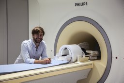 Henrik Lundell合作论文数8Freddy Ståhlberg合作论文数Department of Diagnostic Radiology, Lund University8
 Bruno Medronho合作论文数Department of Chemistry, University of Coimbra8
 Omar Narvaez合作论文数7João P de Almeida Martins合作论文数7
 Enamul Haque Mojumdar合作论文数Leiden Academic Center for Drug Research, University of Leiden7Carin Malmborg合作论文数7Lätt Jimmy合作论文数Ctr Med Imaging & Physiol, Skane Univ Hosp6
 Håkan Wennerström合作论文数Department of Chemistry, Division of Physical Chemistry, Faculty of Science, Lund University6
 Stefanie Eriksson合作论文数6
 Dan Benjamini合作论文数NIH, National Institute of Child Health and Human Development;The Iby and Aladar Fleischman Faculty of Engineering, Tel-Aviv University;National Institute of Child Health and Human Development, Tel-Aviv University6
 Björklund Sebastian合作论文数The Center for Chemistry and Chemical Engineering, Lund University6Cecilia Leal合作论文数Carle Illinois College of Medicine, University of Illinois at Urbana-Champaign6
 Derek Jones合作论文数Brain Research Imaging Centre, School of Psychology, Cardiff University5
 Chantal Tax合作论文数School of Physics and Astronomy, Cardiff University;UMC Utrecht5
 Rachel W Martin合作论文数Department of Chemistry, Yale University5
 Joke Bouwstra合作论文数Division of BioTherapeutics, Leiden University;Leiden Academic Centre for Drug Research, Leiden University5
 Danielle van Westen合作论文数Department of Diagnostic Radiology, Lund University5
 Luis Andres Amorim Alves合作论文数5
 Dimitrios Sakellariou合作论文数4thomas steglich合作论文数Chalmers, Dept Chem & Chem Engn, S-41296 Gothenburg, Sweden4
 Huan Li合作论文数4Eppu Manninen合作论文数Applied Physics, University of Eastern Finland4Leo Svenningsson合作论文数4Göran Carlström合作论文数4杨一鸿合作论文数美国国家药物成瘾研究所4
 Magnus Nydén合作论文数Department of Chemical and Biological Engineering, Chalmers University of Technology4
 Haibo Xu (徐海波)合作论文数Zhongnan Hospital of Wuhan University4
 Lennart Piculell合作论文数Department of Chemistry, Division of Physical Chemistry, Faculty of Science, Lund University4
 Hans Knutsson合作论文数Short CV Link?0?2ping University;Department of Biomedical Engineering4
 Frederik B. Laun合作论文数Department of Medical Physics in Radiology, German Cancer Research Center4




