Evolutionary Computation (EC) and Swarm Intelligence (SI) include some of the most successful meta-heuristics for global optimization. In practical applications, selecting the most suitable EC or SI meta-heuristics as well as the proper setting of its hyper-parameters is usually expensive in terms of computational resources and might be troublesome for novice users. To overcome these downsides, we propose a novel global optimization method, called “Hybrid CMA-PSO” (HyCAPS), which does not demand any complicated procedure for the identification of the hyper-parameters values as it exploits two settings-free algorithms: covariance matrix adaptation evolution strategy (CMA-ES) from EC, and Fuzzy Self-Tuning Particle Swarm Optimization (FST-PSO) from SI. HyCAPS is structured as a two-islands algorithm that evolves one population running CMA-ES and another running FST-PSO, which cooperate by exchanging the fittest individuals at a given frequency. We show that HyCAPS outperforms four optimization methods – FST-PSO, jSO, NL-SHADE-RSP, NL-SHADE-LBC – and competes with CMA-ES and SHADE-ILS on many high-dimensional, continuous, and bounded optimization problems taken from the IEEE CEC’05 and CEC’17 benchmark suites. Our results also highlight that the cooperation between the islands limits the risk of getting stuck in local optima and enhances the search space exploration. We believe that HyCAPS will represent an advantageous meta-heuristics in real-case scenarios where no information about the optimization problem is generally available.
Real-world optimization problems often exhibit multimodality, non-convexity, and stochasticity, which limit the effectiveness of classical algorithms. Gradient-free bio-inspired metaheuristics evolve a population of candidate solutions to address such challenges and find the global optimum. Particle Swarm Optimization (PSO) leverages a swarm of particles (i.e., candidate solutions) that move in the search space attracted by their personal best and the global best solutions. However, similar to other metaheuristics, PSO is prone to stalling, whereby particles fail to escape local minima and reach more promising basins of attraction. We address this issue through Local Bubble Dilation Functions (LBDFs), which locally reshape the landscape around any particle that might have lost its exploration capability. A velocity-based criterion first identifies the stalling particles, distinguishing them from the exploratory ones; then, an LBDF compresses the region around a stalling particle, pushing it away from the bubble center. We integrate this stall remedy—Particle Stall Avoidance through Landscape Manipulation (PSALM)—into Fuzzy Self-Tuning PSO (FST-PSO). We evaluate its effectiveness on the Rastrigin function and on the suite of benchmark functions CEC2017, showing that PSALM improves the search capabilities of FST-PSO by escaping overexploited—and possibly unpromising—areas of the search space, with statistically significant improvements in the best solutions found.
Learning the parameters of Partially Observable Markov Decision Processes (POMDPs) from limited data is a significant challenge. We introduce the Fuzzy MAP EM algorithm, a novel approach that incorporates expert knowledge into the parameter estimation process by enriching the Expectation Maximization (EM) framework with fuzzy pseudo-counts derived from an expert-defined fuzzy model. This integration naturally reformulates the problem as a Maximum A Posteriori (MAP) estimation, effectively guiding learning in environments with limited data. In synthetic medical simulations, our method consistently outperforms the standard EM algorithm under both low-data and high-noise conditions. Furthermore, a case study on Myasthenia Gravis illustrates the ability of the Fuzzy MAP EM algorithm to recover a clinically coherent POMDP, demonstrating its potential as a practical tool for data-efficient modeling in healthcare.
Particle Swarm Optimization (PSO) is a swarm intelligence meta-heuristics whose performance highly depends on the selection of its hyper-parameters, which control the particles’ exploration and exploitation capabilities during the search process. Since the tuning of the hyper-parameters is problem-dependent, settings-free methods are preferable. Fuzzy Self-Tuning PSO (FST-PSO) is a settings-free variant of PSO that exploits a Fuzzy Rule-Based System to determine the best hyper-parameter values for each particle dynamically. Despite this advantage, the optimization process might get stuck in local optima. Here, we propose a what-if strategy to generate and explore alternate swarm histories: what happens if we could go back in time and change something in the past so that one particle “was” different at the beginning of the optimization? Specifically, whenever the global best particle does not improve for a number of iterations, it is terminated and re-initialized, meanwhile sending the rest of the swarm back to the initial configuration. This approach, called “time travel” FST-PSO (FTT-PSO), works under the assumption that the convergence of any particle towards an optimum is strongly related to the influence of the global best particle. We compare the performance of FTT-PSO against FST-PSO on the benchmark suites used in IEEE CEC and GECCO competitions. Our results show that time traveling allows for outperforming both the standard and the multistart versions of FST-PSO.
The histological identification of papillary thyroid carcinoma (PTC) is straightforward for experienced endocrine pathologists. The increase in radical thyroidectomies led to a raise in the rate of postoperative incidental subcentimeter PTC foci and the recent introduction of the Non-Invasive Follicular Thyroid Neoplasm with Papillary-like Nuclear Features (NIFTP) as a less aggressive mimicker of PTC, which significantly complicated the histology screening of thyroid histology specimens. Artificial Intelligence (AI) applied to Whole Slide Images (WSI) can speed up these processes, aiding pathologists to improve diagnostic accuracy and turnaround times. Here we present a computational pathology pipeline for the identification of Microscopic foci of papillary Thyroid Carcinoma-like nuclear features using Artificial intelligence (MiThyCA). This algorithm relies on a tandem architecture consisting of a Convolutional Neural Network (CNN) designed to identify neoplastic areas within thyroid specimens, and a Vision Transformer (TinyViT) focused on detecting PTC-like areas within the neoplastic regions identified by the first model. The study was conducted on a multi-institutional cohort of 73 WSIs from 67 patients with normal thyroid tissue (n = 22 patients, 33
The prevalence of malignant cells in clinical specimens, or tumour purity, is affected by both intrinsic biological factors and extrinsic sampling bias. Molecular characterization of large clinical cohorts is typically performed on bulk samples; data analysis and interpretation can be biased by tumour purity variability. Transcription-based strategies to estimate tumour purity have been proposed, but no breast cancer specific method is available yet. We interrogated over 6000 expression profiles from 10 breast cancer datasets to develop and validate a 9-gene Breast Cancer Purity Score (BCPS). BCPS outperformed existing methods for estimating tumour content. Adjusting transcriptomic profiles using the BCPS reduces sampling bias and aids data interpretation. BCPS-estimated tumour purity improved prognostication in luminal breast cancer, correlated with pathologic complete response in on-treatment biopsies from triple-negative breast cancer patients undergoing neoadjuvant treatment and effectively stratified the risk of relapse in HER2+ residual disease post-neoadjuvant treatment.
The diagnostic assessment of thyroid nodules is hampered by the persistence of uncertainty in borderline cases and further complicated by the inclusion of noninvasive follicular tumor with papillary-like nuclear features (NIFTP) as a less aggressive alternative to papillary thyroid carcinoma (PTC). In this setting, computational methods might facilitate the diagnostic process by unmasking key nuclear characteristics of NIFTP. The main aims of this work were to (1) identify morphometric features of NIFTP and PTC that are interpretable for the human eye and (2) develop a deep learning model for multiclass segmentation as a support tool to reduce diagnostic variability. Our findings confirmed that nuclei in NIFTP and PTC share multiple characteristics, setting them apart from hyperplastic nodules (HP). The morphometric analysis identified 15 features that can be translated into nuclear alterations readily understandable by pathologists, such as a remarkable internuclear homogeneity for HP in contrast to a major complexity in the chromatin texture of NIFTP and to the peculiar pattern of nuclear texture variability of PTC. A few NIFTP cases with available next-generation sequencing data were also analyzed to initially explore the impact of RAS-related mutations on nuclear morphometry. Finally, a pixel-based deep learning model was trained and tested on whole-slide images of NIFTP, PTC, and HP cases. The model, named NUTSHELL (NUclei from Thyroid tumors Segmentation to Highlight Encapsulated Low-malignant Lesions), successfully detected and classified the majority of nuclei in all whole-slide image tiles, showing comparable results with already well-established pathology nuclear scores. NUTSHELL provides an immediate overview of NIFTP areas and can be used to detect microfoci of PTC within extensive glandular samples or identify lymph node metastases. NUTSHELL can be run inside WSInfer with an easy rendering in QuPath, thus facilitating the democratization of digital pathology.
Abstract Funding Acknowledgements Type of funding sources: Public grant(s) – National budget only. Main funding source(s): This research was partly funded by the Italian Ministry of Health Introduction With the worldwide diffusion of cardiac magnetic resonance (CMR), demand on image quality has grown. CMR late gadolinium enhancement (LGE) imaging provides critical diagnostic and prognostic information, and guides management. The identification of optimal Inversion Time (TI), a time-sensitive parameter closely linked to contrast kinetics, is pivotal for correct myocardium nulling. However, determining the optimal TI can be challenging in some diseases and for less experienced operators. Purpose To develop and test an artificial intelligence tool to automatically predict the personalised optimal TI in LGE imaging. Methods The tool, named THAITI, consists of a Random Forest regression model. It considers, as input parameters, patient-specific TI determinants (age, gender, weight, height, kidney function, heart rate) and CMR scan-specific TI determinants (B0, contrast type and dose, time elapsed from contrast injection). THAITI was trained on 219 patients (3585 images) with mixed conditions who underwent CMR (1.5T; Gadobutrol; averaged, MOCO, free-breathing true-FISP IR [1]) for clinical reasons. The dataset was split with a 90–10 policy: 90% of data for training, and 10% for testing. THAITI’s hyperparameters were optimised by embedding k-fold cross validation into an evolutionary computation algorithm, and the best performing model was finally evaluated on the test set. A graphical user interface was also developed. Clinical validation was performed on 55 consecutive patients, randomised to experimental (THAITI-set TI) vs control (operator-set TI) group. Image quality was assessed blindly by 2 independent experienced operators by a 4-points Likert scale, and by means of the contrast/enhancement ratio (CER) (i.e., signal intensity of enhanced/remote myocardium ratio). Results In the testing set, the TI predicted by THAITI differed from the ground truth by ≥ 5ms in 16% of cases. At clinical validation, myocardial nulling quality did not differ between the experimental vs the control group either by CER or visual assessment, with an overall "optimal" or "good" nulling in 96% vs 93%, respectively. Conclusions Using main determinants of contrast kinetics, THAITI efficiently predicted the optimal TI for CMR-LGE imaging. The tool works as a stand-alone on laptops/mobile devices, not requiring adjunctive scanner technology and thus has great potential for diffusion, including in small or recently opened CMR services, and in low-resource settings. Additional development is ongoing to increase generalisability (multi-vendor, multi-sequence, multi-contrast) and to test its potential to further improve CMR-LGE image quality and reduce the need for repeated imaging for inexperienced operators. Figure 1. Top: THAITI interface. Bottom: examples of experimental group CMR-LGE imaging. Table 1. Control vs experimental group. Data expressed as absolute number (%), mean ± SD, median [IQR]. ⧧ T-test; * Chi-square.
Introduction: Oxford Nanopore Technologies (ONT) is a third generation sequencing approach that allows the analysis of individual, full-length nucleic acids. ONT records the alterations of an ionic current flowing across a nano-scaled pore while a DNA or RNA strand is threading through the pore. Basecalling methods are then leveraged to translate the recorded signal back to the nucleic acid sequence. However, basecall generally introduces errors that hinder the process of barcode demultiplexing, a pivotal task in single-cell RNA sequencing that allows for separating the sequenced transcripts on the basis of their cell of origin. Methods: To solve this issue, we present a novel framework, called UNPLEX, designed to tackle the barcode demultiplexing problem by operating directly on the recorded signals. UNPLEX combines two unsupervised machine learning methods: autoencoders and self-organizing maps (SOM). The autoencoders extract compact, latent representations of the recorded signals that are then clustered by the SOM. Results and Discussion: Our results, obtained on two datasets composed of in silico generated ONT-like signals, show that UNPLEX represents a promising starting point for the development of effective tools to cluster the signals corresponding to the same cell.
Tumor recognition by T cells is essential for antitumor immunity. A comprehensive characterization of T cell diversity may be key to understanding the success of immunomodulatory drugs and failure of PD-1 blockade in tumors such as multiple myeloma (MM). Here, we use single-cell RNA and T cell receptor sequencing to characterize bone marrow T cells from healthy adults ( n = 4) and patients with precursor ( n = 8) and full-blown MM ( n = 10). Large T cell clones from patients with MM expressed multiple immune checkpoints, suggesting a potentially dysfunctional phenotype. Dual targeting of PD-1 + LAG3 or PD-1 + TIGIT partially restored their function in mice with MM. We identify phenotypic hallmarks of large intratumoral T cell clones, and demonstrate that the CD27 − and CD27 + T cell ratio, measured by flow cytometry, may serve as a surrogate of clonal T cell expansions and an independent prognostic factor in 543 patients with MM treated with lenalidomide-based treatment combinations.
Artificial intelligence is getting a foothold in medicine for disease screening and diagnosis. While typical machine learning methods require large labeled datasets for training and validation, their application is limited in clinical fields since ground truth information can hardly be obtained on a sizeable cohort of patients. Unsupervised neural networks – such as Self-Organizing Maps (SOMs) – represent an alternative approach to identifying hidden patterns in biomedical data. Here we investigate the feasibility of SOMs for the identification of malignant and non-malignant regions in liquid biopsies of thyroid nodules, on a patient-specific basis. MALDI-ToF (Matrix Assisted Laser Desorption Ionization - Time of Flight) mass spectrometry-imaging (MSI) was used to measure the spectral profile of bioptic samples. SOMs were then applied for the analysis of MALDI-MSI data of individual patients' samples, also testing various pre-processing and agglomerative clustering methods to investigate their impact on SOMs' discrimination efficacy. The final clustering was compared against the sample's probability to be malignant, hyperplastic or related to Hashimoto thyroiditis as quantified by multinomial regression with LASSO. Our results show that SOMs are effective in separating the areas of a sample containing benign cells from those containing malignant cells. Moreover, they allow to overlap the different areas of cytological glass slides with the corresponding proteomic profile image, and inspect the specific weight of every cellular component in bioptic samples. We envision that this approach could represent an effective means to assist pathologists in diagnostic tasks, avoiding the need to manually annotate cytological images and the effort in creating labeled datasets.
Multi-objective optimization algorithms might struggle in finding optimal dominating solutions, especially in real-case scenarios where problems are generally characterized by non-separability, non-differentiability, and multi-modality issues. An effective strategy that already showed to improve the outcome of optimization algorithms consists in manipulating the search space, in order to explore its most promising areas. In this work, starting from a Pareto front identified by an optimization strategy, we exploit Local Bubble Dilation Functions (LBDFs) to manipulate a locally bounded region of the search space containing non-dominated solutions. We tested our approach on the benchmark functions included in the DTLZ and WFG suites, showing that the Pareto front obtained after the application of LBDFs is most of the time characterized by an increased hyper-volume value. Our results confirm that LBDFs are an effective means to identify additional non-dominated solutions that can improve the quality of the Pareto front.
Calcium homeostasis and signaling processes in Saccharomyces cerevisiae, as well as in any eukaryotic organism, depend on various transporters and channels located on both the plasma and intracellular membranes. The activity of these proteins is regulated by a number of feedback mechanisms that act through the calmodulin-calcineurin pathway. When exposed to hypotonic shock (HTS), yeast cells respond with an increased cytosolic calcium transient, which seems to be conditioned by the opening of stretch-activated channels. To better understand the role of each channel and transporter involved in the generation and recovery of the calcium transient-and of their feedback regulations-we defined and analyzed a mathematical model of the calcium signaling response to HTS in yeast cells. The model was validated by comparing the simulation outcomes with calcium concentration variations before and during the HTS response, which were observed experimentally in both wild-type and mutant strains. Our results show that calcium normally enters the cell through the High Affinity Calcium influx System and mechanosensitive channels. The increase of the plasma membrane tension, caused by HTS, boosts the opening probability of mechanosensitive channels. This event causes a sudden calcium pulse that is rapidly dissipated by the activity of the vacuolar transporter Pmc1. According to model simulations, the role of another vacuolar transporter, Vcx1, is instead marginal, unless calcineurin is inhibited or removed. Our results also suggest that the mechanosensitive channels are subject to a calcium-dependent feedback inhibition, possibly involving calmodulin. Noteworthy, the model predictions are in accordance with literature results concerning some aspects of calcium homeostasis and signaling that were not specifically addressed within the model itself, suggesting that it actually depicts all the main cellular components and interactions that constitute the HTS calcium pathway, and thus can correctly reproduce the shaping of the calcium signature by calmodulin- and calcineurin-dependent complex regulations. The model predictions also allowed to provide an interpretation of different regulatory schemes involved in calcium handling in both wild-type and mutants yeast strains. The model could be easily extended to represent different calcium signals in other eukaryotic cells.
Raw data (original images, labelled masks) from "An accurate and time-efficient deep learning-based system for automated segmentation and reporting of cardiac magnetic resonance-detected ischemic scar"; https://doi.org/10.1016/j.cmpb.2022.107321
Several software tools for the simulation and analysis of biochemical reaction networks have been developed in the last decades; however, assessing and comparing their computational performance in executing the typical tasks of computational systems biology can be limited by the lack of a standardized benchmarking approach. To overcome these limitations, we propose here a novel tool, named SMGen, designed to automatically generate synthetic models of reaction networks that, by construction, are characterized by relevant features (e.g., system connectivity and reaction discreteness) and non-trivial emergent dynamics of real biochemical networks. The generation of synthetic models in SMGen is based on the definition of an undirected graph consisting of a single connected component that, generally, results in a computationally demanding task; to speed up the overall process, SMGen exploits a main–worker paradigm. SMGen is also provided with a user-friendly graphical user interface, which allows the user to easily set up all the parameters required to generate a set of synthetic models with any number of reactions and species. We analysed the computational performance of SMGen by generating batches of symmetric and asymmetric reaction-based models (RBMs) of increasing size, showing how a different number of reactions and/or species affects the generation time. Our results show that when the number of reactions is higher than the number of species, SMGen has to identify and correct a large number of errors during the creation process of the RBMs, a circumstance that increases the running time. Still, SMGen can generate synthetic models with hundreds of species and reactions in less than 7 s.
Solving optimization problems is one of the most complex and widespread task in Computer Science. In many scenarios, finding the global optimum of a function is hampered by several features that characterize the fitness landscapes, such as noisiness, multi-modality, non-convexity, non-separability, and non-differentiability. In order to facilitate the optimization process, a variety of methods have been proposed to manipulate either the search space or the fitness landscape. Among these, Dilation Functions (DFs) were introduced to expand regions of the search space that are characterized by promising fitness values. In this work, we extend the family of DFs by introducing Local Bubble Dilation Functions (LBDFs), a novel approach that generates local distortions bounded by hyper-spheres. By performing an appropriate mapping of the search space, LBDFs can improve the optimization performance, since they expand and reveal the promising regions around the global optimum, while leaving the rest of the fitness landscape untouched. The additional advantage of LBDFs, with respect to DFs, is that different dilations can be applied to each dimension of the search space, which is useful in the case of asymmetric landscapes. In order to show the benefits of local dilations, we executed several tests on the Michalewicz benchmark function, with different settings for the LBDFs. Our results show that a properly designed LBDF can lead to statistically significant better results than using vanilla optimization. Finally, we investigated the use of LBDFs to facilitate the solution of the parameter estimation problem in Systems Biology by analyzing the landscape related to a stochastic model of enzyme kinetics.
Abstract Introduction Identifying the optimal Inversion Time (TI) is pivotal to null the myocardium and obtain high quality cardiac magnetic resonance (CMR) late gadolinium enhancement (LGE) imaging. Setting the optimal TI can be challenging in some diseases and for less experienced operators. We propose an Artificial Intelligence (AI) tool to automatically predict the optimal TI in CMR-LGE imaging. Methods The AI tool, named THAITI, consists of a Random Forest regression model whose hyperparametrs were optimized by means of evolutionary computation. The model considers as input parameters patient-specific TI determinants, such as age, gender, weight, height, kidney function, heart rate, contrast dose, and time from injection to image acquisition. THAITI was trained on 155 patients (2588 CMR-LGE images) with mixed cardiac conditions who underwent CMR (1.5T Siemens AvantoFit; Gadovist; averaged, motion-corrected, free-breathing true-FISP IR). Clinical testing was performed on 55 matched patients, randomized to experimental (THAITI-set TI) vs control (experienced operator-set TI) group. A user interface was developed for clinical testing. Image quality was assessed blindly by 2 independent experienced operators. Results THAITI Mean Squared Error (MSE) in the validation set was 4.7 and percentage of mispredicted TI of 4.5%. During clinical testing, LGE quality did not differ between the experimental vs control group: quality was “optimal” or “good” in 96% vs 93%, “poor” in 4% vs 7%. The average number of LGE images acquired and LGE imaging duration were similar (experimental vs control group: 17 ± 3 vs 16 ± 3 LGE images per patient; 12:14 vs 12:20 mm:ss, respectively). Conclusion THAITI efficiently predicts optimal TI for CMR-LGE imaging. Further development is needed to increase generalizability (multi-vendor, multi-sequence, multi-contrast) and to test its potential to improve LGE image quality and reduce the need for repeated imaging for inexperienced operators. Figure 1. Top panel: THAITI interface. Bottom panel: examples of experimental group LGE imaging. Table 1. Control vs experimental group. Data expressed as absolute number (%), mean ±SD
Acute myeloid leukemia (AML) is a highly frequent hematological malignancy, characterized by clinical and biological diversity, along with high relapse and mortality rates. The inherent functional and genetic intra-tumor heterogeneity in AML is thought to play an important role in disease recurrence and resistance to chemotherapy. Patient-derived xenograft (PDX) models preserve important features of the original tumor, allowing, at the same time, experimental manipulation and in vivo amplification of the human cells. Here we present a detailed protocol for the generation of fluorescently labeled AML PDX models to monitor cell proliferation kinetics in vivo, at the single-cell level. Although experimental protocols for cell proliferation studies are well established and widespread, they are not easily applicable to in vivo contexts, and the analysis of related time-series data is often complex to achieve. To overcome these limitations, model-driven approaches can be exploited to investigate different aspects of cell population dynamics. Among the existing approaches, the ProCell framework is able to perform detailed and accurate stochastic simulations of cell proliferation, relying on flow cytometry data. In particular, by providing an initial and a target fluorescence histogram, ProCell automatically assesses the validity of any user-defined scenario of intra-tumor heterogeneity, that is, it is able to infer the proportion of various cell subpopulations (including quiescent cells) and the division interval of proliferating cells. Here we explain the protocol in detail, providing a description of our methodology for the conditional expression of H2B-GFP in human AML xenografts, data processing by flow cytometry, and the final elaboration in ProCell.
Combination therapies proved to be a valuable strategy in the fight against cancer, thanks to their increased efficacy in inducing tumor cell death and in reducing tumor growth, metastatic potential, and the risk of developing drug resistance. The identification of effective combinations of drug targets generally relies on costly and time consuming processes based on in vitro experiments. Here, we present a novel computational approach that, by integrating dynamic fuzzy modeling with multi-objective optimization, allows to efficiently identify novel combination cancer therapies, with a relevant saving in working time and costs. We tested this approach on a model of oncogenic K-ras cancer cells characterized by a marked Warburg effect. The computational approach was validated by its capability in finding out therapies already known in the literature for this type of cancer cell. More importantly, our results show that this method can suggest potential therapies consisting in a small number of molecular targets. In the model of oncogenic K-ras cancer cells, for instance, we identified combination of up to three targets, which affect different cellular pathways that are crucial for cancer proliferation and survival.
 Paolo Cazzaniga合作论文数Dipartimento di Informatica, Sistemistica e Comunicazione, Università degli Studi di Milano-Bicocca100
 Giancarlo Mauri合作论文数Department of Informatics, Systems and Communication, Universita degli Studi di Milano-Bicocca82
 Marco Salvatore Nobile合作论文数Dipartimento di Informatica, Sistemistica e Comunicazione, Università degli Studi di Milano-Bicocca74
 Dario Pescini合作论文数Dipartimento di Informatica, Sistemistica e Comunicazione, Università degli Studi di Milano-Bicocca42
 Andrea Tangherloni合作论文数Univ Milano Bicocca, Dept Informat Syst & Commun, I-20126 Milan, Italy26
 Simone Spolaor合作论文数University of Milano-Bicocca25Claudio Zandron合作论文数Dipartimento di Informatica, Sistemistica e Comunicazione (DISCo)15
 Daniele M. Papetti合作论文数University of Milano-Bicocca13
 Leonardo Rundo合作论文数University of Salerno11
 Luca Manzoni合作论文数Dipartimento di Informatica, Sistemistica e Comunicazione, University of Milano-Bicocca8
 Riccardo Colombo合作论文数7
 Enzo Martegani合作论文数Sezione di Biochimica Comparata, Università degli Studi di Milano7
 Sonia Colombo合作论文数Dipartimento di Biotecnologie e Bioscienze, Università degli Studi di Milano-Bicocca7
 Claudio Ferretti合作论文数Dipartimento di Informatica Sistemistica e Comunicazione, Università degli Studi di Milano-Bicocca6Matteo Bellini合作论文数4
 Thalia Vlachou合作论文数IEO, European Institute of Oncology IRCCS, Department of Experimental Oncology, Via Adamello 16, 20139 Milan, Italy4
 Pier Giuseppe Pelicci合作论文数Department of Experimental Oncology, Istituto Europeo di Oncologia;Institute of Medical Pathology, Faculty of Medicine and Surgery, University of Parma4
 Ioan Ardelean合作论文数Technical University of Cluj-Napoca4Nadia Busi合作论文数4
 Ivan Merelli合作论文数the National Research Council, Institute for Biomedical Technologies4M. S. Nobile合作论文数3Vasco Coelho合作论文数3
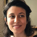 Giulia Capitoli合作论文数Bicocca Bioinformatics Biostatistics and Bioimaging Centre - B4, University of Milano-Bicocca3
 Matteo Dugo合作论文数Fondazione Centro San Raffaele;Department of Medical Oncology, IRCCS Ospedale San Raffaele3
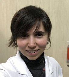 Torlasco Camilla合作论文数S. Luca Hospital, University of Milano-Bicocca3
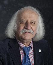 Grzegorz Rozenberg合作论文数Department of Computer Science, Leiden University;Department of Computer Science, University of Colorado at Boulder3
 Parati Gianfranco合作论文数Dipartimento di Medicina e Chirurgia, Scuola di Specializzazione in Malattie Cardiovascolari, Università degli Studi di Milano-Bicocca;Unità di Cardiologia, Metaboliche dell'Ospedale San Luca, Istituto Auxologico Italiano, IRCCS;Dipartimento di Scienze Cardiovascolari, Neurologiche e Metaboliche dell'Ospedale San Luca, Istituto Auxologico Italiano, IRCCS3
 Carmelo Militello合作论文数Department of Aerospace Engineering Sciences and Center for Space Structures and Controls, University of Colorado3Peter Kellman合作论文数3
 Marzio Pennisi合作论文数University of Catania3Stefania Galimberti合作论文数School of Medicine, University of Milano-Bicocca3
 Luigi P. Badano合作论文数IstitutoAuxologico;School of medicine and surgery, Università Milano-Bicocca3
 Alberto Leporati合作论文数Dipartimento di Informatica, Sistemistica e Comunicazione, Università degli Studi di Milano-Bicocca;Registri Digitali Srl3
 Pietro Liò合作论文数Department of Computer Science and Technology, University of Cambridge;Cambridge Centre for AI in Medicine, University of Cambridge3
 Marco Beccuti合作论文数Dipartimento di Informatica, Universita di Torino, Italy3
 Daniel Ashlock合作论文数Department of Mathematics and Statistics, College of Engineering and Physical Sciences, University of Guelph3
 Lilia Alberghina合作论文数SYSBIO - Centre of Systems Biology, University of Milano-Bicocca3
 Leonardo Vanneschi合作论文数Instituto Superior de Estatística e Gestão de Informação, Nova Information Management School, Universidade Nova de Lisboa3
 Fabio Pagni合作论文数Department of Pathology, Desio Hospital3
 Nicola Fusco合作论文数2
 Carlos Federico Lopez合作论文数2
 Giorgio Cazzaniga合作论文数2Kirsten Van Abeelen合作论文数2Simone G. Riva合作论文数Weatherall Institute of Molecular Medicine, University of Oxford2Niccoló Totis合作论文数2
 Caro Fuchs合作论文数Eindhoven Univ Technol, Informat Syst Grp, Sch Ind Engn, Eindhoven, Netherlands2
 Vincenzo L'Imperio合作论文数San Gerardo Hosp, Univ Milano Bicocca2
 Giuseppe Muscogiuri合作论文数Department of Radiology and Radiological Science, Medical University of South Carolina;Department of Medical-Surgical Sciences and Translational Medicine, University of Rome;Sapienza", Rome, Italy.;Medical University of South Carolina, University of Rome "Sapienza"2
 Castelletti Silvia合作论文数2




