Core myopathies are congenital diseases with clinical, pathological and genetic heterogeneity. Main histological features are fiber “cores” showing a focally reduced oxidative enzyme activity. Dusty Core Disease (DuCD) differs from Central Core Myopathy for the presence of irregular areas, without clear borders and round/ovoidal shape, and myofibrillar disorganization characterized by reddish purple granular material depositions. This disorder is defined clinically by severe phenotypes with early onset of disease and molecularly by low level of RyR1 in muscle. Until now DuCD was associated only to biallelic recessive RYR1 mutations. We analyzed the clinical aspects, pathological features and mutational spectrum of four DuCD patients, belonging to our cohort of Congenital Myopathy probands. Molecular analysis detected 5 different RYR1 pathogenic variants, two of them so far unreported. Patients presented a heterogeneous phenotype ranging from severe recessive infantile forms to moderate dominant adult-onset presentations. Histological, immunological and ultrastructural techniques were employed to validate these dominant cases, which expand our knowledge on the inheritance of this subgroup of diseases.
Inclusion body myositis (IBM) is a slowly progressive disorder belonging to the idiopathic inflammatory myopathies, and it represents the most common adult-onset acquired myopathy. The main clinical features include proximal or distal muscular asymmetric weakness, with major involvement of long finger flexors and knee extensors. The main histological findings are the presence of fiber infiltrations, rimmed vacuoles, and amyloid inclusions. The etiopathogenesis is a challenge because both environmental and genetic factors are implicated in muscle degeneration and a distinction has been made previously between sporadic and hereditary forms. Here, we describe an Italian patient affected with a hereditary form of IBM with onset in his mid-forties. Next-generation sequencing analysis disclosed a heterozygous mutation c.76C>T (p.Pro26Ser) in the PDZ motif of the LDB3/ZASP gene, a mutation already described in a family with a late-onset myopathy and highly heterogenous degree of skeletal muscle weakness. In the proband’s muscle biopsy, the expression of ZASP, myotilin, and desmin were increased. In our family, in addition to the earlier age of onset, the clinical picture is even more peculiar given the evidence, in one of the affected family members, of complete ophthalmoplegia in the vertical gaze. These findings help extend our knowledge of the clinical and genetic background associated with inclusion body myopathic disorders.
BackgroundCongenital myopathies are a group of heterogeneous inherited disorders, mainly characterized by early-onset hypotonia and muscle weakness. The spectrum of clinical phenotype can be highly variable, going from very mild to severe presentations. The course also varies broadly resulting in a fatal outcome in the most severe cases but can either be benign or lead to an amelioration even in severe presentations. Muscle biopsy analysis is crucial for the identification of pathognomonic morphological features, such as core areas, nemaline bodies or rods, nuclear centralizations and congenital type 1 fibers disproportion. However, multiple abnormalities in the same muscle can be observed, making more complex the myopathological scenario.Case presentationHere, we describe an Italian newborn presenting with severe hypotonia, respiratory insufficiency, inability to suck and swallow, requiring mechanical ventilation and gastrostomy feeding. Muscle biopsy analyzed by light microscopy showed the presence of vacuoles filled with glycogen, suggesting a metabolic myopathy, but also fuchsinophilic inclusions. Ultrastructural studies confirmed the presence of normally structured glycogen, and the presence of minirods, directing the diagnostic hypothesis toward a nemaline myopathy. An expanded Next Generation Sequencing analysis targeting congenital myopathies genes revealed the presence of a novel heterozygous c.965 T > A p. (Leu322Gln) variant in the ACTA1 gene, which encodes the skeletal muscle alpha-actin.ConclusionOur case expands the repertoire of molecular and pathological features observed in actinopathies. We highlight the value of ultrastructural examination to investigate the abnormalities detected at the histological level. We also emphasized the use of expanded gene panels in the molecular analysis of neuromuscular patients, especially for those ones presenting multiple bioptic alterations.
Isolated mitochondrial respiratory chain Complex IV (Cytochrome c Oxidase or COX) deficiency is the second most frequent isolated respiratory chain defect. Causative mutations are mainly identified in structural COX subunits or in proteins involved in the maturation and assembly of the COX holocomplex. We describe an Italian familial case of mitochondrial myopathy due to a variant in the COX assembly factor 8 gene (COA8). Patient 1 is a 52-year-old woman who presented generalized epilepsy and retinitis pigmentosa at 10 years of age. From her early adulthood she complained about cramps and myalgia after exercise, and bilateral hearing loss emerged. Last neurological examination (52 years of age) showed bilateral ptosis, muscle weakness, peripheral neuropathy, mild dysarthria and dysphonia, cognitive impairment. Muscle biopsy had shown the presence of ragged-red fibers. Patient 2 (Patient 1’s sister) is a 53-year-old woman presenting fatigability, myalgia, and hearing loss. Neurological examination showed ptosis and muscle weakness. Muscle biopsy displayed a diffuse reduction of COX activity staining and ragged-red fibers. Both sisters presented secondary amenorrhea. After ruling out mtDNA mutations, Whole Exome Sequencing analysis identified the novel homozygous COA8 defect c.170_173dupGACC, p.(Pro59fs) in the probands. Loss-of-function COA8 mutations have been associated with cavitating leukoencephalopathy with COX deficiency in 9 reported individuals. Disease course shows an early-onset rapid clinical deterioration, affecting both cognitive and motor functions over months, followed by stabilization and slow improvement over several years. Our findings expand the clinical spectrum of COA8-related disease. We confirm the benign course of this rare disorder, highlighting its (intrafamilial) clinical variability.
Collagen VI is a heterotrimeric protein expressed in several tissues and involved in the maintenance of cell integrity. It localizes at the cell surface, creating a microfilamentous network that links the cytoskeleton to the extracellular matrix. The heterotrimer consists of three chains encoded by COL6A1, COL6A2 and COL6A3 genes. Recessive and dominant molecular defects cause two main disorders, the severe Ullrich congenital muscular dystrophy and the relatively mild and slowly progressive Bethlem myopathy. We analyzed the clinical aspects, pathological features and mutational spectrum of 15 COL6-mutated patients belonging to our cohort of muscular dystrophy probands. Patients presented a heterogeneous phenotype ranging from severe forms to mild adult-onset presentations. Molecular analysis by NGS detected 14 different pathogenic variants, three of them so far unreported. Two changes, localized in the triple-helical domain of COL6A1, were associated with a more severe phenotype. Histological, immunological and ultrastructural techniques were employed for the validation of the genetic variants; they documented the high variability in COL6 distribution and the extracellular matrix disorganization, highlighting the clinical heterogeneity of our cohort. The combined use of these different technologies is pivotal in the diagnosis of COL6 patients.
Mitochondrial encephalomyopathy, lactic acidosis, and stroke-like episodes (MELAS) syndrome is a systemic disorder in which multi-organ dysfunction may occur from mitochondrial metabolism failure. Maternally inherited mutations in the MT-TL1 gene are the most frequent causes for this disorder. Clinical manifestations may include stroke-like episodes, epilepsy, dementia, headache and myopathy. Among these, acute visual failure, usually in association with cortical blindness, can occur because of stroke-like episodes affecting the occipital cortex or the visual pathways. Vision loss due to optic neuropathy is otherwise considered a typical manifestation of other mitochondrial diseases such as Leber hereditary optic neuropathy (LHON). Here we describe a 55-year-old woman, sister of a previously described patient with MELAS harbouring the m.3243A > G (p.0, MT-TL1) mutation, with otherwise unremarkable medical history, that presented with subacute, painful visual impairment of one eye, accompanied by proximal muscular pain and headache. Over the next weeks, she developed severe and progressive vision loss limited to one eye. Ocular examination confirmed unilateral swelling of the optic nerve head; fluorescein angiography showed segmental perfusion delay in the optic disc and papillary leakage. Neuroimaging, blood and CSF examination and temporal artery biopsy ruled out neuroinflammatory disorders and giant cell arteritis (GCA). Mitochondrial sequencing analysis confirmed the m.3243A > G transition, and excluded the three most common LHON mutations, as well as the m.3376G > A LHON/MELAS overlap syndrome mutation. Based on the constellation of clinical symptoms and signs presented in our patient, including the muscular involvement, and the results of the investigations, the diagnosis of optic neuropathy as a stroke-like event affecting the optic disc was performed. L-arginine and ubidecarenone therapies were started with the aim to improve stroke-like episode symptoms and prevention. The visual defect remained stable with no further progression or outbreak of new symptoms. Atypical clinical presentations must be always considered in mitochondrial disorders, even in well-described phenotypes and when mutational load in peripheral tissue is low. Mitotic segregation of mitochondrial DNA (mtDNA) does not allow to know the exact degree of heteroplasmy existent within different tissue, such as retina and optic nerve. Important therapeutic implications arise from a correct diagnosis of atypical presentation of mitochondrial disorders.
Pathogenic variants impacting upon assembly of mitochondrial respiratory chain Complex IV (Cytochrome c Oxidase or COX) predominantly result in early onset mitochondrial disorders often leading to CNS, skeletal and cardiac muscle manifestations. The aim of this study is to describe a molecular defect in the COX assembly factor gene COX18 as the likely cause of a neonatal form of mitochondrial encephalo-cardio-myopathy and axonal sensory neuropathy. The proband is a 19-months old female displaying hypertrophic cardiomyopathy at birth and myopathy with axonal sensory neuropathy and failure to thrive developing in the first months of life. Serum lactate was consistently increased. Whole exome sequencing allowed the prioritization of the unreported homozygous substitution NM_001297732.2:c.667 G > C p.(Asp223His) in COX18. Patient’s muscle biopsy revealed severe and diffuse COX deficiency and striking mitochondrial abnormalities. Biochemical and enzymatic studies in patient’s myoblasts and in HEK293 cells after COX18 silencing showed a severe impairment of both COX activity and assembly. The biochemical defect was partially rescued by delivery of wild-type COX18 cDNA into patient’s myoblasts. Our study identifies a novel defect of COX assembly and expands the number of nuclear genes involved in a mitochondrial disorder due to isolated COX deficiency.
Mitochondrial DNA (mtDNA) depletion syndromes are disorders characterized by infantile-onset, severe progression, and the drastic loss of mtDNA content in affected tissues. In a patient who showed severe hypotonia, proximal tubulopathy and sensorineural hearing loss after birth, we observed severe mtDNA depletion and impaired respiratory chain activity in muscle due to heterozygous variants c.686G > T and c.551-2A > G in RRM2B, encoding the p53R2 subunit of the ribonucleotide reductase.
Mitochondrial DNA (mtDNA) maintenance disorders embrace a broad range of clinical syndromes distinguished by the evidence of mtDNA depletion and/or deletions in affected tissues. Among the nuclear genes associated with mtDNA maintenance disorders, RNASEH1 mutations produce a homogeneous phenotype, with progressive external ophthalmoplegia (PEO), ptosis, limb weakness, cerebellar ataxia, and dysphagia. The encoded enzyme, ribonuclease H1, is involved in mtDNA replication, whose impairment leads to an increase in replication intermediates resulting from mtDNA replication slowdown. Here, we describe two unrelated Italian probands (Patient 1 and Patient 2) affected by chronic PEO, ptosis, and muscle weakness. Cerebellar features and severe dysphagia requiring enteral feeding were observed in one patient. In both cases, muscle biopsy revealed diffuse mitochondrial abnormalities and multiple mtDNA deletions. A targeted next-generation sequencing analysis revealed the homozygous RNASEH1 mutations c.129-3C>G and c.424G>A in patients 1 and 2, respectively. The c.129-3C>G substitution has never been described as disease-related and resulted in the loss of exon 2 in Patient 1 muscle RNASEH1 transcript. Overall, we recommend implementing the use of high-throughput sequencing approaches in the clinical setting to reach genetic diagnosis in case of suspected presentations with impaired mtDNA homeostasis.
Ryanodine receptor type 1-related congenital myopathies are the most represented subgroup among congenital myopathies (CMs), typically presenting a central core or multiminicore muscle histopathology and high clinical heterogeneity. We evaluated a cohort of patients affected with Ryanodine receptor type 1-related congenital myopathy (RYR1-RCM), focusing on four patients who showed a severe congenital phenotype and underwent a comprehensive characterization at few months of life. To date there are few reports on precocious instrumental assessment. In two out of the four patients, a muscle biopsy was performed in the first days of life (day 5 and 37, respectively) and electron microscopy was carried out in two patients detecting typical features of congenital myopathy. Two patients underwent brain MRI in the first months of life (15 days and 2 months, respectively), one also a fetal brain MRI. In three children electromyography was performed in the first week of life and neurogenic signs were excluded. Muscle MRI obtained within the first years of life showed a typical pattern of RYR1-CM. The diagnosis was confirmed through genetic analysis in three out of four cases using Next Generation Sequencing (NGS) panels. The development of a correct and rapid diagnosis is a priority and may lead to prompt medical management and helps optimize inclusion in future clinical trials.
We present the case of an Italian family, whose affected members showed different degrees of muscle involvement and histopathological features compatible with congenital myopathies (CM), in the presence of a rare CACNA1S heterozygous mutation. CMs are a heterogeneous group of genetic muscle disorders ranging from severe and life-threatening neonatal manifestations to adult-onset milder forms [ [1] Schorling D.C. Kirschner J. Bönnemann C.G. Congenital muscular dystrophies and myopathies: an overview and update. Neuropediatrics. 2017; 48: 247-261 Crossref PubMed Scopus (28) Google Scholar ] and classified according to specific histological and ultrastructural muscle biopsy features [ [2] Ravenscroft G. Laing N.G. Bönnemann C.G. Pathophysiological concepts in the congenital myopathies: blurring the boundaries, sharpening the focus. Brain. 2015; 138: 246-268 Crossref PubMed Scopus (62) Google Scholar ]. Main clinical characteristics include hypotonia and weakness of proximal and facial muscles, often associated with respiratory failure and cardiac involvement [ [1] Schorling D.C. Kirschner J. Bönnemann C.G. Congenital muscular dystrophies and myopathies: an overview and update. Neuropediatrics. 2017; 48: 247-261 Crossref PubMed Scopus (28) Google Scholar ]. Disease course is slowly progressive and serum creatine-kinase (CK) levels are normal to mildly elevated [ [1] Schorling D.C. Kirschner J. Bönnemann C.G. Congenital muscular dystrophies and myopathies: an overview and update. Neuropediatrics. 2017; 48: 247-261 Crossref PubMed Scopus (28) Google Scholar ]. Over the last decades, many disease-causing genes have been associated with CMs [ [2] Ravenscroft G. Laing N.G. Bönnemann C.G. Pathophysiological concepts in the congenital myopathies: blurring the boundaries, sharpening the focus. Brain. 2015; 138: 246-268 Crossref PubMed Scopus (62) Google Scholar ]. Genes belonging to the CACNA1 family encode for tissue-specific alpha subunits of the voltage-gated calcium channel and have been associated with ataxias, hemiplegic migraine, blindness and deafness [ [3] Jurkat-Rott K. Lehmann-Horn F. The impact of splice isoforms on voltage-gated calcium channel alpha1 subunits. J. Physiol. 2004; 554: 609-619 Crossref PubMed Scopus (65) Google Scholar , [4] Wu J. Yan Z. Li Z. Yan C. Lu S. Dong M. et al. Structure of the voltage-gated calcium channel Cav1.1 complex. Science. 2015; 350: 2395 Crossref Scopus (206) Google Scholar ]. CACNA1S expressed in skeletal muscle encodes for Cav1.1 protein, the pore-forming subunit of the dihydropyridine receptor (DHPR) which is coupled to the Ryanodine receptor Ca2+-release channel-1 (RYR1) in muscle excitation-contraction [ [4] Wu J. Yan Z. Li Z. Yan C. Lu S. Dong M. et al. Structure of the voltage-gated calcium channel Cav1.1 complex. Science. 2015; 350: 2395 Crossref Scopus (206) Google Scholar , [5] Jurkat-Rott K. Lehmann-Horn F. Elbaz A. Heine R. Gregg R.G. Hogan K. et al. A calcium channel mutation causing hypokalemic periodic paralysis. Hum. Mol. Genet. 1994; : 1415-1419 Crossref PubMed Scopus (275) Google Scholar ]. CACNA1S mutations have been formerly associated with malignant hyperthermia, hypokalemic and normokaliemic periodic paralysis [ [4] Wu J. Yan Z. Li Z. Yan C. Lu S. Dong M. et al. Structure of the voltage-gated calcium channel Cav1.1 complex. Science. 2015; 350: 2395 Crossref Scopus (206) Google Scholar ], and, more recently CMs [ 6 Schartner V. Romero N.B. Donkervoort S. Treves Munot P. Pierson T.M. et al. Dihydropyridine receptor (DHPR, CACNA1S) congenital myopathy. Acta Neuropathol. 2017; : 517-533 Crossref PubMed Scopus (55) Google Scholar , 7 Yiş U. Hiz S. Güneş S. Diniz G. Baydan F. Töpf A. et al. Dihydropyridine receptor congenital myopathy in a consanguineous Turkish family. J. Neuromuscul. Dis. 2019; 6: 377-384 Crossref PubMed Scopus (4) Google Scholar , 8 Anandan C. Cipriani M.A. Laughlin R.S. Niu Z. Milone M. Rhabdomyolysis and fluctuating asymptomatic hyperCKemia associated with CACNA1S variant. Eur. J. Neurol. 2018 Feb; 25: 417-419 Crossref PubMed Scopus (12) Google Scholar , 9 Edizadeh M. Vazehan R. Javadi F. Dehdahsi S. Fadaee M. Faraji Zonooz M. Parsimehr E. Ahangari F. Abolhassani A. Kalhor Z. Fattahi Z. Beheshtian M. Kariminejad A. Akbari M.R. Najmabadi H. Nafissi S. De novo mutation in CACNA1S gene in a 20-year-old man diagnosed with metabolic myopathy. Arch. Iran Med. 2017 Sep; 20: 617-62029048924 PubMed Google Scholar , 10 Juntas Morales R. Perrin A. Solé G. Lacourt D. Pegeot H. Walther-Louvier U. Cintas P. Cances C. Espil C. Theze C. Zenagui R. Yauy K. Cosset E. Renard D. Rigau V. Maues de Paula A. Uro-Coste E. Arne-Bes M.C. Martin Négrier M.L. Leboucq N. Acket B. Malfatti E. Biancalana V. Metay C. Richard P. Rendu J. Rivier F. Koenig M. Cossée M. An integrated clinical-biological approach to identify interindividual variability and atypical phenotype-genotype correlations in myopathies: Experience on A cohort of 156 families. Genes (Basel). 2021 Jul 31; 12: 1199 Crossref PubMed Scopus (2) Google Scholar ].
Leigh syndrome (LS) or subacute necrotizing encephalomyelopathy is a progressive, lethal, mitochondrial disease mostly presenting in the childhood.
Biallelic TYMP variants result in the mitochondrial neurogastrointestinal encephalomyopathy (MNGIE), a juvenile-onset disorder with progressive course and fatal outcome. Milder late-onset (>40 years) form has been rarely described. Gene panel sequencing in a cohort of 60 patients featuring muscle accumulation of mitochondrial DNA (mtDNA) deletions detected TYMP defects in three subjects (5%), two of them with symptom onset in the fifth decade. One of the patients only displayed ptosis and ophthalmoparesis. Biochemical and molecular studies supported the diagnosis. Screening of TYMP is recommended in adult patients with muscle mtDNA instability, even in the absence of cardinal MNGIE features.
Duchenne and Becker muscular dystrophies are the most common muscle diseases and are both currently incurable. They are caused by mutations in the dystrophin gene, which lead to the absence or reduction/truncation of the encoded protein, with progressive muscle degeneration that clinically manifests in muscle weakness, cardiac and respiratory involvement and early death. The limits of animal models to exactly reproduce human muscle disease and to predict clinically relevant treatment effects has prompted the development of more accurate in vitro skeletal muscle models. However, the challenge of effectively obtaining mature skeletal muscle cells or satellite stem cells as primary cultures has hampered the development of in vitro models. Here, we discuss the recently developed technologies that enable the differentiation of skeletal muscle from human induced pluripotent stem cells (iPSCs) of Duchenne and Becker patients. These systems recapitulate key disease features including inflammation and scarce regenerative myogenic capacity that are partially rescued by genetic and pharmacological therapies and can provide a useful platform to study and realize future therapeutic treatments. Implementation of this model also takes advantage of the developing genome editing field, which is a promising approach not only for correcting dystrophin, but also for modulating the underlying mechanisms of skeletal muscle development, regeneration and disease. These data prove the possibility of creating an accurate Duchenne and Becker in vitro model starting from iPSCs, to be used for pathogenetic studies and for drug screening to identify strategies capable of stopping or reversing muscular dystrophinopathies and other muscle diseases.
The maintenance of mitochondrial DNA (mtDNA) relies on proteins encoded by nuclear genes. Mutations in their coding sequences result in heterogenous clinical presentations featuring mtDNA instability in affected tissues. DNA2 is a multi-catalytic protein involved in the removal of single strand DNA during mtDNA replication or Long Patch Base Excision Repair pathway. We have previously described DNA2 mutations in adult patients affected with familial and sporadic forms of mitochondrial myopathy. Here we describe four novel probands presenting with limb weakness associated with novel DNA2 molecular defects. Biochemical assays were established to investigate the functional effects of these variants.
Mitochondrial encephalomyopathy, lactic acidosis, and stroke-like episodes (MELAS) syndrome is a maternally inherited mitochondrial disorder that is most commonly caused by the m. 3243A>G mutation in the MT-TL1 mitochondrial DNA gene, resulting in impairment of mitochondrial energy metabolism. Although childhood is the typical age of onset, a small fraction (1-6%) of individuals manifest the disease after 40 years of age and usually have a less aggressive disease course. The clinical manifestations are variable and mainly depend on the degree of heteroplasmy in the patient's tissues and organs. They include muscle weakness, diabetes, lactic acidemia, gastrointestinal disturbances, and stroke-like episodes, which are the most commonly observed symptom. We describe the case of a 50-year-old male patient who presented with relapsing intestinal pseudo-obstruction (IPO) episodes, which led to a late diagnosis of MELAS. After diagnosis, he presented several stroke-like episodes in a short time period and developed a rapidly progressive cognitive decline, which unfortunately resulted in his death. We describe the variable clinical manifestations of MELAS syndrome in this atypical and relatively old patient, with a focus on paralytic ileus and stroke-like episodes; the first symptom may have driven the others, leading to a relentless decline. Moreover, we provide a brief revision of previous reports of IPO occurrence in MELAS patients with the m.3243A>G mutation, and we investigate its relationship with stroke-like episodes. Our findings underscore the importance of recognizing gastrointestinal disturbance to prevent neurological comorbidities.
Sporadic cases account for 90–95% of all patients with Parkinson's Disease (PD). Atypical Parkinsonism comprises approximately 20% of all patients with parkinsonism. Progressive Supranuclear Palsy (PSP) belongs to the atypical parkinsonian diseases and is histopathologically classified as a tauopathy. Here, we report that mesenchymal stem cells (MSCs) derived from the bone marrow of patients with PSP exhibit mitochondrial dysfunction in the form of decreased membrane potential and inhibited NADH-dependent respiration. Furthermore, mitochondrial dysfunction in PSP-MSCs led to a significant increase in mitochondrial ROS generation and oxidative stress, which resulted in decrease of major cellular antioxidant GSH. Additionally, higher basal rate of mitochondrial degradation and lower levels of biogenesis were found in PSP-MSCs, together leading to a reduction in mitochondrial mass. This phenotype was biologically relevant to MSC stemness properties, as it heavily impaired their differentiation into adipocytes, which mostly rely on mitochondrial metabolism for their bioenergetic demand. The defect in adipogenic differentiation was detected as a significant impairment of intracellular lipid droplet formation in PSP-MSCs. This result was corroborated at the transcriptional level by a significant reduction of PPARγ and FABP4 expression, two key genes involved in the adipogenic molecular network. Our findings in PSP-MSCs provide new insights into the etiology of ‘idiopathic’ parkinsonism, and confirm that mitochondrial dysfunction is important to the development of parkinsonism, independent of the type of the cell.
1 IRCCS Institute of Neurological Sciences of Bologna, Bellaria Hospital, Bologna, Italy 2 Department of Neuroscience, University of Padova, Padova, Italy 3 Neurology Unit, Department of Biomedical and Neuromotor Sciences (DIBINEM), University of Bologna, Bologna, Italy 4 Nuclear Medicine Unit, Department of Medicine DIMED, University-Hospital of Padova, Italy 5 Padova Neuroscience Center, University of Padova, Italy
BackgroundLeber's hereditary optic neuropathy (LHON) is a mitochondrial disease characterized by visual loss consequent to optic nerve atrophy. In some cases, LHON is associated with heterogeneous neurological extraocular manifestations and is referred to as Leber plus disease; rarely it is associated with a multiple sclerosis (MS)-like syndrome known as Harding disease, but no pediatric extraocular acute spinal onset is reported.Case presentationWe describe the case of a 5-year-old girl carrying the G3460A mtDNA mutation who was referred to clinical examination for bilateral upper and lower limb weakness with no sign of optic neuropathy. Spinal cord MRI showed hyperintense signal alterations in T2-weighted and restricted diffusion in DWI sequences in the anterior portion of the cervical and dorsal spinal cord resembling a spinal cord vascular injury. No association between this mutation and pediatric spinal cord lesions has previously been reported. Alternative diagnostic hypotheses, including infective, ischemic and inflammatory disorders, were not substantiated by clinical and instrumental investigations.ConclusionsOur case reports a novel pediatric clinical manifestation associated with the m.3460G>A mtDNA mutation, broadening the clinical spectrum of this disease. Early identification of new cases and monitoring of carriers beginning in childhood is important to prevent neurological deterioration and preserve long-term function.
 Giacomo Pietro Comi合作论文数Department of Pathophysiology and Transplantation, University of Milan;Dino Ferrari Center, University of Milan;Faculty of Medicine, University of Milan;UOC Neurology Unit, Fondazione I.R.C.C.S. Ca’ Granda Ospedale Maggiore Policlinico;Motor Neuron Center, Foundation I.R.C.C.S. “Ca’Granda” Ospedale Maggiore Policlinico Hospital22
 Stefania Corti合作论文数21
 Dario Ronchi合作论文数21Francesca Magri合作论文数16Monica Sciacco合作论文数12
 Nereo Bresolin合作论文数University of Milan11Simona Zanotti合作论文数8
 Michela Ripolone合作论文数8Patrizia Ciscato合作论文数7Maurizio Moggio合作论文数6Laura Napoli合作论文数6Sabrina Salani合作论文数6Francesco Fortunato合作论文数6Megi Meneri合作论文数5
 Eleonora Mauri合作论文数5Pagliarani Serena合作论文数5Velardo D合作论文数Division of Neuroscience, San Raffaele Scientific Institute5
 Alessandra Govoni合作论文数5
 Bertini Enrico Silvio合作论文数Laboratorio di Medicina Molecolare, Unità di Malattie Neuromuscolari e Neurodegenerative, Dipartimento di Neuroscienze e Neuroriabilitazione, Ospedale Pediatrico Bambino Gesù;Università di Tor Vergata4
 Delia Gagliardi合作论文数4D'Angelo Maria Grazia合作论文数Department of Neurorehabilitation, Istituto di Ricerca e Cura a Carattere Scientifico Eugenio Medea4Roberta Brusa合作论文数4Marina Mora合作论文数4
 Vincenzo Nigro合作论文数Istituto di Patologia Generale ed Oncologia, Seconda Università degli Studi di Napoli4Denise Cassandrini合作论文数4Robertino Dilena合作论文数Fondazione IRCCS Ospedale Maggiore Policlinico4Antonio Toscano合作论文数4Arianna Manini合作论文数3
 Caporali Leonardo合作论文数Bellaria Hospital, IRCCS Institute of Neurological Sciences of Bologna3
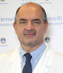 Eugenio Maria Mercuri合作论文数Department of Women, Children and Public Health Sciences, Fondazione Policlinico Universitario Agostino Gemelli IRCCS;Università Cattolica del Sacro Cuore Sede di Roma3
 Valerio Carelli合作论文数Laboratory of Neurogenetics, Department of Biomedical and Neuromotor Sciences, School of Medicine, University of Bologna3Claudio Bruno合作论文数Muscular and Neurodegenerative Disease Unit, Giannina Gaslini Institute3Claudia M. Cinnante合作论文数3Isabella Moroni合作论文数3
 Triulzi Fabio Maria合作论文数Dipartimento di Fisiopatologia Medico-Chirurgica e dei Trapianti, Università degli Studi di Milano3Martina Rimoldi合作论文数2Lucia Valente合作论文数2Manuela Garbellini合作论文数2
 Elena Abati合作论文数2Adele D'Amico合作论文数2
 Antonella Spinazzola合作论文数Department of Clinical and Movement Neurosciences, Queen Square Institute of Neurology, Faculty of Brain Sciences, University College London2
 Maria Francesca Bedeschi合作论文数Fondazione IRCCS Ca’ Granda Ospedale Maggiore Policlinico2
 Irene Faravelli合作论文数2
 Francesca Tagliavini合作论文数2
 Alessandro Santini合作论文数2
 Fabio Mosca合作论文数University of Milan;Fondazione IRCCS Ospedale Maggiore Policlinico2Graziella Uziel合作论文数Department of Child Neurology, Istituto Nazionale Neurologico “C. Besta”,2Lucia Morandi合作论文数2
 Massimo Zeviani合作论文数Department of Neurosciences, University of Padova;Veneto Institute for Molecular Medicine2




