The hallmarks of cancer, first proposed in 2000, have since provided a unified framework for understanding the complexity of carcinogenesis. This conceptual model has profoundly influenced the treatment landscape of primary liver cancer, which includes hepatocellular carcinoma (HCC, ∼85%) and intrahepatic cholangiocarcinoma (iCCA, 10%)—malignancies with high mortality. Key hallmarks exhibited by HCC include sustaining proliferative signaling, inducing or accessing vasculature, and avoiding immune detection. Over the past two decades, outcomes for patients with advanced HCC have significantly improved with immunotherapies. iCCA is characterized by hallmarks such as sustaining proliferative signaling, deregulating cellular metabolism, and avoiding immune detection. Unlike HCC, roughly 45% of iCCA harbor alterations amenable to precision oncology approaches, including fibroblast growth factor receptor 2 (FGFR2) fusions, isocitrate dehydrogenase 1 (IDH1) mutations, ERBB2 alterations, and BRAF mutations. In this review, we explore how this framework has reshaped liver cancer care and discuss the resulting breakthroughs in management and emerging directions that may further improve therapeutic strategies.
The incidence of breast cancer shows its largest peak around the age of 65 but also a peak around 45 indicating a bimodal distribution. While some potential explanations, such as age-related enrichment in specific breast cancer sub-types and BRCA1/2 mutations were proposed, they do not explain why the peaks are at 45 and 65, raising the possibility that the way the breast ages may play a role. Here using two mouse models, we show two distinct aging patterns of the mammary gland; one being progressive, the other bimodal. In the bimodal model, waves of overlapping genes and proteins associated with breast cancer pathways are observed at 11 and 19 months but are absent at 3 and 14 months. Further, in the bimodal model, the mammary glands at 11 and 19 months are more permissive to the growth of cancer cells but not at 14 or > 22 months, while in the progressive model, the growth of cancer cells increases starting at 14 months of age. Using scRNAseq, we established a bimodal aging signature. Since 11 and 19 months in mice correlate with 45 and 65 in humans, we tested the mammary gland-derived bimodal signature in two breast cancer databases and found that the signature is enriched in women diagnosed at 45 and 65. Therefore, our study raises the possibility that distinct patterns of aging of the breast exist and that they may contribute to the bimodal distribution of breast cancer. Further, our study adds to the growing evidence of non-linear aging.
Cholangiocarcinoma (CCA) is an adenocarcinoma of the hepatobiliary system that has recently risen in incidence and mortality with unsatisfactory treatment options. Claudin-1 (CLDN1) is a transmembrane protein expressed in tight junctions and exposed on the cell surface in liver fibrosis and cancer. Using single-cell RNA sequencing (scRNA-seq) and spatial transcriptomics of tissues of patients with CCA, we show that CLDN1 expression is up-regulated in cancer cells and is associated with stemness and cell fate. Genetic gain-of-function studies in CCA orthotopic in vivo mouse models showed decreased survival and enhanced tumor growth, unraveling a functional role of CLDN1 as an oncogenic driver. Targeting exposed nonjunctional CLDN1 using highly specific CLDN1 monoclonal antibodies (mAbs) inhibited tumor growth across CCA CDX and PDX mouse models and patient-derived CCA organoids, including tumors with medium or low CLDN1 expression. Moreover, antibody treatment inhibited tumor cell migration, invasion, and extrahepatic metastasis. Mechanistically, targeting exposed cell surface CLDN1 on CCA tumors using mAbs inhibited Notch1 and TROP2/STAT3 signaling pathways, resulting in decreased cancer cell stemness and epithelial-to-mesenchymal transition. Loss-of-function studies using CRISPR-Cas9 and RNAi combined with rescue and confocal imaging studies confirmed the functional and mechanistic role of these pathways. In conclusion, these results uncover CLDN1 as a previously undiscovered CCA driver and therapeutic target, paving the way for the clinical development of CLDN1 mAbs to improve the dismal outcome of patients with advanced CCA.
Non-linear aging, identified through oscillations in human plasma proteomics around 44 and 60, is postulated to contribute to the onset of diseases. Incidence of breast cancer follows such bimodal pattern. We show that mitochondria-nucleus matching in mice results in two mammary gland aging patterns, including a bimodal pattern associated with susceptibility to mammary tumor and that this bimodal signature is enriched in breast cancer patients diagnosed at 45 and 65.
Abstract Background and Aims: Atezolizumab (anti-PDL1) plus bevacizumab (anti-VEGFA) (atezo+bev) is one first-line standard-of-care (SOC) treatment for advanced hepatocellular carcinoma (aHCC) with objective responses in ∼30% of cases. Clinical benefit is hindered in non-viral HCC, including metabolic dysfunction-associated steatohepatitis (MASH)-related cases, a raising HCC etiology. MASH-HCC tumors are enriched in TGFß signaling, which has been associated with resistance to atezo+bev. The bioavailability and the Treg-related immunosuppressive effect of the TGFß1 ligand is controlled by the TGFβ-anchoring receptor GARP. Hence, we hypothesize that GARP blockade might revert immune suppression and overcome resistance to aPDL1+aVEGF (SOC) in MASH-HCC. Method: We used an immune-genetic MASH-HCC model that combines diet-induced steatohepatitis with MYC and CTNNB1 overexpression (via hydrodynamic gene delivery), previously shown to drive atezo+bev resistance. After tumor onset, animals were treated with SOC+anti-GARP (triple combination), SOC alone or placebo until the end of the study. Spectral cytometry was conducted at day 14 to assess changes in the tumor microenvironment (n=5/arm). Remaining animals were followed for overall survival (OS) analysis (n=7/arm). GARP expression was correlated with molecular/immune features in three independent cohorts of HCC patients (n=559). Results: SOC+anti-GARP significantly extended OS compared to placebo (median of 68 vs 59 days, p=0.045), as opposed to SOC alone (p=0.21). From the immune perspective, the triple combination induced a shift from central memory (CD62L+CD127+) to tumor-resident (CD62L-CD69+) CD8+ T cells (p=0.031) accompanied by an increase in total CD8+ (p=0.0028) and CD4+ (p=0.016) T cells compared to placebo, suggesting a more locally active T-cell response. Accordingly, tumors from SOC+anti-GARP-treated mice presented increased infiltration of both effector (CD44+PD1+TIM3-) (p=0.036) and exhausted (PD1+TIM3+) CD8+ T cells (p=0.007). Among CD4+ T cells, the triple combination also increased antigen-experienced CD44+ (p=0.045) and showed a trend of the ratio between tissue-resident memory (CD69+CD103+) and Tregs (CD25+FOXP3+) (p=0.053) when compared to the rest. None of these changes were observed with SOC alone. In HCC patients, high GARP expression significantly correlated with immune exhaustion (fold-change vs rest >1.5 in 87% of cases), with an increase of Treg infiltration compared with other patients, highlighting a subset of patients that may benefit from this combination. Conclusions: Our findings highlight GARP as a promising therapeutic target whose blockade restores antitumor immune infiltration and extends survival OS when added to SOC in an immunotherapy-resistant murine model of MASH-HCC. Citation Format: Julia Huguet-Pradell, David Camell-Raventos, Elisa Fernández-Martínez, Anthony Lozano, Tiago de Castro, Marcus Zeitlhoefler, Ugne Balaseviciute, Albert Gris-Oliver, Daniela Sia, Roser Pinyol, Josep M Llovet. Anti-GARP reverts immune suppression and extends overall survival in a MASH-HCC murine model resistant to anti-PDL1+anti-VEGFA therapy [abstract]. In: Proceedings of the American Association for Cancer Research Annual Meeting 2026; Part 2 (Late-Breaking, Clinical Trial, and Invited Abstracts); 2026 Apr 17-22; San Diego, CA. Philadelphia (PA): AACR; Cancer Res 2026;86(8_Suppl):Abstract nr LB154.
Intrahepatic cholangiocarcinoma (iCCA) is one of the deadliest malignancies, with an overall 5-year survival rate of approximately 10%. For decades, surgery and chemotherapy have represented the only treatment options for early- and late-stage disease, respectively. More recently, characterisation of the genomic landscape of iCCA has identified several "druggable" oncogenic drivers and led to the FDA approval of the first targeted therapies, including FGFR and IDH inhibitors, for second-line treatment in genetically defined patient subsets. Nonetheless, most patients treated with these therapies rapidly develop resistance and eventually experience disease progression. At a time when immune checkpoint inhibitors (ICIs) are profoundly reshaping cancer treatment, their use in combination with chemotherapy has yielded only modest survival benefits in iCCA, with more than two-thirds of patients exhibiting intrinsic resistance. The aggressive and refractory nature of this cancer is often attributed to its intricate tumour microenvironment (TME); however, the complex interplay between tumour cells and other TME components (i.e. immune cells, cancer-associated fibroblasts, and endothelial cells), as well as the molecular and cellular mechanisms driving tumour progression and therapeutic resistance, remain poorly understood. In this review, we discuss the critical role of the stromal and immune TME in iCCA and how its characterisation has informed patient stratification and molecular classification. We also describe recent findings supporting distinct genotype-immunophenotype relationships in iCCA, as well as the existence of functionally heterogeneous subsets of cancer-associated fibroblasts. Ultimately, this review aims to provide a comprehensive overview of current knowledge while stimulating discussion on therapeutic implications and future research directions.
Abstract Tumor-reactive T cells (TRTs) are critical for anti-tumor immunity but are incompletely captured by current assays, which fail to reproduce tumor-specific antigen diversity. Here, we show that multiplex functional profiling of patient-derived tumor organoid-T cell co-cultures (PDOTs) enables robust identification of TRTs across CD8, CD4, and double-negative (DN) T cell populations. Single activation markers underestimated TRT responses, whereas integrated analysis revealed broader functional repertoire. MHCI blockade abrogated CD8 and DN TRT responses while preserving CD4 reactivity, supporting antigen-dependent recognition across T cell lineages. Tumor PDO expressed MHCI and MHCII, and PDOTs enabled generation and detection of TRTs from peripheral blood. PD1 blockade induced heterogeneous responses, enhancing CD8 and DN activity and unexpectedly augmenting CD4 reactivity. PDOTs further identified additional inhibitory pathways whose therapeutic targeting in combination with PD1 blockade increased TRT responses. These findings establish PDOTs as a platform to identify TRTs and functionally stratify patient-specific tumor-T cell responses to checkpoint immunotherapy.
Abstract Background Intrahepatic cholangiocarcinoma (iCCA) is a rare, but deadly biliary tract cancer, increasing in incidence globally. One in five patients harbor a genetic activation of FGFR2 (fibroblast growth factor 2), where gene fusions enhance neoplastic growth. Inhibitors of FGFRs have displayed sub-optimal results—patients with advanced stage iCCA acquire drug resistance. Therefore, novel, alternative therapeutics for FGFR-driven iCCAs are crucial. A major limitation is that current preclinical models do not mimic the immunobiology of human disease. Methods We generated a mouse model harboring the FGFR2-PPHLN1 fusion (or its murine homolog mFgfr2-Pphln1) and activated YAP1 via hydrodynamic tail vein injection, and established cell lines and organoids. Additionally, the V565F resistance mutation was introduced in the FGFR2 fusion gene. We characterized tumors using RNA-sequencing (n = 3 per group) and spectral cytometry (n = 3-7 per group) and performed cross-species analysis using published human datasets [OEP001105, (n = 262); GSE33327, (n = 149)] and molecular characterization by GSEA. In vitro and in vivo drug treatment with reversible and irreversible FGFR inhibitors was performed. Results FGFR2-driven murine tumors were molecularly and immunologically similar to human iCCA carrying FGFR2 alterations, with accumulation of tumor-associated neutrophils (TANs, 60% of all CD45+) and impaired T cell infiltration. Organoids reproduced clinical responses to reversible and irreversible FGFR inhibitors, while also showing a sustained response to the reversible inhibitor derazantinib in the FGFR2V565F-PPHLN1 organoids. Mice exhibited response to derazantinib but no synergy was observed when combined with immune checkpoint inhibitor PD-L1. Conclusion Our mouse model and it’s in vitro derivatives recapitulate the immunobiology and therapeutic sensitivities of human disease. Inter-species and cross-species analyses suggest a critical role of FGFR2 fusions in shaping a cold tumor microenvironment with accumulation of TANs in both humans and mice. Poor sensitivity to ICI in vivo suggests the need for novel therapeutic strategies harnessing the immunosuppressive nature of these tumors.
Abstract Background: Atezolizumab+bevacizumab (atezo+bev) is a first-line therapy for advanced hepatocellular carcinoma (aHCC) with ∼30% objective responses. Single-cell studies identified responses linked to CD8⁺ T effector cells (CD8_Tex and Temra) and CXCL10⁺ macrophages (Macro_CXCL10), and resistance to TREM2⁺ macrophages (Macro_TREM2) and CD14⁺ monocytes (Mono_CD14). We hypothesized that, beyond cell composition, immune spatial organization within the tumor microenvironment (TME) determines response to atezo+bev. Methods: Single-cell spatial transcriptomics (ST) was performed on 27 aHCCs (9 atezo+bev responders and 18 resistant) using the Xenium Prime 5.000-gene panel (10x Genomics) customized with 100 additional genes. For local tissue organization, distances between cells were computed using the dbscan package [radius(r)=300µm]. Immune cell clusters were defined as groups of ≥3 immune cells within 100 µm, and were identified through a density-based algorithm. This spatial clustering was assessed using both ST and multiplexed immunohistochemistry (mIHC) data. Cell-to-cell interactions (neighborhoods) were analyzed (r=25µm). Ligand-receptor interactions were evaluated using a spatial neighborhood-based approach. Results: ST of 27 aHCCs yielded 4.6 million cells. In responders, CD8_Tex and Temra, and Macro_CXCL10 cells localized within the tumor and were closer to cancer cells (p=0.02, p=0.08, and p=0.1, respectively). Furthermore, in responders, tumor hepatocytes exhibited a significantly enriched lymphoid neighborhood composition and significantly higher rates of immune-recruting interactions, including PDL1-PD1, CXCL9-CXCR3 and CXCL10-CXCR3 (p<0.05). Non-responders were enriched in TREM2⁺ macrophage-containing clusters. Specifically, they showed Macro_TREM2-pure clusters (10% vs 0%, p=0.009), mixed Macro_TREM2-Mono_CD14 clusters (27% vs 5%, p=0.03), and mixed Macro_TREM2-CD8⁺ T-cell clusters (38% vs 22%, p=0.04). This was accompanied by enrichment of the CXCL12-CXCR4 chemokine axis, consistent with an immunosuppressive TME. Using mIHC, we confirmed enrichment of Macro_TREM2-pure (p=0.02) and Macro_TREM2-CD8⁺ mixed clusters (p=0.03). Conclusions: Atezo+bev response in HCC is marked by intratumoral effector CD8⁺ T cells and CXCL10_Macro, tumor hepatocytes enriched in lymphoid neighborhoods, and immune-recruiting ligand-receptor interactions, whereas resistance is driven by TREM2_Macro clusters and CXCL12-CXCR4 signaling. These findings identify spatial immune organization as a key determinant of response to atezo+bev. Citation Format: Anna Vila-Escoda, Marta Piqué-Gili, Marta Casado-Pelaez, Roser Pinyol, Ana Hernández de Sande, Verónica Davalos, Albert Gris-Oliver, Carla Montironi, Judit Peix, Daniela Grases, Ezequiel Mauro, Guillem Cano-Segarra, Sarah Cappuyns, Igor Figueiredo, Giorgio Ioannou, Edgar Gonzalez-Kozlova, Tim Meyer, Anja Lachenmayer, Jens U Marquardt, Helen L Reeves, Julien Edeline, Fabian Finkelmeier, Jörg Trojan, Sacha Gnjatic, Jean-Frederic Blanc, Richard A Hubner, Matthias Pinter, Tom Luedde, Arndt Vogel, Daniela Sia, Vincenzo Mazzaferro, Manel Esteller, Jeroen Dekervel, Eduard Porta-Pardo, Josep M Llovet. Spatial immune architecture at single-cell resolution predicts response to atezolizumab plus bevacizumab in advanced HCC [abstract]. In: Proceedings of the American Association for Cancer Research Annual Meeting 2026; Part 2 (Late-Breaking, Clinical Trial, and Invited Abstracts); 2026 Apr 17-22; San Diego, CA. Philadelphia (PA): AACR; Cancer Res 2026;86(8_Suppl):Abstract nr LB123.
Steatotic liver diseases (SLDs), including metabolic dysfunction-associated steatotic liver disease (MASLD; formerly known as non-alcoholic fatty liver disease) and alcoholic liver disease, are the leading causes of chronic hepatitis, liver dysfunction, cirrhosis and liver cancer development. Severe manifestations of MASLD and alcoholic liver disease include metabolic dysfunction-associated steatohepatitis (MASH; formerly known as non-alcoholic steatohepatitis) and alcoholic steatohepatitis (ASH) or a combination thereof, termed metabolic dysfunction and alcohol-associated liver disease. While MASH-associated and ASH-associated liver cancers display common histopathological features, the underlying cellular and molecular pathophysiological mechanisms of each are distinct. Recent studies indicate that SLDs encompass previously unrecognized spectra of heterogeneous, metabolic, immunological and genetic diseases that, in combination with lifestyle measures and individual patient comorbidities, affect disease pathogenesis and therapy response. Here, we review the current knowledge of the molecular, genetic and cellular mechanisms underlying the transition of MASH or ASH to liver cancer as well as novel developments in liver cancer risk assessment in SLDs. We further discuss possible obstacles of differential diagnosis and outline current developments in the therapeutic management of both MASH-related and ASH-related liver cancer.
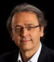 Josep M. Llovet合作论文数Faculty of Medicine, University of Barcelona111
 Swan N Thung合作论文数Icahn School of Medicine, Mount Sinai49
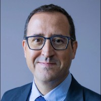 Augusto Villanueva合作论文数NYU Langone Health48
 Roser Pinyol合作论文数43
 Myron E Schwartz合作论文数Recanati/Miller Transplantation Institute, Mount Sinai;The Mount Sinai Medical Center;Icahn School of Medicine at Mount Sinai38
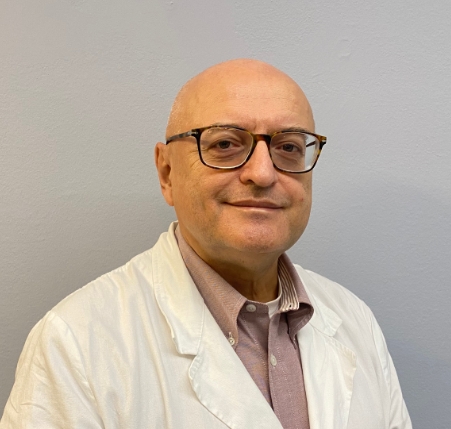 Mazzaferro Vincenzo Maria合作论文数Department of Oncology and Hematology-Oncology, Università degli Studi di Milano37Miguel Torres-Martin合作论文数32Moeini Agrin合作论文数30Judit Peix合作论文数29Yujin Hoshida合作论文数28Carla Montironi合作论文数Institut d'Investigacions Biomèdiques August Pi i Sunyer (IDIBAPS),, Universitat de Barcelona25Sara Torrecilla合作论文数24
 Sasan Roayaie合作论文数White Plains Hospital24
 Helena Cornella合作论文数22
 Amaia Lujambio合作论文数Icahn Sch Med Mt Sinai, Dept Oncol Sci, New York, NY 10029 USA20Laia Cabellos合作论文数20
 Genis Camprecios合作论文数18
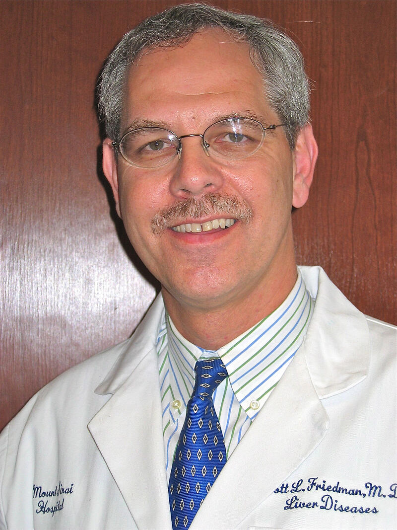 Scott L. Friedman合作论文数Division of Liver Diseases, Mount Sinai School of Medicine18Philipp K Haber合作论文数17
 Zhang Zhongyang合作论文数Icahn Institute for Genomics and Multiscale Biology, Icahn School of Medicine at Mount Sinai16Victoria Tovar合作论文数16Miho Maeda合作论文数15Roger Esteban-Fabró合作论文数Liver Unit, Hosp Clin Barcelona15Carmen Andreu-Oller合作论文数15
 Alsinet Clara合作论文数HCC Translat Res Lab, Univ Barcelona15
 Wei-Qiang Leow合作论文数Department of Pathology, Singapore General Hospital15Katherine E. Lindblad合作论文数14
 Bassaganyas Laia合作论文数14Bojan Losic合作论文数Ambry Genetics14Puigvehí M合作论文数14Anja Lachenmayer合作论文数14
 Marina Barcena-Varela合作论文数Icahn Sch Med Mt Sinai, Div Liver Dis, Dept Med, Tisch Canc Inst,Liver Canc Program, New York, NY 10029 USA13
 Laura Torrens合作论文数Institut d'Investigacions Biomèdiques August Pi i Sunyer (IDIBAPS),, Universitat de Barcelona12
 Andrew V Uzilov合作论文数Sema4, Stamford, CT USA12
 Ke Hao (郝柯)合作论文数College of Environmental Science and Engineering, Tongji University12Robert Montal合作论文数11Fiel M Isabel合作论文数11
 Jordi Bruix合作论文数Barcelona Clinic Liver Cancer Group, Hospital Clinic of Barcelona;University of Barcelona11Beatriz Minguez合作论文数Tisch Cancer Institute, Transplant Institute11
 Oriana Miltiadous合作论文数10Jordi Abril-Fornaguera合作论文数Institut d'Investigacions Biomèdiques August Pi i Sunyer (IDIBAPS),, Universitat de Barcelona10Iris Martinez-Quetglas合作论文数10
 Carlos Villacorta-Martin合作论文数Boston Univ, Ctr Regenerat Med, Boston, MA 02118 USA10
 Ismail Labgaa合作论文数Department of Visceral Surgery, CHUV University Hospital10Manel Solé合作论文数Clínic Barcelona10Patricia Taik合作论文数9Marta Piqué-Gili合作论文数9
 Nicholas K Akers合作论文数Icahn Sch Med Mt Sinai, New York, NY 10029 USA9
 Manel Esteller合作论文数Cancer Epigenetics Group, Josep Carreras Leukaemia Research Institute;Faculty of Medicine and Health Sciences, University of Barcelona;Catalan Institution for Research and Advanced Studies9




