Membrane lipid regulation of cell function is poorly understood. In early development, sterol efflux and the ganglioside GM1 regulate sperm acrosome exocytosis (AE) and fertilization competence through unknown mechanisms. Here, we show that sterol efflux and focal enrichment of GM1 trigger Ca2+ influx necessary for AE through CaV2.3, whose activity has been highly controversial in sperm. Sperm lacking CaV2.3’s pore-forming α1E subunit showed altered Ca2+ responses, reduced AE, and a strong subfertility phenotype. Surprisingly, AE depended on spatiotemporal information encoded by flux through CaV2.3, not merely the presence/amplitude of Ca2+ waves. Using studies in both sperm and voltage clamp of Xenopus oocytes, we define a molecular mechanism for GM1/CaV2.3 regulatory interaction, requiring GM1’s lipid and sugar components and CaV2.3’s α1E and α2δ subunits. Our results provide a mechanistic understanding of membrane lipid regulation of Ca2+ flux and therefore Ca2+-dependent cellular and developmental processes such as exocytosis and fertilization.
Despite enormous interest in membrane raft micro-domains, no studies in any cell type have defined the relative compositions of the raft fractions on the basis of their major components--sterols, phospholipids, and proteins--or additional raft-associating lipids such as the ganglioside, G(M1). Our previous localization data in live sperm showed that the plasma membrane overlying the acrosome represents a stabilized platform enriched in G(M1) and sterols. These findings, along with the physiological requirement for sterol efflux for sperm to function, prompted us to characterize sperm membrane fractions biochemically. After confirming limitations of commonly used detergent-based approaches, we utilized a non-detergent-based method, separating membrane fractions that were reproducibly distinct based on sterol, G(M1), phospholipid, and protein compositions (both mass amounts and molar ratios). Based on fraction buoyancy and biochemical composition, we identified at least three highly reproducible sub-types of membrane raft. Electron microscopy revealed that raft fractions were free of visible contaminants and were separated by buoyancy rather than morphology. Quantitative proteomic comparisons and fluorescence localization of lipids suggested that different organelles contributed differentially to individual raft sub-types, but that multiple membrane micro-domain sub-types could exist within individual domains. This has important implications for scaffolding functions broadly associated with rafts. Most importantly, we show that the common practice of characterizing membrane domains as either "raft" or "non-raft" oversimplifies the actual biochemical complexity of cellular membranes.
We demonstrate for the first time that a stable, micron‐scale segregation of focal enrichments of sterols exists at physiological temperature in the plasma membrane of live murine and human sperm. These enrichments of sterols represent microheterogeneities within this membrane domain overlying the acrosome. Previously, we showed that cholera toxin subunit B (CTB), which binds the glycosphingolipid, GM1, localizes to this same domain in live sperm. Interestingly, the GM1 undergoes an unexplained redistribution upon cell death. We now demonstrate that GM1 is also enriched in the acrosome, an exocytotic vesicle. Transfer of lipids between this and the plasma membrane occurs at cell death, increasing GM1 in the plasma membrane without apparent release of acrosomal contents. This finding provides corroborative support for an emerging model of regulated exocytosis in which membrane communications might occur without triggering the “acrosome reaction.” Comparison of the dynamics of CTB‐bound endogenous GM1 and exogenous BODIPY–GM1 in live murine sperm demonstrate that the sub‐acrosomal ring (SAR) functions as a specialized diffusion barrier segregating specific lipids within the sperm head plasma membrane. Our data show significant differences between endogenous lipids and exogenous lipid probes in terms of lateral diffusion. Based on these studies, we propose a hierarchical model to explain the segregation of this sterol‐ and GM1‐enriched domain in live sperm, which is positioned to regulate sperm fertilization competence and mediate interactions with the oocyte. Moreover, our data suggest potential origins of subtypes of membrane raft microdomains enriched in sterols and/or GM1 that can be separated biochemically. J. Cell. Physiol. 218: 522–536, 2009. © 2008 Wiley‐Liss, Inc.
We previously showed that in live murine and bovine sperm heads, the ganglioside GM1 localizes to the sterol-rich plasma membrane overlying the acrosome (APM). Labeling GM1 using the pentameric cholera toxin subunit B (CTB) induced a dramatic redistribution of signal from the APM to the sterol-poor post-acrosomal plasma membrane (PAPM) upon sperm death. We now show a similar phenomenon in the flagellum where CTB induced GM1 redistribution to sterol-poor membrane sub-domains of the annulus and flagellar zipper. Because sterol efflux from the plasma membrane is required for capacitation, we next examined whether GM1 localization might be useful to detect membrane changes associated with capacitation and/or acrosomal exocytosis. First, incubation of murine sperm with their respective stimuli for capacitation did not change GM1 distribution in live cells. However, incubation with specific stimuli for capacitation, followed by the use of specific fixation conditions, induced reproducible, stimulus-specific patterns of GM1 distribution. By assessing changes in GM1 distribution in response to progesterone-induced acrosomal exocytosis, we show that these patterns reflect the response of sperm populations to capacitating stimuli. These data suggest that GM1 localization can be used as a diagnostic tool for evaluating sperm response to stimuli for capacitation and/or acrosomal exocytosis. Such information could be useful when deciding between technologies of assisted reproduction, or when screening for male fertility. Furthermore, stimulus-specific changes in GM1 distribution showed that sperm could respond to NaHCO3 or mediators of sterol efflux independently, thereby refining existing models of capacitation. (poster)
We previously showed that in live murine and bovine sperm heads, the ganglioside G(M1) localizes to the sterol-rich plasma membrane overlying the acrosome (APM). Labeling G(M1) using the pentameric cholera toxin subunit B (CTB) induced a dramatic redistribution of signal from the APM to the sterol-poor postacrosomal plasma membrane (PAPM) upon sperm death. We now show a similar phenomenon in the flagellum where CTB induces G(M1) redistribution to sterol-poor membrane subdomains of the annulus and flagellar zipper. Because sterol efflux from the plasma membrane is required for capacitation, we examined whether G(M1) localization might be useful to detect membrane changes associated with capacitation and/or acrosomal exocytosis. First, incubation of murine and bovine sperm with their respective stimuli for capacitation did not change G(M1) distribution in live cells. However, incubation of sperm of both species with specific stimuli for capacitation, followed by the use of specific fixation conditions, induced reproducible, stimulus-specific patterns of G(M1) distribution. By assessing changes in G(M1) distribution in response to progesterone-induced AE, we show that these patterns reflect the response of murine sperm populations to capacitating stimuli. These data suggest that G(M1) localization can be used as a diagnostic tool for evaluating sperm response to stimuli for capacitation and/or AE. Such information could be useful when deciding between technologies of assisted reproduction or when screening for male fertility. Furthermore, stimulus-specific changes in G(M1) distribution showed that sperm could respond to NaHCO(3) or mediators of sterol efflux independently, thereby refining existing models of capacitation.
Lipid rafts, membrane sub-domains enriched in sterols and sphingolipids, are controversial because demonstrations of rafts have often utilized fixed cells. We showed in living sperm that the ganglioside G(M1) localized to a micron-scale membrane sub-domain in the plasma membrane overlying the acrosome. We investigated four models proposed for membrane sub-domain maintenance. G(M1) segregation was maintained in live sperm incubated under non-capacitating conditions, and after sterol efflux, a membrane alteration necessary for capacitation. The complete lack of G(M1) diffusion to the post-acrosomal plasma membrane (PAPM) in live cells argued against the transient confinement zone model. However, within seconds after cessation of sperm motility, G(M1) dramatically redistributed several microns from the acrosomal sub-domain to the post-acrosomal, non-raft sub-domain. This redistribution was not accompanied by movement of sterols, and was induced by the pentameric cholera toxin subunit B (CTB). These data argued against a lipid-lipid interaction model for sub-domain maintenance. Although impossible to rule out a lipid shell model definitively, mice lacking caveolin-1 maintained segregation of both sterols and G(M1), arguing against a role for lipid shells surrounding caveolin-1 in sub-domain maintenance. Scanning electron microscopy of sperm freeze-dried without fixation identified cytoskeletal structures at the sub-domain boundary. Although drugs used to disrupt actin and intermediate filaments had no effect on the segregation of G(M1), we found that disulfide-bonded proteins played a significant role in sub-domain segregation. Together, these data provide an example of membrane sub-domains extreme in terms of size and stability of lipid segregation, and implicate a protein-based membrane compartmentation mechanism.
The organization of membrane subdomains in mammalian sperm has recently generated controversy, with several reports describing widely differing localization patterns for the ganglioside GM1. Using the pentameric B subunit of cholera toxin (CTB), we found GM1 to be restricted to the plasma membrane overlying the acrosome in the heads of live murine sperm. Interestingly, CTB had minimal binding to live bovine and human sperm. To investigate whether this difference in GM1 localization was because of species differences or differences between collection from the epididymis (mouse) or an ejaculate (bull, human), we examined epididymal bovine and human sperm. We found that GM1 localized to the plasma membrane overlying the acrosome in sperm from these species. To determine whether some component of seminal plasma was interfering with the ability of CTB to access GM1, we incubated epididymal mouse sperm with fluid from murine seminal vesicles and epididymal bull sperm with bovine seminal plasma. This treatment largely abolished the ability of the CTB to bind to GM1, producing a fluorescence pattern similar to that reported for the human. The most abundant seminal plasma protein, PDC-109, was not responsible for this loss. As demonstration that the seminal plasma was not removing GM1, sperm exposed to seminal plasma were fixed before CTB addition, and again displayed fluorescence over the acrosome. These observations reconcile inconsistencies reported for the localization of GM1 in sperm of different species, and provide evidence for the segregation of GM1 to a stable subdomain in the plasma membrane overlying the acrosome.
 Alexander Travis合作论文数Center for Sustainable Global Enterprise, Cornell SC Johnson College of Business, Cornell University7
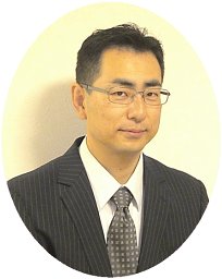 Atsushi Asano合作论文数Dept Appl Chem, Natl Def Acad6
 Jacquelyn L. Nelson合作论文数6
 Vimal Selvaraj合作论文数College of Agriculture and Life Sciences, Cornell University5Gary R. Hunnicutt合作论文数3Klaus Angela V合作论文数2
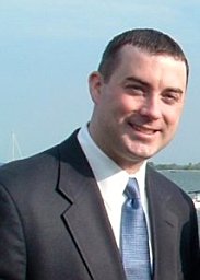 John L. Mcelwee合作论文数Cornell University, University of Pennsylvania Medical Center2Collin A. Wolff合作论文数2James E. Evans合作论文数1Karin M. Green合作论文数1
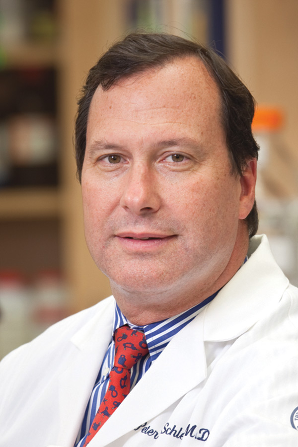 Peter N. Schlegel合作论文数New York Men’s Health Medical, PLLC1
 Robert Weiss合作论文数Biomedical Sciences, College of Veterinary Medicine, Cornell University1
 Richard Joel Miller合作论文数Department of Pharmacology, Feinberg School of Medicine, Northwestern University1
 Chinatsu Mukai合作论文数1
 Moshe Cohen-Kutner合作论文数1
 Daphne Atlas合作论文数Department of Biological Chemistry, The Hebrew University of Jerusalem1
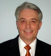 Gregory S. Kopf合作论文数FHI 3601
 Roy Cohen合作论文数The Institute of Life Sciences and the Otto Loewi Center, The Hebrew University of Jerusalem1Alex W. Cohen合作论文数1
 George H. Rothblat合作论文数Division of Gastroenterology, Hepatology and Nutrition, The Children's Hospital of Philadelphia1Dongjun Ren合作论文数1Tanya Merdiushev合作论文数1Miguel W. Fornés合作论文数1
 Prabuddha Sengupta合作论文数FDA1
 Michael Lisanti合作论文数School of Science, Engineering and Environment, University of Salford1




