Fig.S1 effects of PRH KD Fig.S2 effects of PRH OE Fig.S3 Gene expression and gene ontology Fig.S4 Heat maps Fig.S5 ChIP sequencing Fig.S6 Dose response curves Fig. S7 Western blotting Rb expression
Abstract Aberrant Notch and Wnt signaling are known drivers of cholangiocarcinoma (CCA), but the underlying factors that initiate and maintain these pathways are not known. Here, we show that the proline-rich homeodomain protein/hematopoietically expressed homeobox (PRH/HHEX) transcription factor forms a positive transcriptional feedback loop with Notch3 that is critical in CCA. PRH/HHEX expression is elevated in CCA, and depletion of PRH reduces CCA tumor growth in a xenograft model. Overexpression of PRH in primary human biliary epithelial cells is sufficient to increase cell proliferation and produce an invasive phenotype. Interrogation of the gene networks regulated by PRH and Notch3 reveals that unlike Notch3, PRH directly activates canonical Wnt signaling. These data indicate that hyperactivation of Notch and Wnt signaling is independent of the underlying mutational landscape and has a common origin in dysregulation of PRH. Moreover, they suggest new therapeutic options based on the dependence of specific Wnt, Notch, and CDK4/6 inhibitors on PRH activity. Significance: The PRH/HHEX transcription factor is an oncogenic driver in cholangiocarcinoma that confers sensitivity to CDK4/6 inhibitors.
Supramolecular signaling assemblies are of interest for their unique signaling properties. A µm scale signaling assembly, the central supramolecular signaling cluster (cSMAC), forms at the center of the interface of T cells activated by antigen-presenting cells. We have determined that it is composed of multiple complexes of a supramolecular volume of up to 0.5 µm3 and associated with extensive membrane undulations. To determine cSMAC function, we have systematically manipulated the localization of three adaptor proteins, LAT, SLP-76, and Grb2. cSMAC localization varied between the adaptors and was diminished upon blockade of the costimulatory receptor CD28 and deficiency of the signal amplifying kinase Itk. Reconstitution of cSMAC localization restored IL-2 secretion which is a key T cell effector function as dependent on reconstitution dynamics. Our data suggest that the cSMAC enhances early signaling by facilitating signaling interactions and attenuates signaling thereafter through sequestration of a more limited set of signaling intermediates.
Introduction Proline-Rich Homeodomain protein/Haematopoietically Expressed Homeobox (PRH/HHEX) is a transcription factor that regulates cell proliferation, migration, and differentiation in multiple tissues. PRH is essential for embryonic development of the liver and bile ducts and has tumour suppressor activity in hepatocellular carcinoma (HCC). Mis-regulation of PRH is associated with several cancers including HCC, breast cancer, prostate cancer, and leukaemia. Our objective is to determine whether PRH has a role in cholangiocarcinoma (cancer of the bile duct). Material and methods PRH was over-expressed in cholangiocarcinoma (CCA) cell lines and in primary bile duct epithelial cells (BECs) using an adenovirus expressing myc-tagged PRH and knockdown of PRH was achieved by stable expression of PRH shRNA. Cell proliferation was measured by EdU incorporation. RNA sequencing (RNA-seq) was used to determine genes and pathways regulated by PRH in CCA. Quantitative RT-PCR (qPCR) was used to confirm these changes and Western blotting and immunohistochemistry was used to examine the expression and localisation of the corresponding proteins. Mining of TCGA transcriptomics and genomics data was performed using UCSC Xena. Results and discussions Western blotting and immunohistochemical staining reveals that PRH protein is elevated in CCA compared to cholangiocytes and TCGA expression data indicate that PRH mRNA is commonly upregulated in CCA. Over-expression (OE) of PRH increases the proliferation of CCLP1 and CCSW1 CCA cell lines and BECs. Conversely, PRH KD decreases CCLP1 proliferation and alters the morphological phenotype of CCLP1 cells from mesenchymal to epithelial. Xenograft experiments with CCLP1 PRH knockdown (KD) cells compared to control cells shows that depletion of PRH significantly decreases tumour growth. Gene ontology and gene set enrichment analysis of RNA-seq data from CCLP1 PRH KD and OE cells indicates differential expression of genes involved in proliferation, adhesion, and migration including genes associated with Wnt signalling, and epithelial- mesenchymal transition (EMT). Western blotting confirms changes in protein expression in the KD cells in accord with the RNA-seq data. Examination of β-catenin subcellular localisation shows that PRH KD cells have decreased nuclear β-catenin. Conclusion Our data suggest that the PRH protein is intimately involved in the development of CCA.
Notch is a critical regulator of T cell differentiation and is activated through proteolytic cleavage in response to ligand engagement. Using murine myelin-reactive CD4 T cells, we demonstrate that proximal T cell signaling modulates Notch activation by a spatiotemporally constrained mechanism. The protein kinase PKCθ is a critical mediator of signaling by the T cell antigen receptor and the principal costimulatory receptor CD28. PKCθ selectively inactivates the negative regulator of F-actin generation, Coronin 1A, at the center of the T cell interface with the antigen presenting cell (APC). This allows for effective generation of the large actin-based lamellum required for recruitment of the Notch-processing membrane metalloproteinase ADAM10. Such enhancement of Notch activation is critical for efficient T cell proliferation and Th17 differentiation. We reveal a novel mechanism that, through modulation of the cytoskeleton, controls Notch activation at the T cell:APC interface thereby linking T cell receptor and Notch signaling pathways.
Fluorescence microscopy is one of the most important tools in cell biology research because it provides spatial and temporal information to investigate regulatory systems inside cells. This technique can generate data in the form of signal intensities at thousands of positions resolved inside individual live cells. However, given extensive cell-to-cell variation, these data cannot be readily assembled into three- or four-dimensional maps of protein concentration that can be compared across different cells and conditions. We have developed a method to enable comparison of imaging data from many cells and applied it to investigate actin dynamics in T cell activation. Antigen recognition in T cells by the T cell receptor (TCR) is amplified by engagement of the costimulatory receptor CD28. We imaged actin and eight core actin regulators to generate over a thousand movies of T cells under conditions in which CD28 was either engaged or blocked in the context of a strong TCR signal. Our computational analysis showed that the primary effect of costimulation blockade was to decrease recruitment of the activator of actin nucleation WAVE2 (Wiskott-Aldrich syndrome protein family verprolin-homologous protein 2) and the actin-severing protein cofilin to F-actin. Reconstitution of WAVE2 and cofilin activity restored the defect in actin signaling dynamics caused by costimulation blockade. Thus, we have developed and validated an approach to quantify protein distributions in time and space for the analysis of complex regulatory systems.
Dynamic subcellular distributions of signaling system components are critical regulators of cellular signal transduction through their control of molecular interactions. Understanding how signaling activity depends on such distributions and the cellular structures driving them is required for comprehensive insight into signal transduction. In the activation of primary murine T cells by antigen presenting cells (APC) signaling intermediates associate with various subcellular structures, prominently a transient, wide, and actin-associated lamellum extending from an interdigitated T cell:APC interface several micrometers into the T cell. While actin dynamics are well established as general regulators of cellular organization, their role in controlling signaling organization in primary T cell:APC couples and the specific cellular structures driving it is unresolved. Using modest interference with actin dynamics with a low concentration of Jasplakinolide as corroborated by costimulation blockade we show that T cell actin preferentially controls lamellal signaling localization and activity leading downstream to calcium signaling. Lamellal localization repeatedly related to efficient T cell function. This suggests that the transient lamellal actin matrix regulates T cell signaling associations that facilitate T cell activation.
 Ka Ying Lee合作论文数4Sheela Jayaraman合作论文数4
 Kevin Gaston合作论文数School of Medicine, Faculty of Medicine and Health Sciences, The University of Nottingham4
 Jomnarong Lertsuwan合作论文数4Christoph Wuelfing合作论文数4
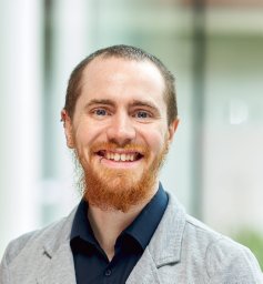 Philip Kitchen合作论文数Molecular Organisation and Assembly in Cells Doctoral Training Centre, University of Warwick4
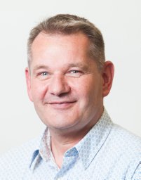 Paul Verkade合作论文数School of Medical Sciences, University Walk4
 Sebastian Oltean合作论文数Inst Biomed & Clin Sci, Univ Exeter4Jutamaad Satayavivad合作论文数4
 Anyaporn Sawasdichai合作论文数3Nikki Lau合作论文数3
 Simon Afford合作论文数Institute of Immunology and Immunotherapy, College of Medical and Dental Sciences, University of Birmingham3Xiongtao Ruan合作论文数2Rachel Ambler合作论文数2
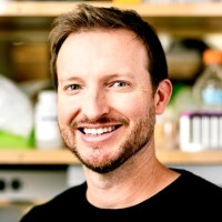 Kole Roybal合作论文数Department of Microbiology and Immunology, School of Medicine, University of California, San Francisco;Parker Institute for Cancer Immunotherapy, University of California, San Francisco2Tunbridge Helen M合作论文数School of Cellular and Molecular Medicine, University of Bristol2
 Robert F. Murphy合作论文数School of Computer Science, Carnegie Mellon University;Department of Biological Sciences, College of Science, Carnegie Mellon University2Kentner L Singleton合作论文数1Lea A. Hampton-O'Neil合作论文数1Andrew Herman合作论文数1Minna Du合作论文数1Lidiya Mykhaylechko合作论文数1Clémentine Massoué合作论文数1Harry Thompson合作论文数1Sin Lih Tan合作论文数1
 Kerrie McNally合作论文数School of Biochemistry, University of Bristol1Jianwei Zhang合作论文数1Sabatos-Peyton Catherine A合作论文数1Graham J. Britton合作论文数School of Cellular and Molecular Medicine, University of Bristol;School of Cellular and Molecular Medicine, University of Bristol1
 Bronwen R Burton合作论文数1Alibhai Dominic合作论文数1Buck Taraz E合作论文数1
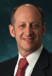 Jordan S. Orange合作论文数Vagelos College of Physicians & Surgeons, Columbia University Irving Medical Center;NewYork-Presbyterian;Morgan Stanley Children’s Hospital1Pamela L. Schwartzberg合作论文数1Alan J. Hedges合作论文数1
 Emily M. Mace合作论文数Department of Pediatrics, Children's Hospital of Philadelphia1
 Baek Hwan Cho合作论文数Department of Biomedical Engineering, Hanyang University1
 David Cameron Wraith合作论文数Institute of Immunology and Immunotherapy, University of Birmingham1Elaine V. Hill合作论文数1A. Sawasdichai合作论文数1




