We tackle the challenging problem of single-source domain generalization (DG) for medical image segmentation, where we train a network on one domain (e.g., CT) and directly apply it to a different domain (e.g., MR) without adapting the model and without requiring images or annotations from the new domain during training. Our method diversifies the source domain through semantic-aware random convolution, where different regions of a source image are augmented differently at training-time, based on their annotation labels. At test-time, we complement the randomization of the training domain via mapping the intensity of target domain images, making them similar to source domain data. We perform a comprehensive evaluation on a variety of cross-modality and cross-center generalization settings for abdominal, whole-heart and prostate segmentation, where we outperform previous DG techniques in a vast majority of experiments. Additionally, we also investigate our method when training on whole-heart CT or MR data and testing on the diastolic and systolic phase of cine MR data captured with different scanner hardware. Overall, our evaluation shows that our method achieves new state-of-the-art performance in DG for medical image segmentation, even matching the performance of the in-domain baseline in several settings. Code is available at: https://github.com/imigraz/SRCSM_Domain_Generalization
Deep learning has shown remarkable success in medical image analysis over the last decade; however, many contributions focused on supervised methods which learn exclusively from labeled training samples. Acquiring expert-level annotations in large quantities is time-consuming and costly, even more so in medical image segmentation, where annotations are required on a pixel level and often in 3D. As a result, available labeled training data and consequently performance is often limited. Frequently, however, additional unlabeled data are available and can be readily integrated into model training, paving the way for semi- or self-supervised learning (SSL). In this work, we investigate popular SSL strategies in more detail, namely Transformation Consistency, Student–Teacher and Pseudo-Labeling, as well as exhaustive combinations thereof. We comprehensively evaluate these methods on two 2D and 3D cardiac Magnetic Resonance datasets (ACDC, MMWHS) for which several different multi-compartment segmentation labels are available. To assess performance in limited dataset scenarios, different setups with a decreasing amount of patients in the labeled dataset are investigated. We identify cascaded Self-Supervision as the best methodology, where we propose to employ Pseudo-Labeling and a self-supervised cascaded Student–Teacher model simultaneously. Our evaluation shows that in all scenarios, all investigated SSL methods outperform the respective low-data supervised baseline as well as state-of-the-art self-supervised approaches. This is most prominent in the very-low-labeled data regime, where for our proposed method we demonstrate 10.17% and 6.72% improvement in Dice Similarity Coefficient (DSC) for ACDC and MMWHS, respectively, compared with the low-data supervised approach, as well as 2.47% and 7.64% DSC improvement, respectively, when compared with related work. Moreover, in most experiments, our proposed method is able to greatly decrease the performance gap when compared to the fully supervised scenario, where all available labeled samples are used. We conclude that it is always beneficial to incorporate unlabeled data in cardiac MRI segmentation whenever it is present.
As the leading cause of death worldwide, cardiovascular diseases motivate the development of more sophisticated methods to analyze the heart and its substructures from medical images like Computed Tomography (CT) and Magnetic Resonance (MR). Semantic segmentations of important cardiac structures that represent the whole heart are useful to assess patient-specific cardiac morphology and pathology. Furthermore, accurate semantic segmentations can be used to generate cardiac digital twin models which allows e.g. electrophysiological simulation and personalized therapy planning. Even though deep learning-based methods for medical image segmentation achieved great advancements over the last decade, retaining good performance under domain shift - i.e. when training and test data are sampled from different data distributions - remains challenging. In order to perform well on domains known at training-time, we employ a (1) balanced joint training approach that utilizes CT and MR data in equal amounts from different source domains. Further, aiming to alleviate domain shift towards domains only encountered at test-time, we rely on (2) strong intensity and spatial augmentation techniques to greatly diversify the available training data. Our proposed whole heart segmentation method, a 5-fold ensemble with our contributions, achieves the best performance for MR data overall and a performance similar to the best performance for CT data when compared to a model trained solely on CT. With 93.33
Atrial fibrillation (AF) represents the most prevalent type of cardiac arrhythmia for which treatment may require patients to undergo ablation therapy. In this surgery cardiac tissues are locally scarred on purpose to prevent electrical signals from causing arrhythmia. Patient-specific cardiac digital twin models show great potential for personalized ablation therapy, however, they demand accurate semantic segmentation of healthy and scarred tissue typically obtained from late gadolinium enhanced (LGE) magnetic resonance (MR) scans. In this work we propose the Left Atrial Cascading Refinement CNN (LA-CaRe-CNN), which aims to accurately segment the left atrium as well as left atrial scar tissue from LGE MR scans. LA-CaRe-CNN is a 2-stage CNN cascade that is trained end-to-end in 3D, where Stage 1 generates a prediction for the left atrium, which is then refined in Stage 2 in conjunction with the original image information to obtain a prediction for the left atrial scar tissue. To account for domain shift towards domains unknown during training, we employ strong intensity and spatial augmentation to increase the diversity of the training dataset. Our proposed method based on a 5-fold ensemble achieves great segmentation results, namely, 89.21
The localization of teeth and segmentation of periapical lesions in cone-beam computed tomography (CBCT) images are crucial tasks for clinical diagnosis and treatment planning, which are often time-consuming and require a high level of expertise. However, automating these tasks is challenging due to variations in shape, size, and orientation of lesions, as well as similar topologies among teeth. Moreover, the small volumes occupied by lesions in CBCT images pose a class imbalance problem that needs to be addressed. In this study, we propose a deep learning-based method utilizing two convolutional neural networks: the SpatialConfiguration-Net (SCN) and a modified version of the U-Net. The SCN accurately predicts the coordinates of all teeth present in an image, enabling precise cropping of teeth volumes that are then fed into the U-Net which detects lesions via segmentation. To address class imbalance, we compare the performance of three reweighting loss functions. After evaluation on 144 CBCT images, our method achieves a 97.3% accuracy for teeth localization, along with a promising sensitivity and specificity of 0.97 and 0.88, respectively, for subsequent lesion detection.
Myocardial infarction (MI) is one of the most prevalent cardiovascular diseases and consequently, a major cause for mortality and morbidity worldwide. Accurate assessment of myocardial tissue viability for post-MI patients is critical for diagnosis and treatment planning, e.g. allowing surgical revascularization, or to determine the risk of adverse cardiovascular events in the future. Fine-grained analysis of the myocardium and its surrounding anatomical structures can be performed by combining the information obtained from complementary medical imaging techniques. In this work, we use late gadolinium enhanced (LGE) magnetic resonance (MR), T2-weighted (T2) MR and balanced steady-state free precession (bSSFP) cine MR in order to semantically segment the left and right ventricle, healthy and scarred myocardial tissue, as well as edema. To this end, we propose the Multi-Sequence Cascading Refinement CNN (MS-CaRe-CNN), a 2-stage CNN cascade that receives multi-sequence data and generates predictions of the anatomical structures of interest without considering tissue viability at Stage 1. The prediction of Stage 1 is then further refined in Stage 2, where the model additionally distinguishes myocardial tissue based on viability, i.e. healthy, scarred and edema regions. Our proposed method is set up as a 5-fold ensemble and semantically segments scar tissue achieving 62.31 and 87.69 results for such small and challenging structures confirm that MS-CaRe-CNN is well-suited to generate semantic segmentations to assess the viability of myocardial tissue, enabling downstream tasks like personalized therapy planning.
The risen complexity of automotive systems requires new development strategies and methods to master the upcoming challenges. Traditional methods need thus to be changed by an increased level of automation, and a faster continuous improvement cycle. In this context, current vehicle performance tests represent a very time-consuming and expensive task due to the need to perform the tests in real driving conditions. As a consequence, agile/iterative processes like DevOps are largely hindered by the necessity of triggering frequent tests. This paper reports on a practical experience of developing an AI-augmented solution based on Machine Learning and Model-based Engineering to support continuous vehicle development and testing. In particular, historical data collected in real driving conditions is leveraged to synthesize a high-fidelity driving simulator and hence enable performance tests in virtual environments. Based on this practical experience, this paper also proposes a conceptual framework to support predictions based on real driving behavior.
The aim of this validation study was to comprehensively evaluate the performance and generalization capability of a deep learning-based periapical lesion detection algorithm on a clinically representative cone-beam computed tomography (CBCT) dataset and test for non-inferiority. The evaluation involved 195 CBCT images of adult upper and lower jaws, where sensitivity and specificity metrics were calculated for all teeth, stratified by jaw, and stratified by tooth type. Furthermore, each lesion was assigned a periapical index score based on its size to enable a score-based evaluation. Non-inferiority tests were conducted with proportions of 90% for sensitivity and 82% for specificity. The algorithm achieved an overall sensitivity of 86.7% and a specificity of 84.3%. The non-inferiority test indicated the rejection of the null hypothesis for specificity but not for sensitivity. However, when excluding lesions with a periapical index score of one (i.e., very small lesions), the sensitivity improved to 90.4%. Despite the challenges posed by the dataset, the algorithm demonstrated promising results. Nevertheless, further improvements are needed to enhance the algorithm’s robustness, particularly in detecting very small lesions and the handling of artifacts and outliers commonly encountered in real-world clinical scenarios.
Cardiac digital twins (Cardiac Digital Twin (CDT)s) of human electrophysiology (Electrophysiology (EP)) are digital replicas of patient hearts derived from clinical data that match like-for-like all available clinical observations. Due to their inherent predictive potential, CDTs show high promise as a complementary modality aiding in clinical decision making and also in the cost-effective, safe and ethical testing of novel EP device therapies. However, current workflows for both the anatomical and functional twinning phases within CDT generation, referring to the inference of model anatomy and parameters from clinical data, are not sufficiently efficient, robust and accurate for advanced clinical and industrial applications. Our study addresses three primary limitations impeding the routine generation of high-fidelity CDTs by introducing; a comprehensive parameter vector encapsulating all factors relating to the ventricular EP; an abstract reference frame within the model allowing the unattended manipulation of model parameter fields; a novel fast-forward electrocardiogram (Electrocardiogram (ECG)) model for efficient and bio-physically-detailed simulation required for parameter inference. A novel workflow for the generation of CDTs is then introduced as an initial proof of concept. Anatomical twinning was performed within a reasonable time compatible with clinical workflows (<4h) for 12 subjects from clinically-attained magnetic resonance images. After assessment of the underlying fast forward ECG model against a gold standard bidomain ECG model, functional twinning of optimal parameters according to a clinically-attained 12 lead ECG was then performed using a forward Saltelli sampling approach for a single subject. The achieved results in terms of efficiency and fidelity demonstrate that our workflow is well-suited and viable for generating biophysically-detailed CDTs at scale.
Automatic segmentation of abdominal organs in CT scans plays an important role in clinical practice. However, most existing benchmarks and datasets only focus on segmentation accuracy, while the model efficiency and its accuracy on the testing cases from different medical centers have not been evaluated. To comprehensively benchmark abdominal organ segmentation methods, we organized the first Fast and Low GPU memory Abdominal oRgan sEgmentation (FLARE) challenge, where the segmentation methods were encouraged to achieve high accuracy on the testing cases from different medical centers, fast inference speed, and low GPU memory consumption, simultaneously. The winning method surpassed the existing state-of-the-art method, achieving a 19× faster inference speed and reducing the GPU memory consumption by 60% with comparable accuracy. We provide a summary of the top methods, make their code and Docker containers publicly available, and give practical suggestions on building accurate and efficient abdominal organ segmentation models. The FLARE challenge remains open for future submissions through a live platform for benchmarking further methodology developments at https://flare.grand-challenge.org/.
INTRODUCTION:Cone-beam computed tomography (CBCT) is an essential diagnostic tool in oral radiology. Radiolucent periapical lesions (PALs) represent the most frequent jaw lesions. However, the description, interpretation, and documentation of radiological findings, especially incidental findings, are time-consuming and resource-intensive, requiring a high degree of expertise. To improve quality, dentists may use artificial intelligence in the form of deep learning tools. This study was conducted to develop and validate a deep convolutional neuronal network for the automated detection of osteolytic PALs in CBCT data sets. METHODS:CBCT data sets from routine clinical operations (maxilla, mandible, or both) performed from January to October 2020 were retrospectively screened and selected. A 2-step approach was used for automatic PAL detection. First, tooth localization and identification were performed using the SpatialConfiguration-Net based on heatmap regression. Second, binary segmentation of lesions was performed using a modified U-Net architecture. A total of 144 CBCT images were used to train and test the networks. The method was evaluated using the 4-fold cross-validation technique. RESULTS:The success detection rate of the tooth localization network ranged between 72.6% and 97.3%, whereas the sensitivity and specificity values of lesion detection were 97.1% and 88.0%, respectively. CONCLUSIONS:Although PALs showed variations in appearance, size, and shape in the CBCT data set and a high imbalance existed between teeth with and without PALs, the proposed fully automated method provided excellent results compared with related literature.
An important factor for the development of spinal degeneration, pain and the outcome of spinal surgery is known to be the balance of the spine. It must be analyzed in an upright, standing position to ensure physiological loading conditions and visualize load-dependent deformations. Despite the complex 3D shape of the spine, this analysis is currently performed using 2D radiographs, as all frequently used 3D imaging techniques require the patient to be scanned in a prone position. To overcome this limitation, we propose a deep neural network to reconstruct the 3D spinal pose in an upright standing position, loaded naturally. Specifically, we propose a novel neural network architecture, which takes orthogonal 2D radiographs and infers the spine’s 3D posture using vertebral shape priors. In this work, we define vertebral shape priors using an atlas and a spine shape prior, incorporating both into our proposed network architecture. We validate our architecture on digitally reconstructed radiographs, achieving a 3D reconstruction Dice of 0.95, indicating an almost perfect 2D-to-3D domain translation. Validating the reconstruction accuracy of a 3D standing spine on real data is infeasible due to the lack of a valid ground truth. Hence, we design a novel experiment for this purpose, using an orientation invariant distance metric, to evaluate our model’s ability to synthesize full-3D, upright, and patient-specific spine models. We compare the synthesized spine shapes from clinical upright standing radiographs to the same patient’s 3D spinal posture in the prone position from CT.
We propose a reinforcement learning (RL) based approach for anatomical landmark localization in medical images, where the agent can move in arbitrary directions with a variable step size. Using a continuous action space reduces the average number of steps required to locate a landmark by more than 30 times compared to localization using discrete actions. Our approach outperforms a state-of-the-art RL method based on a discrete action space and is inline with state-of-the-art supervised regression based methods. Furthermore, we extend our approach to a multi-agent setting, where we allow collaboration between agents to enable learning of the landmarks’ spatial configuration. The results of the multi-agent RL based approach show that the position of occluded landmarks can be successfully estimated based on the relative position predicted for the visible landmarks.
Brain ageing is a complex neurobiological process associated with morphological changes that can be assessed on MRI scans. Recently, Deep learning (DL)-based approaches have been proposed for the prediction of chronological brain age from MR images yielding high accuracy. These approaches, however, usually do not address quantification of uncertainty and, therefore, intrinsic physiological variability. Considering uncertainty is essential for the interpretation of the difference between predicted and chronological age. In addition, DL-based models lack in explainability compared to classical approaches like voxel-based morphometry. In this study, we aim to address both, modeling uncertainty and providing visual explanations to explore physiological patterns in brain ageing. T1-weighted brain MRI datasets of 10691 participants of the German National Cohort Study, drawn from the general population, were included in this study (chronological age from 20 to 72 years). A regression model based on a 3D Convolutional Neural Network taking into account aleatoric noise was implemented for global as well as regional brain age estimation. We observed high overall accuracy of global brain age estimation with a mean absolute error of 3.2 ± 2.5 years and mean uncertainty of 2.9 ± 0.6 years. Regional brain age estimation revealed higher estimation accuracy and lower uncertainty in central compared to peripheral brain regions. Visual explanations illustrating the importance of brain sub-regions were generated using Grad-CAM: the derived saliency maps showed a high relevance of the lateral and third ventricles, the insular lobe as well as parts of the basal ganglia and the internal capsule.
In landmark localization, due to ambiguities in defining their exact position, landmark annotations may suffer from large observer variabilities, which result in uncertain annotations. To model the annotation ambiguities of the training dataset, we propose to learn anisotropic Gaussian parameters modeling the shape of the target heatmap during optimization. Furthermore, our method models the prediction uncertainty of individual samples by fitting anisotropic Gaussian functions to the predicted heatmaps during inference. Besides state-of-the-art results, our experiments on datasets of hand radiographs and lateral cephalograms also show that Gaussian functions are correlated with both localization accuracy and observer variability. As a final experiment, we show the importance of integrating the uncertainty into decision making by measuring the influence of the predicted location uncertainty on the classification of anatomical abnormalities in lateral cephalograms.
Even though many semantic segmentation methods exist that are able to perform well on many medical datasets, often, they are not designed for direct use in clinical practice. The two main concerns are generalization to unseen data with a different visual appearance, e.g., images acquired using a different scanner, and efficiency in terms of computation time and required Graphics Processing Unit (GPU) memory. In this work, we employ a multi-organ segmentation model based on the SpatialConfiguration-Net (SCN), which integrates prior knowledge of the spatial configuration among the labelled organs to resolve spurious responses in the network outputs. Furthermore, we modified the architecture of the segmentation model to reduce its memory footprint as much as possible without drastically impacting the quality of the predictions. Lastly, we implemented a minimal inference script for which we optimized both, execution time and required GPU memory.
Vertebral labelling and segmentation are two fundamental tasks in an automated spine processing pipeline. Reliable and accurate processing of spine images is expected to benefit clinical decision-support systems for diagnosis, surgery planning, and population-based analysis on spine and bone health. However, designing automated algorithms for spine processing is challenging predominantly due to considerable variations in anatomy and acquisition protocols and due to a severe shortage of publicly available data. Addressing these limitations, the Large Scale Vertebrae Segmentation Challenge (VerSe) was organised in conjunction with the International Conference on Medical Image Computing and Computer Assisted Intervention (MICCAI) in 2019 and 2020, with a call for algorithms towards labelling and segmentation of vertebrae. Two datasets containing a total of 374 multi-detector CT scans from 355 patients were prepared and 4505 vertebrae have individually been annotated at voxel-level by a human-machine hybrid algorithm (https://osf.io/nqjyw/, https://osf.io/t98fz/). A total of 25 algorithms were benchmarked on these datasets. In this work, we present the the results of this evaluation and further investigate the performance-variation at vertebra-level, scan-level, and at different fields-of-view. We also evaluate the generalisability of the approaches to an implicit domain shift in data by evaluating the top performing algorithms of one challenge iteration on data from the other iteration. The principal takeaway from VerSe: the performance of an algorithm in labelling and segmenting a spine scan hinges on its ability to correctly identify vertebrae in cases of rare anatomical variations. The content and code concerning VerSe can be accessed at: https://github.com/anjany/verse.
: Localization and segmentation of vertebral bodies from spine CT volumes are crucial for pathological diagnosis, surgical planning, and postoperative assessment. However, fully automatic analysis of spine CT volumes is difficult due to the anatomical variation of pathologies, noise caused by screws and implants, and the large range of different field-of-views. We propose a fully automatic coarse to fine approach for vertebrae localization and segmentation based on fully convolutional CNNs. In a three-step approach, at first, a U-Net localizes the rough position of the spine. Then, the SpatialConfiguration-Net performs vertebrae localization and identification using heatmap regression. Finally, a U-Net performs binary segmentation of each identified vertebrae in a high resolution, before merging the individual predictions into the resulting multi-label vertebrae segmentation. The evaluation shows top performance of our approach, ranking first place and winning the MICCAI 2019 Large Scale Vertebrae Segmentation Challenge (VerSe 2019).
In landmark localization, due to ambiguities in defining their exact position, landmark annotations may suffer from both large inter- and intra-observer variabilites, which result in uncertain annotations. Therefore, predicting a single coordinate for a landmark is not sufficient for modeling the distribution of possible landmark locations. We propose to learn the Gaussian covariances of target heatmaps, such that covariances for pointed heatmaps correspond to more certain landmarks and covariances for flat heatmaps to more uncertain or ambiguous landmarks. By fitting Gaussian functions to the predicted heatmaps, our method is able to obtain landmark location distributions, which model location uncertainties. We show on a dataset of left hand radiographs and on a dataset of lateral cephalograms that the predicted uncertainties correlate with the landmark error, as well as inter-observer variabilities.
 Martin Urschler合作论文数Institute for Medical Informatics, Statistics and Documentation, Medical University of Graz51
 Christian Payer合作论文数Graz Univ Technol, Inst Comp Graph & Vis, Graz, Austria27
 Horst Bischof合作论文数Institute of Computer Graphics and Vision, Graz University of Technology14
 Tomaž Vrtovec合作论文数Faculty of Electrical Engineering, University of Ljubljana11
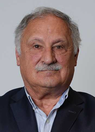 Franjo Pernuš合作论文数Department of Electrical Engineering, Faculty of Electrical Engineering, University of Ljubljana10
 Bostjan Likar合作论文数Laboratory of Imaging Technologies, Faculty of Electrical Engineering, University of Ljubljana10
 Thomas Ebner合作论文数9Franz Thaler合作论文数Department of Mathematics, Nanjing University7
 Gernot Plank合作论文数Computational Cardiology Laboratory, Medical University of Graz;NumeriCor;University of Oxford5
 Neff, T.合作论文数FibroGen Inc4Sabine Grassegger合作论文数4
 Vesna Njagulj合作论文数3Arnela Hadzic合作论文数3
 Amirhossein Bayat合作论文数Department of Informatics, Technical University of Munich, Munich, Germany.3
 Kerstin Hammernik合作论文数Graz University of Technology3
 Anjany Sekuboyina合作论文数Dept Informat Munich Germany, Tech Univ Munich3
 Jan S Kirschke合作论文数School of Medicine, Technical University of Munich3
 Barbara Kirnbauer合作论文数3
 Thomas Pock合作论文数Institute of Visual Computing, Graz University of Technology3
 Bjoern Menze合作论文数Department of Quantitative Biomedicine, University of Zurich;Faculty of Science, University of Zurich3
 Johannes C. Paetzold合作论文数Tech Univ Munich, Dept Informat, Munich, Germany2Ehammer Thomas合作论文数2
 Guoyan Zheng (郑国焱)合作论文数School of Biomedical Engineering, Shanghai Jiao Tong University2
 Chunliang Wang (王春亮)合作论文数Division of Biomedical Imaging, Department of Biomedical Engineering and Health Systems, School of Engineering Sciences in Chemistry, Biotechnology and Health, KTH Royal Institute of Technology2Robert Korez合作论文数2
 Ben Glocker合作论文数Department of Computing, Imperial College London2
 Pheng Ann Heng (王平安)合作论文数Department of Computer Science and Engineering, The Chinese University of Hong Kong2
 Matthias Ruther合作论文数Institute for Computer Graphics and Vision, Graz University of Technology2Price Jackson合作论文数1Miran Buermen合作论文数1Dalong Cheng合作论文数1Alexandre Kirszenberg合作论文数1Daniel Pucher合作论文数1
 Alexey Novikov合作论文数1
 Mattias Paul Heinrich合作论文数Institute of Medical Informatics, University of Lübeck1Zhu Qiongjie合作论文数1
 Hao Chen (陳浩)合作论文数Department of Computer Science and Engineering, School of Engineering, The Hong Kong University of Science and Technology;Division of Life Science, School of Science, The Hong Kong University of Science and Technology;Department of Chemical and Biological Engineering, School of Engineering, The Hong Kong University of Science and Technology1
 Mohanasankar Sivaprakasam合作论文数1
 Julien Oster合作论文数1David Martinčič合作论文数1Yinan Xu合作论文数1Chenchen Sun合作论文数1Dong Guoqiang合作论文数1Christoph Angerman合作论文数1Walter Unterpirker合作论文数1Anna Gostler合作论文数1Maodong Chen合作论文数1Shangliang Xu合作论文数1XinJun Ma合作论文数1




