PURPOSE:We developed a novel approach to treat newly diagnosed glioblastoma (GBM) using genetically modified gamma-delta (γδ) T cells following the forced upregulation of stress-associated targets on tumor cells. We leveraged the temozolomide (TMZ)-induced activation of the DNA damage response pathway to transiently upregulate the natural killer ligand (NKG2D-L) targets on GBM. Manufactured γδ T cells are engineered to be resistant to alkylating chemotherapies, including TMZ, through insertion of a methylguanine-DNA methyltransferase (MGMT)-expressing lentivector (DeltEx drug-resistant immunotherapy-DRI). METHODS:A total of 23 patients were enrolled, and 13 were treated (62% male; median age 66 years [range, 21-75]; 92% isocitrate dehydrogenase wild type (IDH-WT), 54% MGMT unmethylated, 46% subtotal resection). Cohorts 1, 2, and 3 received 1, 3, or up to 6 doses, respectively (1 × 10⁷ DRI cells/dose), using a Rickham catheter, which was placed into the resection cavity. The DRI cells were dosed in combination with 150 mg/m2 intravenous (IV) TMZ once per day on Day (D) 1 of each maintenance cycle, which was followed by 4 days of oral TMZ. RESULTS:No dose-limiting toxicities were seen nor were any occurrences of cytokine release syndrome (CRS) or neurotoxicity (immune effector cell-associated neurotoxicity syndrome) observed. The median follow-up of patients who received DRI γδ T cells was 15.6 months. For Cohort 1 patients who received a single dose of DRI γδ T cells, the median progression-free survival (mPFS) was 8.0 months; the median PFS was 9.9 months for all patients and 16.1 months for patients who received repeated doses in Cohorts 2 and 3. The median overall survival for all patients was 15.6 months. CONCLUSION:To date, all patients had manageable toxicity with outpatient treatment and a continued encouraging trend in outcomes from repeated investigational treatments with intracranially delivered, longitudinal DRI γδ T cells.
Proteome and transcriptome data combined can help assess the relevance of non-coding germline variants. Here, we combine germline Structural Variants (SVs) with mass spectrometry-based proteomics on tumors from 1637 cancer patients spanning various tumor tissues of origin to determine the extent SV breakpoint patterns involve differential protein expression of nearby genes. Rare and singleton SVs disrupting protein expression of known cancer susceptibility genes collectively involve 6% of patients. About 24% of the hundreds of genes with SV-associated non-coding cis-regulatory alterations at the mRNA level are similarly associated at the protein level. Both rare and common SVs may associate with differential protein expression within a specific tumor type or across multiple tissue types, including SVs differentially represented by patient ancestry. SVs involving altered methylation of CpG Islands or enhancers are also implicated in differential protein expression. Our results emphasize the contribution of germline SVs to cancer heterogeneity at the proteome level.
Clear cell adenocarcinoma of the urinary tract (CCA-UT) is a rare, potentially aggressive tumor with very limited information regarding its clinicopathologic characteristics and molecular alterations. This study aimed to elucidate the clinicopathologic features and molecular landscape of one of the largest cohorts (35 cases) of this tumor, to identify genomic alterations and potential therapeutic targets. Seventy-nine percent of the patients were women, with a median age of 61 years. The urethra was the most common site (18; 51%), and all cases were ≥pT2 (pT2:15; pT3:11; pT4:8). Twenty-nine percent of the patients died of their disease on follow-up. On whole-exome sequencing, pathogenic/oncogenic alterations were identified in 91% (32/35) cases. These alterations, most frequently involved chromatin modifiers (66% cases), including ATRX , KMT2C , ARID1A , and ARID1B . Other frequently mutated genes included ATM , NF1 , and ERBB2 . Ninety-seven percent (34/35) of cases were microsatellite stable, and tumor mutational burden (TMB) was >10 mut/Mb in 9% (3/35) of cases. Five cases were homologous recombinant-deficient on ScarHRD analysis, and 3 cases showed BRCA mutations. Recurrent copy number loss events in Chr 1(p36.33-p35.3) were the most common copy number alterations (80%; n=28 cases). RNA-sequencing data analysis revealed numerous differentially expressed genes and enrichment of the epithelial-to-mesenchymal transition gene signature in individual samples. However, there was no statistical significance in the progression-free survival between cases with epithelial and mesenchymal phenotypes. CCA-UT are aggressive tumors with a heterogeneous molecular profile, underscoring the role of molecular analysis in identifying potential therapeutic options for the treatment of this pernicious tumor.
Clear cell adenocarcinoma of the urinary tract (CCA-UT) is a rare, potentially aggressive tumor with very limited information regarding its clinicopathologic characteristics and molecular alterations. This study aimed to elucidate the clinicopathologic features and molecular landscape of one of the largest cohorts (35 cases) of this tumor, to identify genomic alterations and potential therapeutic targets. Seventy-nine percent of the patients were women, with a median age of 61 years. The urethra was the most common site (18; 51%), and all cases were ≥pT2 (pT2:15; pT3:11; pT4:8). Twenty-nine percent of the patients died of their disease on follow-up. On whole-exome sequencing, pathogenic/oncogenic alterations were identified in 91% (32/35) cases. These alterations, most frequently involved chromatin modifiers (66% cases), including ATRX , KMT2C , ARID1A , and ARID1B . Other frequently mutated genes included ATM , NF1 , and ERBB2 . Ninety-seven percent (34/35) of cases were microsatellite stable, and tumor mutational burden (TMB) was >10 mut/Mb in 9% (3/35) of cases. Five cases were homologous recombinant-deficient on ScarHRD analysis, and 3 cases showed BRCA mutations. Recurrent copy number loss events in Chr 1(p36.33-p35.3) were the most common copy number alterations (80%; n=28 cases). RNA-sequencing data analysis revealed numerous differentially expressed genes and enrichment of the epithelial-to-mesenchymal transition gene signature in individual samples. However, there was no statistical significance in the progression-free survival between cases with epithelial and mesenchymal phenotypes. CCA-UT are aggressive tumors with a heterogeneous molecular profile, underscoring the role of molecular analysis in identifying potential therapeutic options for the treatment of this pernicious tumor.
PURPOSE:In the United States, African Americans (AA) have higher Pancreatic ductal adenocarcinoma (PDAC) incidence and mortality rates than Caucasian Americans (CA). This study aimed to identify distinct gene expression signatures and differentially regulated pathways in AA and CA PDACs. METHODS:Transcriptomic analyses were conducted on FFPE sections of PDACs (n = 40) from AA (9 PDACs/3 normal) and CA (31 PDACs/5 normal) tissues to evaluate the differential expression and signaling pathways within and between racial groups and to identify distinctive and common genes/pathways. RESULTS:We identified unique differentially expressed genes in both racial groups. Distinct set genes were modulated in AA and CA PDACs, compared to their respective normal tissues. Thirteen genes (seven upregulated and six downregulated) were differentially modulated in AA PDACs vs. CA PDACs. CIBERSORT analysis revealed distinct immune cell composition, with increased resting NK cells and activated mast cells, in AA PDACs, and higher CD4 memory T cells present in CA PDACs. Canonical subtype analyses indicated a more heterogenous subtype distribution in AA PDACs, whereas CA PDACs showed a predominance of classical subtypes. Using a publicly available database, we analyzed the top 25 upregulated genes (normal vs. tumor) for AA and CA racial groups and seven differentially upregulated genes in AA PDACs vs. CA PDACs comparison for associations with survival outcomes. Eight genes (CHST15, PARP15, NUDT16, SERPINB3, PADI1, H3C8, ZNF488, and LETM2) correlated with poor patient survival. CONCLUSION:These findings show distinct gene expression profiles and modulated pathways in AA and CA PDACs, supporting development of race-based therapeutic targets.
Microsatellite instability (MSI) is a vital genomic biomarker in endometrial cancer (EC), playing a key role in guiding patient management decisions. Microsatellite status can be assessed using various methods, including immunohistochemistry (IHC), polymerase chain reaction (PCR), and next-generation sequencing (NGS). However, accurately determining microsatellite status in EC can be challenging due to the typically weak MSI phenotype observed in this cancer type. This study aims to enhance microsatellite analysis in EC by combining EC mutation profiling with MSI detection techniques into a unified analytical workflow. We performed comprehensive genomic profiling on 43 EC samples using the PGDx elio™ tissue complete (ETC), an FDA-cleared NGS test targeting 505 solid tumor related genes. In addition to mutation profile, we obtained microsatellite status and tumor mutational burden for each case. Mismatch repair (MMR) status were also determined for all 43 ECs using IHC (MLH1, PMS2, MSH2 and MSH6). ‘maftools’ R package was utilized for data processing and visualization, while genomic classifier was developed using ‘randomForest’ R package. IHC analysis led to identification of 28 proficient MMR (pMMR) and 15 deficient MMR (dMMR) EC samples. We observed 100% (28/28) concordance for pMMR samples and 73.3% (11/15) concordance for dMMR samples between IHC analysis and NGS assay. Four discordant samples showed low TMB and low-grade instability in 68 mononucleotide tracts considered by NGS assay indicative of weak MSI phenotype. We also identified 15 genes (including ERBB4, PTPRT, ARID1A, ARID1B, and BRD4) significantly altered in dMMR samples compared to pMMR samples. Random forest classifier was developed considering 88 distinct parameters (including mutation profile of 15 genes, 5 clinicopathologic features and 68-mononucleotide tract status) for all EC samples. We observed varying degrees of concordance (100% specificity and 73.3% sensitivity) between MSI status by NGS based ETC and MMR status by IHC in a cohort of EC. Targeted genomic profiling via ETC identified significantly altered genes in dMMR endometrial tumors and led to development of random forest classifier to predict MSI EC tumor [which showed AUC value of 0.875, 87.5% specificity and 100% sensitivity for test set]. Integration of mutational profiling with MSI detection can be leveraged to increase sensitivity for MSI detection on the ETC platform. Darshan Shimoga Chandrashekar, João Lobo, Mehenaz Hanbazazh, Kanako Okamoto, Jennifer B. Jackson, Shuko Harada, Alexander C. Mackinnon. Characterization of microsatellite status in endometrial tumors using comprehensive genomic profiling via the PGDx elio tissue complete. [abstract]. In: Proceedings of the American Association for Cancer Research Annual Meeting 2025; Part 1 (Regular Abstracts); 2025 Apr 25-30; Chicago, IL. Philadelphia (PA): AACR; Cancer Res 2025;85(8_Suppl_1):Abstract nr 688.
Cancer is a major cause of death worldwide. Various genomic and proteomic alterations in cells results in initiation of cancer, disease progression, and tumor metastasis. Large scale molecular data has been generated recently that can help identify these molecular changes. These genomic, transcriptomic, proteomic and epigenetic data can be utilized to evaluate and identify cancer biomarkers and sub-class specific precision targets. In order to analyze the large scale data, user friendly tools are needed. We had earlier developed UALCAN, an integrative pan-cancer cancer data analysis platform that allows users to evaluate the expression of each genes, microRNAs and long-noncoding RNAs and identify changes between normal and cancer tissues. Previously, we have described the development and release of the UALCAN Mobile application (app) that provides cancer transcriptomic data from The Cancer Genome Atlas (TCGA) to evaluate gene expression based on cancer subtypes. Here, we describe the update to the UALCAN mobile, which now provides data analysis option for epigenetic changes due to DNA promoter methylation and Clinical Proteomic Tumor Analysis Consortium (CPTAC) cancer proteomic data. This app, which is suitable for tablets and mobile devices, provides access to large cancer molecular datasets at the fingertips of the researchers. To find changes in expression of causative genes and to identify biomarkers and therapeutic targets, app will be extremely valuable. The app is free and can be downloaded from both iOS/Apple and Android play store. Citation Format: Sooryanarayana Varambally, David Rubey, Darshan Shimoga Chandrashekar, Ahmedur Rahman Shovon, Gopi Chand Puli, Santhosh Kumar Karthikeyan, Upender Manne, Chad J. Creighton, Sidarth Kumar. UALCAN Mobile app, an update to the cancer multi-omic data analysis application [abstract]. In: Proceedings of the AACR Special Conference in Cancer Research: Functional and Genomic Precision Medicine in Cancer: Different Perspectives, Common Goals; 2025 Mar 11-13; Boston, MA. Philadelphia (PA): AACR; Cancer Res 2025;85(5 Suppl):Abstract nr A019.
Cancer is a complex disease affecting various organs and is a major cause of death worldwide. During cancer initiation, disease progression, and tumor metastasis, various genomic and proteomic alterations are observed. Recent technological advances have led to the generation of large amounts of molecular data, including genomics and transcriptomics. These large-scale datasets can be utilized to analyze and identify sub-class-specific cancer biomarkers and targets. However, there is a need for the development of user-friendly tools for large-scale data analysis, disseminating the analyzed data in a visualizable format to cancer researchers with no programming skills. We developed UALCAN, a comprehensive platform that allows users to integrate disparate data to better understand the genes, proteins, and pathways perturbed in cancer and make discoveries of potential biomarkers and targets. In the current study, we describe the development of the UALCAN Mobile application (app) that will provide cancer transcriptomic data obtained from The Cancer Genome Atlas (TCGA) project to evaluate protein-coding gene expression based on various stratifications, including stage, grade, race, gender, and molecular-subtypes across over 30 types of cancers. In addition, the UALCAN mobile provides data analysis options for epigenetic changes due to DNA promoter methylation and Clinical Proteomic Tumor Analysis Consortium (CPTAC) cancer proteomic data. The app provides access to large cancer molecular datasets on the go. To find changes in the expression of causative genes and proteins and to identify biomarkers and therapeutic targets, UALCAN mobile app will be extremely valuable. The "UALCAN Mobile" app is free to use and can be downloaded from both the iOS/Apple and the Android Play Store and has been downloaded over 100 times in each of iOS and android app stores.
Accurate classification of somatic variations from high-throughput sequencing data has become integral to diagnostics and prognostics across various cancers. However, the classification of these variations remains highly manual, inherently variable, and largely inaccessible outside specialized laboratories. Here, we introduce Azurify - a computational tool that integrates machine learning, public resources recommended by professional societies, and clinically annotated data to classify the pathogenicity of variations in precision cancer medicine. Trained on over 15,000 clinically classified variants from 8,202 patients across 138 cancer phenotypes, Azurify achieves 99.1% classification accuracy for concordant pathogenic variants in data from two external clinical laboratories. Additionally, Azurify reliably performs precise molecular profiling in leukemia cases. Azurify’s unified, scalable, and modular framework can be easily deployed within bioinformatics pipelines and retrained as new data emerges. In addition to supporting clinical workflows, Azurify offers a high-throughput screening solution for research, enabling genomic studies to identify meaningful variant-disease associations with greater efficiency and consistency. ### Competing Interest Statement The authors have declared no competing interest. National Cancer Institute, , R01-CA230800, R01-CA248041
Dogs share features in prostate gland anatomy, physiology, and pathology with men. However, human and canine prostate carcinoma (PC) have histologic and molecular differences. Particularly, the histogenesis of canine PC (cPC) is unclear. This study investigated the origin of cPC using histopathology and transcriptomics with comparison to men. Prostate glands retrospectively and prospectively collected from 445 dogs (approximately 95 % autopsy samples) were surveyed for early carcinomas and preneoplastic lesions, particularly high-grade prostatic intraepithelial neoplasia (HGPIN) due to its role in the pathogenesis of PC in men. Lineage gene signatures defining prostate luminal epithelium and urothelium were identified for inter- and intraspecies RNA-sequencing comparisons, including between cPC and canine urinary bladder urothelial carcinoma (UC). Postmortem prostate lesion frequencies were similar to previously reported canine studies. Intraductal/intra-acinar growth (31/35; 88.6 %) was common in representative samples of cPC. Prostate epithelial changes consistent with HGPIN in men were not observed. Proliferative lesions and early carcinomas were rare (7/445; 1.6 %). Patterns in prostate and urothelium marker gene expression signatures differed between human and canine PC. Compared to non-neoplastic prostate gland, cPC had significantly decreased prostate-specific and increased urothelium gene signatures. The results suggest many cases diagnosed as cPC are UC or have urothelial differentiation and thus differ from PC in men, with important implications for canine tumor classification and translational studies.
Breast cancer (BCa), a leading malignancy among women, is characterized by morphological and molecular heterogeneity. While early-stage, hormone receptor, and HER2-positive BCa are treatable, triple-negative BCa and metastatic BCa remains largely untreatable. Advances in sequencing and proteomic technologies have improved our understanding of the molecular alterations that occur during BCa initiation and progression and enabled identification of subclass-specific biomarkers and therapeutic targets. Despite the availability of abundant omics data in public repositories, user-friendly tools for multi-omics data analysis and integration are scarce. To address this, we developed a comprehensive BCa data analysis platform called MammOnc-DB ( http://resource.path.uab.edu/MammOnc-Home.html ), comprising data from more than 20,000 BCa samples. MammOnc-DB facilitates hypothesis generation and testing, biomarker discovery, and therapeutic targets identification. The platform also includes pre- and post-treatment data, which can help users identify treatment resistance markers and support combination therapy strategies, offering researchers and clinicians a comprehensive tool for BCa data analysis and visualization.
Clear cell renal cell carcinoma (ccRCC), the most common subtype of kidney cancer, exhibits notable metabolic reprogramming. We previously reported elevated HDAC7, a class II histone deacetylase, in ccRCC. Here, we demonstrate that HDAC7 promotes aggressive phenotypes and in vivo tumor progression in RCC. HDAC7 suppresses the expression of genes mediating branched-chain amino acid (BCAA) catabolism. Notably, lower expression of BCAA catabolism genes is strongly associated with worsened survival in ccRCC. Suppression of BCAA catabolism promotes expression of SNAIL1, a central mediator of aggressive phenotypes including migration and invasion. HDAC7-mediated suppression of the BCAA catabolic program promotes SNAI1 messenger RNA transcription via NOTCH signaling activation. Collectively, our findings provide innovative insights into the role of metabolic remodeling in ccRCC tumor progression.
Arsenicals are deadly chemical warfare agents that primarily cause death through systemic capillary fluid leakage and hypovolemic shock. Arsenical exposure is also known to cause acute kidney injury, a condition that contributes to arsenical-associated death due to the necessity of the kidney in maintaining whole-body fluid homeostasis. Because of the global health risk that arsenicals pose, a nuanced understanding of how arsenical exposure can lead to kidney injury is needed. We used a nontargeted transcriptional approach to evaluate the effects of cutaneous exposure to phenylarsine oxide, a common arsenical, in a murine model. Here we identified an upregulation of metabolic pathways such as fatty acid oxidation, fatty acid biosynthesis, and peroxisome proliferator-activated receptor (PPAR)-α signaling in proximal tubule epithelial cell and endothelial cell clusters. We also revealed highly upregulated genes such as Zbtb16, Cyp4a14, and Pdk4, which are involved in metabolism and metabolic switching and may serve as future therapeutic targets. The ability of arsenicals to inhibit enzymes such as pyruvate dehydrogenase has been previously described in vitro. This, along with our own data, led us to conclude that arsenical-induced acute kidney injury may be due to a metabolic impairment in proximal tubule and endothelial cells and that ameliorating these metabolic effects may lead to the development of life-saving therapies. SIGNIFICANCE STATEMENT: In this study, we demonstrate that cutaneous arsenical exposure leads to a transcriptional shift enhancing fatty acid metabolism in kidney cells, indicating that metabolic alterations might mechanistically link topical arsenical exposure to acute kidney injury. Targeting metabolic pathways may generate promising novel therapeutic approaches in combating arsenical-induced acute kidney injury.
Androgen receptor (AR)-negative triple-negative breast cancer (TNBC), often termed quadruple-negative breast cancer (QNBC), disproportionately impacts women of African descent, leading to poorer overall survival (OS). MiRNAs regulate the expression of gene drivers involved in critical signaling pathways in TNBC, such as the AR gene, and their expression varies across races and breast cancer subtypes. This study investigates whether differentially expressed miRNAs influence AR transcription, potentially contributing to the observed disparities between African American (AA) and European American (EA) QNBC patients. Race-annotated TNBC samples (n = 129) were analyzed for AR expression status and revealed the prevalence of QNBC in AA patients compared to EA (76.6% vs. 57.7%) and a significant association of AR loss with poor survival among AAs. The Cancer Genome Atlas (TCGA) RNA-seq data showed that AAs with TNBC (n = 32) had lower AR mRNA levels than EAs (n = 67). Among TCGA patients in the AR-low group, AAs had significantly poorer OS than EAs. In our cohort, 46 miRNAs exhibited differential expression between AAs and EAs with QNBC. Ten of these miRNAs (miR-1185-5p, miR-1305, miR-3161, miR-3690, miR-494-3p, miR-509-3-5p, miR-619-3p, miR-628-3p, miR-873-5p, and miR-877-5p) were predicted to target the AR gene/signaling. The loss of AR expression is linked to poorer prognoses in AA women. The understanding of the specific miRNAs involved and their regulatory mechanisms on AR expression could provide valuable insights into why AA women are more prone to QNBC.
Abstract Cancer is a complex disease effecting different organs and a major cause of death and a burden on the society. Multiple molecular alteration occur during cancer initiation and disease progression and metastasis. Recent advances in technology led to generation of large amount of molecular data including transcriptome. These large datasets can be used to analyze and identify sub-class specific cancer biomarkers and targets. However, there is a need for the development user friendly tools for its analysis and dissemination of the large scale cancer molecular data. Earlier, we had developed and upgraded web based comprehensive proteogenomic platform UALCAN (ualcan.path.uab.edu), which allows users to analyze and integrate the disparate data to better understand the gene, proteins, and pathways perturbed in cancer and make discoveries. UALCAN web platform enables intuitive analysis and discovery by cancer research community. In the current study, we describe the development of UALCAN Mobile application (App) that will provide cancer transcriptomic data obtained from The Cancer Genome Atlas (TCGA) project, to evaluate protein-coding gene expression based on various stratification including stage, grade, race, gender and molecular-subtypes across over 30 types of cancers. The UALCAN Mobile app aims at providing large cancer dataset on the go. In order to find causative gene expression changes, identify biomarkers and therapeutic targets, UALCAN Mobile will assist as the data will be easily mobile and transportable with this App. Here, we describe the development of UALCAN Mobile and its utility to the cancer research community. UALCAN Mobile App can be found at both iOS/Apple and Android play store and is free to download and use. Citation Format: Sooryanarayana Varambally, Darshan Shimoga Chandrashekar, Gopi Chand Puli, Santhosh Kumar Karthikeyan, Upender Manne, Chad J. Creighton, Sidarth Kumar. UALCAN Mobile, An app for cancer gene expression data analysis [abstract]. In: Proceedings of the American Association for Cancer Research Annual Meeting 2024; Part 1 (Regular Abstracts); 2024 Apr 5-10; San Diego, CA. Philadelphia (PA): AACR; Cancer Res 2024;84(6_Suppl):Abstract nr 2270.
Abstract Introduction: In the US, there are racial disparities in PDAC incidence and mortality, higher among blacks than whites. In addition to socioeconomic status, lifestyles, and age, genetics also contributes to these disparities. Thus, we conducted transcriptomic analyses (RNA-seq) of PDAC samples collected from African American (AA) and Caucasian (CA) patients to identify race/ethnicity-specific gene expression profiles and their related pathways to find determinants that contribute to the aggressive phenotypes of PDACs. This study is relevant to the UAB catchment area, since about 30% of patients with PDAC are AAs. Methodology: Histologically confirmed PDACs (n=40) from AA (9 PDAC and 3 matching normal tissues) and CA (31 PDAC and 5 matching normal tissues) were included in this study. FFPE sections of PDACs and their corresponding normal tissues were macro-dissected for RNA isolation. Whole transcriptomic sequencing was performed with a NextSeq 500/550 platform. Trimmed reads were mapped to a human reference genome (hg38) using HISAT, and gene level read count data were obtained using HTSeq. Differential expression analysis was performed using the DESeq2 bioconductor package. ClusterProfiler/DOSE R packages were used for gene ontology and KEGG pathway enrichment analyses. Genes with log 2-fold change of ≥1 and adjusted P-value <0.05 were considered as differentially expressed. Results: Among the top upregulated genes altered in only AA PDACs, compared to their normal tissues, were RNF144B, TESPA1, KLHL17, H2AX, MDGA1, CDK20, PHLDA3, SLC6A16, PARVG, GATD3, NUDT16, RENBP, RTL8C, C1QTNF1, HLA−DRB5, PARP15, PPP1R16B, RASGRP2, and CHST15. Of note, targeting CHST15, by an RNA oligonucleotide, STNM01 in Phase I/IIa trial on unresectable PDAC patients showed improved overall survival. Additionally, inhibition of KLHL17, an upstream activator of Ras/MAPK, could be a candidate target in PDAC. The KEGG pathways altered in AA PDACs, were glycerophospholipid metabolism; bile secretion; retinol metabolism; regulation of lipolysis in adipocytes; and pantothenate and CoA biosynthesis. In CAs, the top upregulated genes in PDACs, compared to their normal tissues, were PPY, UGT1A10, GPR20, SDR16C5, KLK7, MYBPC1, ITLN1, PADI1, CLCA1, UGT1A9, and SERPINB3.The KEGG pathways altered in CA PDACs, were cellular senescence; AGE−RAGE signaling pathway in diabetic complication; PD−L1 and PD−1 checkpoint pathway; and central carbon metabolism. There were 13 genes differentially modulated in AA PDACs as compared to CA PDACs. The 6 down-regulated were USP17L1, C2CD4D, MMP13, DCUN1D5, and 2 genes without annotations (ENSG00000276345 and ENSG00000280966); the 7 up-regulated genes were SEMA4A, LETM2, HMOX1, OTUB2, MEDAG, VEGFD, and HBA1. Immunohistochemical validation of these markers is in progress. Conclusions: Findings of this study showed distinct gene expression profiles and differentially modulated pathways in AA and CA PDAC patients. These results will aid in identifying aggressive phenotypes and new targets for developing race/ethnicity-based therapeutic interventions. Citation Format: Prachi Bajpai, Ravi Paluri, Sameer Al Diffalha, Darshan S. Chandrashekar, Farrukh Afaq, Ryan Bash, C. Ryan Miller, Sooryanarayana Varambally, Moh’d Khushman, Upender Manne. Differential gene expression to delineate racial disparities in the molecular landscape of pancreatic ductal adenocarcinoma [abstract]. In: Proceedings of the AACR Special Conference in Cancer Research: Pancreatic Cancer; 2023 Sep 27-30; Boston, Massachusetts. Philadelphia (PA): AACR; Cancer Res 2024;84(2 Suppl):Abstract nr A006.
 Sooryanarayana Varambally合作论文数84
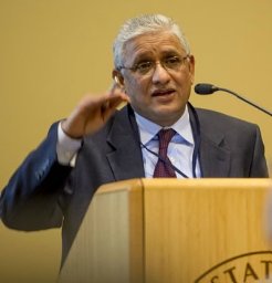 Upender Manne合作论文数the Comprehensive Cancer Center, University of Alabama at Birmingham33
 Sumit Agarwal合作论文数Dept Pathol, Univ Alabama Birmingham26
 Balabhadrapatruni V.S.K. Chakravarthi合作论文数18
 GEORGES J. NETTO合作论文数Department of Pathology, The University of Alabama at Birmingham18
 Sameer Al Diffalha合作论文数Department of Pathology, University of Alabama at Birmingham15Prachi Bajpai合作论文数Cardiovascular Toxicology Division, Indian Institute of Toxicology Research (IITR),15Hyung-Gyoon Kim合作论文数14
 Chad Creighton合作论文数Dan L Duncan Comprehensive Cancer Center, Baylor College of Medicine;Human Genome Sequencing Center, Baylor College of Medicine14
 Kshitish K Acharya合作论文数Institute of Bioinformatics and Applied Biotechnology (IBAB) G-05, ITPL14
 Guru P. Sonpavde合作论文数Department of Medical Oncology, Dana-Farber/Harvard Cancer Center, Dana-Farber Cancer Institute13
 Farrukh Afaq合作论文数Department of Dermatology, School of Medicine, University of Alabama at Birmingham;Comprehensive Cancer Center, University of Alabama at Birmingham12
 Mohd Khushman合作论文数University of South Alabama, Mitchell Cancer Institute11Michael Behring合作论文数11Sai Akshaya Hodigere Balasubramanya合作论文数10
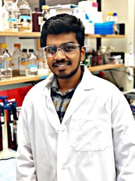 Santhosh Kumar Karthikeyan合作论文数Department of Pathology, University of Alabama at Birmingham9Sunil Sudarshan合作论文数Department of Urology, University of Alabama at Birmingham9
 Anirban Kundu合作论文数8
 Gurudatta Naik合作论文数Comprehensive Cancer Center, University of Alabama at Birmingham;National Cancer Institute, National Institutes of Health8
 Hyeyoung Nam合作论文数Korea Center for Disease Control and Prevention, National Institute of Health8Shajan Peter合作论文数Division of Gastroenterology and Hepatology, University of Alabama8
 Alyncia Robinson合作论文数7Chen Fengju合作论文数7
 Mohammad Athar合作论文数School of Dentistry, The University of Alabama at Birmingham;School of Public Health, The University of Alabama at Birmingham;General Clinical Research Center, The University of Alabama at Birmingham;Department of Dermatology, School of Medicine, The University of Alabama at Birmingham;Nephrology Research & Training Center, The University of Alabama at Birmingham;O'Neal Comprehensive Cancer Center, School of Medicine, The University of Alabama at Birmingham7
 Shuko Harada合作论文数Department of Pathology and Laboratory Medicine, University of Pennsylvania Medical Center7Jennifer B Gordetsky合作论文数Univ Alabama Birmingham7
 Ritu Aneja合作论文数School of Health Professions, The University of Alabama at Birmingham;School of Medicine, The University of Alabama at Birmingham7
 Akhilesh Kumar Bajpai合作论文数Vellore Institute of Technology (VIT) University, Vellore;6
 Sravanthi Davuluri合作论文数Shodhaka Life Sciences Pvt. Ltd.6
 Marie-Lisa Eich合作论文数Institute of Pathology, University Hospital Cologne;Medical Faculty, University of Cologne;University Hospital Cologne, University of Cologne6
 Andreas Seeber合作论文数6C Ryan Miller合作论文数5Maria Del Carmen Rodriguez Pena合作论文数Dept Pathol, Univ Alabama Birmingham5Amr Elkholy合作论文数5Yiqun Zhang合作论文数Baylor Coll Med, Dan L Duncan Canc Ctr, Houston, TX 77030 USA5
 Garrett Brinkley合作论文数5
 Zhaohui (Steve) Qin合作论文数Department of Biostatistics and Bioinformatics, Rollins School of Public Health, Emory University;Winship Cancer Institute, Emory University5
 Nallasivam Palanisamy合作论文数Henry Ford Health System;Saveetha Dental College and Hospital;Michigan State University5
 Sidharth Kumar合作论文数University of Utah5James E Ferguson合作论文数4Amith Varambally合作论文数4Richard L Kirkman合作论文数4
 Neelima Chitturi合作论文数4
 Suman Karki合作论文数Department of Biochemistry and Molecular Genetics, University of Alabama at Birmingham4
 Lakshmi P. Kunju合作论文数Department of Pathology, Medical School, University of Michigan-Ann Arbor4
 Livi Carolina B合作论文数Department of Molecular Medicine and Institute of Biotechnology, University of Texas Health Science Center4Ghatalia Pooja合作论文数4Carskadon Shannon L合作论文数4
 Moloy T Goswami合作论文数4




