Apoptotic cells are immunosuppressive, creating a barrier in cancer treatment. Thus, we investigated immune responses to dying tumor cells after therapy in the tumor draining lymph node (TDLN). A key population responsible for clearing tumor material in the TDLN was medullary sinus macrophages (MSMs). Tumor debris phagocytosis by MSMs induces the cytokine IL-33, and blocking the IL-33 receptor (ST2) or deletion of Il33 in MSMs enhances therapy responses. Mechanistically, IL-33 activates T regulatory cells in TDLNs that migrate to the tumor to suppress CD8+ T cells. Therapeutically combining ST2 blockade, targeted kinase inhibitors, and anti-PD-1 immunotherapy increases CD8+ T cell activity promoting tumor regression. Importantly, we observe similar activity in human macrophages, and IL-33 expression in sentinel lymph nodes correlates with disease stage and survival in melanoma. Thus, our data identifies an IL-33-dependent immune response to therapy that attenuates therapy-induced anti-tumor immunity.
Background Glioblastoma (GBM) is an aggressive malignant brain-tumor that invades adjacent normal brain tissue. Unlike other solid tumors, GBM is infiltrated by various normal brain cells. Methods We analyzed tumor invasion in the murine GSC005 glioma model using both immunodeficient and immunocompetent mice, focusing on the role of host-intrinsic and therapeutic interferon signaling in regulating glioblastoma (GBM) invasion. Results In this study, we observed that mouse GBM tumor GSC005 grown in immunodeficient (RAG1-KO, NSG) mice exhibited a more invasive phenotype compared to those in immunocompetent C57BL/6J mice. Immunofluorescence staining revealed the presence of vimentin + and GFAP + cells at the tumor-border interface. Bulk mRNA-seq analysis showed that GSC005 tumors in NSG mice displayed an upregulated mesenchymal signature, characterized by epithelial-to-mesenchymal transition (EMT), and downregulation of type-I and type-II interferon signaling. Our data further suggests that host-intrinsic and therapeutic type-I interferon promotes, while type-II interferon inhibits, the GBM mesenchymal signature. CD73, a key regulator of the EMT process, was found to be upregulated in GSC005 tumors in NSG mice compared to C57BL/6J mice. Mechanistic studies revealed that type-I interferon increases CD73 expression in both tumor and stromal cells, such as tumor-associated astrocytes (mAS), while type-II interferon suppresses CD73 in mAS. Functional assays indicated that CD73 modulates both type-I and type-II interferon signaling-mediated GBM invasion. Conclusion These findings suggest that therapies inducing type-I or type-II interferon signaling in GBM may reciprocally regulate CD73-mediated mesenchymal transitions, impacting GBM invasion.
Glioblastoma (GBM) is the most frequent malignant brain tumor. We recently discovered that oncolytic herpes simplex virus engineered to disable tumor-intrinsic protein kinase R (PKR) signaling (oHSV-shPKR) could increase oHSV oncolysis and antitumor immune response. However, in this study, we show that disabling tumor-intrinsic PKR signaling can also induce the activation of the indoleamine 2,3-dioxygenase (IDO) signaling pathway. Both GBM tumor progression and oHSV intratumoral therapy increased infiltration of IDO+CD11c+ dendritic cells (DC) into the tumor. The coculture of oHSV-infected human GBM neurospheres with monocyte-derived DCs (MoDC) dramatically increased IDO signaling activation in MoDCs through type-I IFN signaling. Addition of IDO inhibitor (indoximod) in the coculture significantly increased MoDC activation and reduced the consumption of tryptophan. Combining indoximod and oHSV significantly inhibited tumor growth and induced antigen-specific CD8+ T-cell activation. These results suggest that inhibition of the IDO pathway could significantly block feedback immunosuppression during oncolytic virotherapy of GBM.
In naïve mice, a fraction of CD8 T cells displaying high affinity for self-MHC peptide complexes develop into virtual memory T (TVM) cells. Due to self-reactivity, TVM cells are exposed to persistent antigenic stimulation, a condition known to induce T cell exhaustion. However, TVM cells do not exhibit characteristics similar to exhausted CD8 T (TEX) cells. Here, we tested the role of the UFL1, E3 ligase of the ufmylation pathway in TVM cells. We show that UFL1 prevents the acquisition of epigenetic, transcriptional, and phenotypic changes in TVM cells that are similar to TEX cells and thus promote their survival and function. UFL1-deficient TVM cells failed to protect mice against Listeria infection. Epigenetic analysis showed higher BATF activity in UFL1-deficient TVM cells. Deletion of BATF and not PD1 decreased inhibitory molecules expression and restored the survival and function of UFL1-deficient TVM cells. Our findings demonstrate a key role of UFL1 in inhibiting the exhaustion of TVM cells and promoting their survival and function.
TPS2703 Background: In children, brain cancer is the leading cause of cancer related death. The indoleamine 2,3-dioxygenase (IDO) pathway is a metabolic checkpoint, expressed by host myeloid and dendritic cells, that suppresses anti-tumor immune responses following chemotherapy. We recently published results of a first-in-children phase 1 trial (NCT02502708) that showed the oral IDO-pathway inhibitor indoximod was well tolerated and provided meaningful clinical benefit for many patients with progressive childhood brain tumors, when given in combination with oral chemotherapy regimens (1). Using preclinical models, we have also reported the importance of the Bruton’s Tyrosine Kinase (BTK) pathway as a key upstream driver of IDO during chemotherapy (2). Thus, we hypothesize that adding the BTK-inhibitor ibrutinib to the previously studied regimen of investigational indoximod IDO-inhibitor plus oral metronomic cyclophosphamide and etoposide will synergistically enhance anti-tumor immune responses, leading to improvement in Objective Response Rate (ORR) with manageable overlapping toxicity. Methods: The current study (NCT05106296) is an Investigator-Sponsored, open label, single-arm phase 1b trial comprised of a Dose-escalation Cohort (n=18) using a 3+3 dose escalation design to establish safety and dosing of ibrutinib in the combined regimen; followed by an Expansion Cohort (n=19) at the MTD to evaluate efficacy. Patients are treated with the all-oral 4-drug chemo-immunotherapy regimen in 28-day cycles using: (i) ibrutinib [Study Dose once daily on days 1-21]; (ii) indoximod [38.4 mg/kg/day divided twice daily throughout the cycle]; (iii) cyclophosphamide [2.5 mg/kg/dose, maximum dose 100 mg, once daily on days 1-21]; and (iv) etoposide [50 mg/m2/dose once daily on days 1-21]. Eligible patients are age 12 to 25 years with relapsed or refractory brain cancer, MRI evidence of current active disease not recently treated with radiation/proton therapy, ECOG performance score 0-2, and meet standard organ function requirements. Patients with systemic infection, autoimmune disease, recent live-virus vaccination, comorbid conditions that may overlap with expected regimen toxicities, or chronic treatment with anticoagulants or strong CYP3A inhibitors are excluded. Primary Objectives are to: (i) determine the pediatric recommended phase 2 dose (RP2D) of ibrutinib for the combined regimen (Dose-escalation Cohort), and (ii) evaluate preliminary evidence of efficacy for the combined regimen in terms of ORR (Expansion Cohort). Exploratory analyses use single-cell RNA and TCR sequencing (scRNAseq/TCRseq) of cryopreserved serial peripheral blood samples to monitor for treatment-expanded TCR clonotypes and study their phenotype. 1. Neuro-Oncology 26:348-361, 2024. 2. Immunity54:2354-2371, 2021. Clinical trial information: NCT05106296 .
Abstract BACKGROUND Brain tumors are the leading cause of pediatric cancer-related death. METHODS We report interim outcome data from an ongoing multi-site phase 2 study (NCT04049669) of indoximod-based chemo-immunotherapy for patients 3-21 years of age with either recurrent brain cancer (ependymoma, medulloblastoma, high-grade glioma) or newly diagnosed diffuse intrinsic pontine glioma (DIPG). Treatment is oral IDO pathway-inhibitor indoximod (38.4 mg/kg/day, divided BID) plus temozolomide (200 mg/m2/day for 5 days) in 28-day cycles. Patients who may benefit from additional irradiation receive indoximod during the radiation regimen and are analyzed separately. RESULTS Planned accrual is 140; 80 patients have been treated to date. Estimated median follow-up time was 23.5 months (range 0.2 - 42.7 months), and indoximod-based therapy has been well tolerated. Estimated median overall survival (OS) was 23.8 months for recurrent ependymoma (n=31); 11.5 months for recurrent medulloblastoma (n=22); 5.7 months for recurrent HGG (n=18, includes diffuse midline glioma except primary DIPG); and 15.0 months for newly diagnosed DIPG (n=9). Single-cell TCR and RNA sequencing of serially collected blood samples was used to identify expanded CD8+ T cell clones with late-effector phenotypes, and their emergence was associated with significantly improved survival. In patients treated with immunotherapy, radiographic measurement of objective response is challenging, owing to the high incidence of mixed responses and pseudoprogression events. In our recently published phase 1 trial of indoximod (NCT02502708, Neuro-Oncology 26:348-361, 2024) we showed that 41% of relapsed patients had MRI evidence of objective response in at least one tumor using RAPNO criteria, and that these “responder” patients had 3-fold better median OS (25.2 months, p=0.007) than non-responders (7.3 months). To date, the phase 2 data is similar. CONCLUSIONS The GCC1949 phase 2 study is replicating the published phase 1 results, and we will present updated results from the first 80 patients in the GCC1949 trial.
2566 Background: Combination chemo-immunotherapy is a promising strategy, but it is difficult to determine whether clinical responses are associated with on-target immune activation or just due to chemotherapy. The field lacks well-accepted readouts of on-treatment T cell activation/expansion to answer this question. Methods: We analyzed longitudinal blood samples from 30 patients with pediatric brain tumors treated with the IDO-inhibitor drug indoximod combined with either chemotherapy (n=27, NCT02502708, NCT04049669) or chemotherapy plus ibrutinib (n=3, NCT05106296). Patients in this “Training Set” were selected to include multiple trials, various histologies/molecular risk factors, and a range of outcomes (overall survival (OS) range 6-55 months). Longitudinal blood samples (2-11 samples/patient) were obtained over 4-36 months, depending on duration of treatment, and analyzed by single-cell RNA and TCR sequencing (scRNAseq/TCRseq). TCR clonotypes of interest were defined as having at least 2-fold expansion (or appearance de novo) during treatment compared to baseline. To calculate a “Clonal Expansion Index” (CEI), the total number of T cells belonging to treatment-expanded clonotypes was summed for each sample and expressed as a percentage of total T cells. The peak CEI value for each patient was used to stratify subjects into “Immune Responders” vs “Non-responders”, based on an optimized cutoff established by Receiver Operating Characteristic (ROC) analysis with Youden’s J statistic. Results: In these patients, CEI ranged from <1% to >60% of all circulating T cells. The optimized cut-point was found to be a CEI of 8.6%, producing a sensitivity of 91% and specificity of 77% for this dataset. Kaplan-Meier analysis showed a highly significant 3-fold difference in median OS for the CEI-High patients (26.5 months) compared to CEI-Low (8.9 months, p=0.0003). The CEI metric was far superior as a predictor of long-term outcome than radiographic response by RANO, RAPNO or iRANO criteria, which were not predictive. UMAP clustering and Monocle trajectory analysis of the treatment-expanded T cells revealed that the majority arose from a population of early stem-like cells (TCF7+ LEF1+ FOXP1+), progressing through a more activated stem-like population (HOBIT/ZNF683+) to attain proliferation and effector maturation states. Throughout this sequence, the T cells showed minimal markers of exhaustion (PDCD1, HAVCR2, TOX) and acquired a lytic effector phenotype. Conclusions: We hypothesize that clonal expansion of this population of non-exhausted stem-like T cells, as quantitated by the CEI assay, provides a mechanistically based pharmacodynamic readout of T cell response to indoximod-based (and perhaps other) chemo-immunotherapy.
Background. Recurrent brain tumors are the leading cause of cancer death in children. Indoleamine 2,3-dioxygenase (IDO) is a targetable metabolic checkpoint that, in preclinical models, inhibits anti-tumor immunity following chemotherapy.Methods. We conducted a phase I trial (NCT02502708) of the oral IDO-pathway inhibitor indoximod in children with recurrent brain tumors or newly diagnosed diffuse intrinsic pontine glioma (DIPG). Separate dose-finding arms were performed for indoximod in combination with oral temozolomide (200 mg/m2/day x 5 days in 28-day cycles), or with palliative conformal radiation. Blood samples were collected at baseline and monthly for single-cell RNA-sequencing with paired single-cell T cell receptor sequencing.Results. Eighty-one patients were treated with indoximod-based combination therapy. Median follow-up was 52 months (range 39-77 months). Maximum tolerated dose was not reached, and the pediatric dose of indoximod was determined as 19.2 mg/kg/dose, twice daily. Median overall survival was 13.3 months (n = 68, range 0.2-62.7) for all patients with recurrent disease and 14.4 months (n = 13, range 4.7-29.7) for DIPG. The subset of n = 26 patients who showed evidence of objective response (even a partial or mixed response) had over 3-fold longer median OS (25.2 months, range 5.4-61.9, p = 0.006) compared to n = 37 nonresponders (7.3 months, range 0.2-62.7). Four patients remain free of active disease longer than 36 months. Single-cell sequencing confirmed emergence of new circulating CD8 T cell clonotypes with late effector phenotype.Conclusions. Indoximod was well tolerated and could be safely combined with chemotherapy and radiation. Encouraging preliminary evidence of efficacy supports advancing to Phase II/III trials for pediatric brain tumors.
Supplementary Figure S3. β-catenin signaling in DCs limits antitumor immunity by regulating the expression of co-inhibitory and co-stimulatory molecules.
Increasing evidence suggests that many of the effector CD8+ T cells reactivated (‘rejuvenated”) by immunotherapy come from outside the tumor, derived from a circulating pool of “stem-like” memory or “precursor-exhausted” (TPEX) cells. These cells have been characterized in mice, but, despite their importance, circulating counterparts in humans have not yet been identified for study. We hypothesized that immunotherapy designed to enhance immunogenic antigen-presentation during chemotherapy might produce extensive reactivation of these precursor T cells. While the antigen-presentation step occurs in tissues, homing of the rejuvenated T cells to the tumor is via the circulation; thus, we hypothesized that they would be visible in blood. Informative patients were selected from two ongoing clinical trials of children with brain tumors treated with the IDO-inhibitor drug indoximod: a Phase 2 trial (NCT04049669) of indoximod plus chemotherapy; and a Phase 1 trial (NCT05106296) of indoximod, chemotherapy and the BTK-inhibitor ibrutinib, which synergistically destabilizes IDO (Immunity 54:2354-2371, 2021). Patient selection criteria included either (i) massive clonal expansion of activated CD8+ T cells on therapy (expanded clones reaching 16-25% of total CD8+ T cells); or (ii) complete radiographic tumor response (CR) on treatment; or (iii) both. Longitudinal blood samples (4-10 samples per patient) were obtained over a period of 6-24 months and analyzed by single-cell RNA-sequencing (scRNA-seq) with paired single-cell T cell receptor sequencing (scTCR-seq). TCR clonotypes of interest were identified based on robust clonal expansion on-treatment; then each clonotype was traced back through earlier samples to the pre-treatment baseline, or the earliest sample in which that clone could be detected. Clones were pooled, subjected to UMAP clustering, and differential gene-expression and trajectory analysis performed. At earliest appearance, each clonotype was enriched for a phenotype dominated by early transcription factors TCF7 and IKZF2 (Helios). These cells expressed little PDCD1 (PD-1) but showed a “hybrid” combination of genes associated with immaturity/arrest (BACH2, DUSP2, LTB, IL7R, CD160) and effector/memory (NKG7, GZMK, GZMA). Within each responding clone, this “precursor” phenotype could be observed to progressively transition into a mature cytotoxic/effector phenotype (PRF1, GZMB, GZMH, FGFBP2, KLRB1, IFNG). Trajectory analysis of this maturation sequence allowed us to analyze key gene-regulatory networks and transcription factor profiles at each stage. To our knowledge, this is the first study in humans to identify this key stem-like precursor population in circulation, allowing us to sequentially follow the molecular changes in these cells during rejuvenation. Citation Format: Theodore S. Johnson, Rafal Pacholczyk, Zuzana Berrong, Chenbin Huang, Eugene P. Kennedy, Eric Ring, Ramses F. Sadek, Sarthak Satpathy, Beena E. Thomas, Tobey J. MacDonald, Manoj Bhasin, David H. Munn. Indoximod or ibrutinib/indoximod based clinical chemo-immunotherapy drives conversion of extra-tumoral circulating stem-like precursor CD8+ T cells into clonally expanded, rejuvenated effector cells [abstract]. In: Proceedings of the American Association for Cancer Research Annual Meeting 2023; Part 2 (Clinical Trials and Late-Breaking Research); 2023 Apr 14-19; Orlando, FL. Philadelphia (PA): AACR; Cancer Res 2023;83(8_Suppl):Abstract nr LB200.
A subset of primary breast tumors expresses a 308-gene signature that correlated with gene signatures specifically expressed in immune cells.
Cell death is a fundamental aspect of tissue homeostasis that can expose potentially immunogenic cell components that must be removed to prevent inflammatory autoimmunity. We have shown immune suppression in response to apoptotic cell phagocytosis (a process called efferocytosis) is dependent on tissue-resident macrophages (Mf). Indeed, if we disrupt Mf regulatory responses post-efferocytosis, the same apoptotic cells induce inflammation suggesting Mf responses to dying cells are a key determinant of tolerance. We predict that resident macrophages lining the lymphatic sinus inside the lymph nodes are responsible for tolerance against tumor apoptotic cells. Using our mouse melanoma model, we observed that after either chemotherapy or targeted therapy a subpopulation of macrophages of the TDLN, called medullary sinus Mf (MSM), avidly phagocytosed dying tumor cells and acquired a tolerogenic phenotype. RNA sequencing analysis revealed MSM rapidly and exclusively induced expression of the alarmin IL-33 as opposed to other Mf or dendritic cell populations in the TDLN. Importantly, genetic deletion of Il33 in MSM using a novel MSM-specific deletion model (MSM-IL33ko) generated by our laboratory, or blockade of the IL-33 receptor ST2 with IgG, transformed responses to both chemotherapy or targeted therapy with prolonged, enhanced tumor control and reduced cancer reoccurrence. Functionally, MSM-derived IL-33 triggered accumulation and activation of regulatory T cells in the TDLN which then migrated to the tumor limiting intratumoral CD8 +T cell function. Thus, our data revealed a previously undescribed tumor cell death-induced mechanism limiting anti-cancer immunity and therapy efficacy. CIHR, NIH
The promoter region of IDO1 is differentially methylated between breast luminal epithelial and myoepithelial cells in Roadmap Epigenome data.
IDO1 promoter is hypomethylated in ER- and basal-like BC subtypes based on analysis of TCGA data
Supplementary Figure S2. DCs express vitamin A-metabolizing enzymes in response to EG7 tumor.
The cellular and molecular mechanisms underlying tumor cell PD-L1 (tPD-L1) function in tumor immune evasion are incompletely understood. We report here that tPD-L1 does not suppress cytotoxic T lymphocyte (CTL) activity in co-cultures of tumor cells and tumor-specific CTLs and exhibits no effect on primary tumor growth. However, deleting tPD-L1 decreases lung metastasis in a CTL-dependent manner in tumor-bearing mice. Depletion of myeloid cells or knocking out PD-1 in myeloid cells (mPD-1) impairs tPD-L1 promotion of tumor lung metastasis in mice. Single-cell RNA sequencing (scRNA-seq) reveals that tPD-L1 engages mPD-1 to activate SHP2 to antagonize the type I interferon (IFN-I) and STAT1 pathway to repress Cxcl9 and impair CTL recruitment to lung metastases. Human cancer patient response to PD-1 blockade immunotherapy correlates with IFN-I response in myeloid cells. Our findings determine that tPD-L1 engages mPD-1 to activate SHP2 to suppress the IFN-I-STAT1-CXCL9 pathway to impair CTL tumor recruitment in lung metastasis.
Supplemental Figure 1. Inclusion and exclusion criteria for the analysis of human subjects diagnosed with glioblastoma (GBM).
 Andrew Mellor合作论文数Faculty of Medical Sciences, Newcastle University132
 Bruce Blazar合作论文数Division of Pediatric Blood and Marrow Transplantation & Cellular Therapy, Department of Pediatrics, Medical School, University of Minnesota;Center for Translational Medicine, University of Minnesota53
 Theodore Johnson合作论文数41Phillip Chandler合作论文数36Nicholas N. Vahanian合作论文数28
 Ramses Sadek合作论文数25Charles J. Link合作论文数25Rafal Pacholczyk合作论文数25
 Huidong Shi合作论文数Department of Biochemical Engineering and Science, Kyushu Institute of Technology24
 William J. Murphy合作论文数Department of Dermatology, School of Medicine, University of California Davis;Mammalian Comparative Genomics Laboratory, Texas A&M University23
 Jon Serody合作论文数Department of Microbiology and Immunology, School of Medicine, The University of North Carolina at Chapel Hil23Madhav D. Sharma合作论文数23
 Angela Panoskaltsis-Mortari合作论文数Department of Pediatrics, University of Minnesota;Division of Pediatric Blood and Marrow Transplantation & Cellular Therapy, University of Minnesota;Department of Medicine, University of Minnesota23Eugene Kennedy合作论文数20
 Tobey J Macdonald合作论文数20
 Ravindra Kolhe合作论文数Department of Theoretical and Applied Mechanics, Cornell University19Yukai He合作论文数19
 Henrique P Lemos合作论文数Ribeirao Preto School of Medicine, University of Sao Paulo17
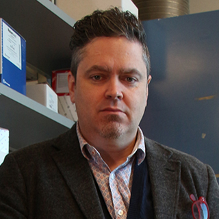 Tracy McGaha合作论文数Department of Immunology, Temerty Faculty of Medicine, University of Toronto;University Health Network16Eun-Joon Lee合作论文数15
 Eric Ring合作论文数14
 Ivan P. Maillard合作论文数Division of Hematology and Oncology, Department of Medicine, Perelman School of Medicine, University of Pennsylvania14
 Bree R Eaton合作论文数Dept Radiat Oncol, Emory Univ14
 George C. Prendergast合作论文数Lankenau Institute for Medical Research, Main Line Health14Berrong Zuzana合作论文数14
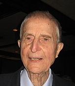 Eugene P. Kennedy合作论文数Harvard Medical School14Lirong Pei合作论文数14
 Geoffrey Hill合作论文数Translational Science and Therapeutics Division, Fred Hutch Cancer Center;Clinical Research Division, Fred Hutch Cancer Center;Division of Hematology and Oncology, School of Medicine, University of Washington14
 Essam A Mohamed合作论文数Composite Materials Department, Central Metallurgical Research and Development Institute (CMRDI),13Aguilera Dolly合作论文数13
 Santhakumar Manicassamy合作论文数Emory Vaccine Ctr13
 Babak Baban合作论文数13
 Muthusamy Thangaraju合作论文数Department of Biochemistry and Molecular Biology, Medical College of Georgia13
 Patricia Taylor合作论文数12
 Lei Huang (黄雷)合作论文数Zhiyuan College, Shanghai Jiao Tong University;Shanghai Jiao Tong University School of Medicine12
 Ryan A. Flynn合作论文数Department of Stem Cell and Regenerative Biology, Harvard University;Boston Children’s Hospital12
 Asim Saha合作论文数the Barrett Cancer Center, University of Cincinnati12James B. Duhadaway合作论文数Lankenau Institute for Medical Research12Yibing Peng合作论文数12
 Pandelakis Koni合作论文数Med Coll Georgia12B Baban合作论文数11Govindarajan Thangavelu合作论文数11
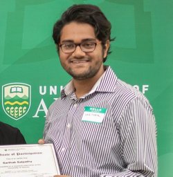 Sarthak Satpathy合作论文数Dept Biomed Informat, Emory Univ11
 Amol Ratnakar Suryawanshi合作论文数College of Veterinary Medicine, Auburn University11
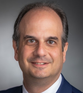 Corey Cutler合作论文数Center for Hematologic Oncology, Dana-Farber Cancer Institute;Division of Hematologic Oncology, Department of Medical Oncology, Dana-Farber Cancer Institute;Harvard Medical School11
 Austin Y Shull合作论文数Department of Biochemistry &Molecular Biology, Georgia Regents University;Department of Biochemistry & Molecular Biology, Georgia Regents University11Manoharan Indumathi合作论文数Georgia Regents University Cancer Center, Georgia Regents University11
 Yousef Zakharia合作论文数11




