The immunomodulatory and anti-tumor activity of Bru-Pel, an aqueous-ether extracted residue of Brucella abortus (strain 456), was investigated. Bru-Pel was administered to C57BL/6 mice intraperitoneally (i.p.) and tested for its effect on natural killer (NK) cell activity in spleen cells, liver, and peritoneal cavity. Three days after injecting 100 micrograms of Bru-Pel i.p., the cytotoxicity of spleen cells against YAC-1 target cells, assessed by LU20 increased by approximately two-fold and nonparenchymal cells of liver by greater than six-fold. The highest stimulatory effect of Bru-Pel was seen with peritoneal exudate cells, and 47-fold augmentation of NK cell activity was observed. Bru-Pel treatment made spleen, liver, and peritoneal exudate cells capable of lysing P815 mastocytoma cells, a tumor cell line highly resistant to lysis by unstimulated NK cells. In vivo, Bru-Pel inhibited the formation of experimental BL6 melanoma metastases; however, there was no significant effect on the eradication of established pulmonary metastatic lesions. These results demonstrate that in addition to its previously described macrophage-activating ability, Bru-Pel is highly efficient in stimulation of NK cell-mediated cytotoxicity in mice.
FEBS LettersVolume 223, Issue 2 p. 207-211 Discussion letterFree Access Conformational aspects of the reaction mechanisms of polysaccharide lyases and epimerases David Sidney Feingold, Corresponding Author David Sidney Feingold Department of Microbiology, Biochemistry and Molecular Biology, School of Medicine, University of Pittsburgh, Pittsburgh, PA 15261, USAD.S. Feingold, Department of Microbiology, Biochemistry and Molecular Biology, School of Medicine, University of Pittsburgh, Pittsburgh, PA 15261, USASearch for more papers by this authorRonald Bentley, Ronald Bentley Department of Biological Sciences, Faculty of Arts and Sciences, University of Pittsburgh, Pittsburgh, PA 15260, USASearch for more papers by this author David Sidney Feingold, Corresponding Author David Sidney Feingold Department of Microbiology, Biochemistry and Molecular Biology, School of Medicine, University of Pittsburgh, Pittsburgh, PA 15261, USAD.S. Feingold, Department of Microbiology, Biochemistry and Molecular Biology, School of Medicine, University of Pittsburgh, Pittsburgh, PA 15261, USASearch for more papers by this authorRonald Bentley, Ronald Bentley Department of Biological Sciences, Faculty of Arts and Sciences, University of Pittsburgh, Pittsburgh, PA 15260, USASearch for more papers by this author First published: November 02, 1987 https://doi.org/10.1016/0014-5793(87)80290-9Citations: 9AboutPDF ToolsRequest permissionExport citationAdd to favoritesTrack citation ShareShare Give accessShare full text accessShare full-text accessPlease review our Terms and Conditions of Use and check box below to share full-text version of article.I have read and accept the Wiley Online Library Terms and Conditions of UseShareable LinkUse the link below to share a full-text version of this article with your friends and colleagues. Learn more.Copy URL References 1 B. Larsen, A. Hang, Carbohydrate Res., 17, (1971), 287– 296. 2 M. Höök, U. Lindahl, G. Bäckström, A. Malmström, L.-Å. Fransson, J. Biol. Chem., 249, (1974), 3908– 3915. 3 H.S. Prihar, P. Campbell, D.S. Feingold, I. Jacobsson, J.W. Jensen, U. Lindahl, L. Rodén, Biochemistry, 19, (1980), 495– 500. 4 I. Jacobsson, G. Bäckström, M. Höök, U. Lindahl, D.S. Feingold, A. Malmström, L. Rodén, J. Biol. Chem., 254, (1979), 2975– 2982. 5 P. Gacesa, FEBS Lett., 212, (1987), 199– 202. 6 J.N. BeMiller, G.V. Kumari, Carbohydrate Res., 25, (1972), 4129– 7 J.F. Stoddardt, Stereochemistry of Carbohydrates (1971), Wiley New York 47– 92. 8 D.A. Rees, Polysaccharide Shapes (1977), Chapman and Hall London 13– 30. 9 J.F. Kennedy, C.A. White, Chemistry Biochemistry and Biology 176, (1983), E. Horwood Chichester 34– 10 R. Bentley, Annu. Rev. Biochem., 41, (1972), 953– 996. 11 B. Larsen, A. Haug, Carbohydrate Res., 20, (1971), 225– 232. 12 K.R. Hanson, J. Am. Chem. Soc., 88, (1966), 2731– 2742. 13 J. Kiss, R.S. Tipson D. Horton Advances in Carbohydrate, Chemistry and Biochemistry 29, (1974), Academic Press New York 229– 303. 14 P.N. Sanderson, T.N. Huckerby, I.A. Nieduszynski, Biochem. J., 243, (1987), 175– 181. 15 D.R. Ferro, A. Provasoli, M. Ragazzi, G. Torri, B. Casu, G. Gatti, J.-C. Jacquinet, P. Sinaÿ, M. Petitou, J. Choay, J. Am. Chem. Soc., 108, (1986), 6773– 6778. 16 G. Skjåk-Bræk, B. Larsen, Carbohydrate Res., 139, (1985), 273– 283. 17 K.H. Min, S.F. Sasaki, Y. Kashiwabara, K. Nisizawa, J. Biochem. (Tokyo), 81, (1977), 547– 553. 18 T. Muramatsu, F. Imasoto, Agric. Biol. Chem., 51, (1987), 1169– 1171. Citing Literature Volume223, Issue2November 02, 1987Pages 207-211 ReferencesRelatedInformation
The year was 1935. In Stockholm, Toronto and Berne preparations were under way for clinical trials to determine whether heparin prevented post-opera- tive thrombosis and could be used in the treatment of established thrombo- embolic disease. Heparin had been dis- covered almost 20 years earlier by McLean 1, who was then a second-year medical student working in Howelrs laboratory at Johns Hopkins University. It had finally been purified in sufficient quantity for testing in humans, thanks to the efforts of Charles and Scott in Toronto and Jorpes in Stockholm. In the summer of 1935, in Stockholm~ Clarence Crafoord began administering heparin to his surgical patients as pro-. phylaxis against post-operative throm-. bosis. Initially, a hefty dose of heparin was given several hours before surge D , but occasional complications in the form of massive hematomas in the wound area made Crafoord settle, somewhat reluctantly, for post-operative treat- ment. What was heparin like as a drug in 1935? There were still problems in arriv- ing at a preparation devoid of side effects. In a paper 2 in 1939, Crafoord pointed out that although heparin
GDPmannose dehydrogenase (EC 1.1.1.132) in a mucoid strain of Pseudomonas aeruginosa isolated from a patient with cystic fibrosis was identified by demonstrating the NAD-linked formation of GDPmannuronate from GDPmannose mediated by a cell extract of the organism. Nonmucoid mutant strains did not contain GDPmannose dehydrogenase, which suggests that the enzyme is involved in the biosynthesis of alginate-like polysaccharide by P. aeruginosa.
The binding of NADH to uridine diphosphate glucose dehydrogenase has been examined by equilibrium dialysis. There is an absolute requirement for the presence of UDP-glucose for the binding of NADH. Other analogs such as UDPxylose, UDPgalactose and UDPglucuronic acid cannot replace UDPglucose as an effector of NADH binding. UDPxylose competes with UDPglucose for the UDP-sugar-binding site, and in so doing releases the bound NADH. The binding of NADH to UDPglucose dehydrogenase in the presence of UDPglucose reaches a saturation limit of 3 mol NADH bound per enzyme hexamer, and displays positive cooperativity, Hill number = 1.34. The effects of UDP-sugars on the fluorescence of UDPglucose dehydrogenase derivatized at the catalytic sites with a fluorophore have also been studied. Two classes of UDPxylose-binding site have been detected. One class has high affinity (Kdiss = 3 microM, determined by equilibrium dialysis) but does not affect fluorophore fluorescence, and the other has lower affinity (Kdiss = 120 microM) and leads to red-shifted fluorescence quenching, presumably by effecting exposure of the fluorophore to solvent. The high-affinity sites are identified as the UDP-sugar subsites of the underivatized catalytic sites, and the low-affinity sites as UDP-sugar subsites of the fluorophore-labeled catalytic sites.
The catalytic-site thiol groups of UDP-glucose dehydrogenase from bovine liver were carboxymethylated with iodo[2-14C]acetate or with iodoacetamidofluorescein. After the residual thiol groups were carboxymethylated with iodoacetate, the proteins were digested with trypsin. The 14C-labelled peptide from the carboxymethylated enzyme was purified to homogeneity by successive thick-layer chromatography on silica gel, paper electrophoresis and chromatography, and column chromatography on Bio-Gel P-6. Homogeneous fluoresceincarboxamidomethylated peptide was prepared from a tryptic digest of fluoresceincarboxamidomethylated enzyme by specific adsorption--desorption from Sephadex G-25. The sequences of either peptide determined by the manual Edman dansyl procedure is: Ala-Ser-Val-Gly-Phe-Gly-Gly-Ser-Cys-Phe-Glx-Glx-Gly-Lys.
Three types of four-electron transfer, pyridine nucleotide-linked dehydrogenases are known: β-methyl-β-hydroxyglutaryl CoA reductase, histidinol dehydrogenase, and nucleoside diphosphate sugar dehydrogenases. Although the reactions catalysed by the latter two classes of enzyme are very similar, the enzymes are structurally different.
A method has been developed for the quantitative determination of the relative proportions of d-mannuronic and l-guluronic acids in alginic acid. To obtain homogeneous reaction conditions the viscosity of the alginic acid sample was first decreased by limited hydrolysis with mineral acid. The carboxyl groups were then esterified by reaction with 1-ethyl-3-[3-(dimethylamino)propyl]-carbodiimide, and reduced with sodium borohydride. The resulting hexosans were converted by acid hydrolysis to d-mannose and an equilibrium mixture of l-gulose and 1,6-anhydro-l-gulose. These were treated with sodium borohydride; the 1,6-anhydro-l-gulose was not reduced whereas d-mannose and l-gulose were converted to d-mannitol and d-glucitol. The hexitols were estimated by gas-liquid chromatography as the n-butane boronic acid esters, and the relative proportions of the uronic acids in the alginic acid were calculated by taking into account the equilibrium ratio of l-gulose and 1,6-anhydro-l-gulose. The method can be used to analyze as little as 2 mg of alginic acid.
Half-of-the-sites reactivity of the catalytic site thiol groups of UDPglucose dehydrogenase (UDPglucose:NAD+ 6-oxidoreductase, EC 1.1.1.22) can be ascribed either to the induction of conformational asymmetry following derivatization of one half of the subunits or to intrinsic conformational differences in the subunits of the native enzyme. If the half-sites reactivity behavior is due to induction effects, the magnitude of the induction could be expected to depend on the nature of the covalent modification. On the other hand, if the half-sites reactivity behavior is due to pre-existing asymmetry and there is no communication between catalytic centers, the properties of unmodified subunits should be independent of the nature of the covalent derivative introduced on the modified subunits. According to the induced asymmetry hypothesis, the catalytic activity of half-sites modified enzyme might be different for different covalent modifications, whereas for the rigid pre-existing asymmetry hypothesis the catalytic activity of half-sites modified enzyme should be the same regardless of the modifying group. During the course of catalytic site thiol group modification by a number of thiol specific reagents, the loss of enzyme activity was equivalent to the degree of modification for most of the reagents employed. However, with iodoacetate and 5-(iodoacetamidoethyl)amimonaphthalene-1-sulfonic acid, half-sites modification of UDPglucose dehydrogenase
ADVERTISEMENT RETURN TO ISSUEPREVArticleNEXTBiosynthesis of heparin. Hydrogen exchange at carbon 5 of the glycuronosyl residuesHarry S. Prihar, Patrick Campbell, David Sidney Feingold, Ingvar Jacobsson, John W. Jensen, Ulf Lindahl, and Lennart RodenCite this: Biochemistry 1980, 19, 3, 495–500Publication Date (Print):February 5, 1980Publication History Published online1 May 2002Published inissue 5 February 1980https://pubs.acs.org/doi/10.1021/bi00544a016https://doi.org/10.1021/bi00544a016research-articleACS PublicationsRequest reuse permissionsArticle Views56Altmetric-Citations31LEARN ABOUT THESE METRICSArticle Views are the COUNTER-compliant sum of full text article downloads since November 2008 (both PDF and HTML) across all institutions and individuals. These metrics are regularly updated to reflect usage leading up to the last few days.Citations are the number of other articles citing this article, calculated by Crossref and updated daily. Find more information about Crossref citation counts.The Altmetric Attention Score is a quantitative measure of the attention that a research article has received online. Clicking on the donut icon will load a page at altmetric.com with additional details about the score and the social media presence for the given article. Find more information on the Altmetric Attention Score and how the score is calculated. Share Add toView InAdd Full Text with ReferenceAdd Description ExportRISCitationCitation and abstractCitation and referencesMore Options Share onFacebookTwitterWechatLinked InRedditEmail Other access optionsGet e-Alertsclose Get e-Alerts
ADVERTISEMENT RETURN TO ISSUEPREVArticleNEXTResonance energy transfer between catalytic sites of bovine liver uridine diphosphoglucose dehydrogenaseJames S. Franzen, Paul S. Marchetti, and David S. FeingoldCite this: Biochemistry 1980, 19, 26, 6080–6089Publication Date (Print):December 1, 1980Publication History Published online1 May 2002Published inissue 1 December 1980https://doi.org/10.1021/bi00567a021RIGHTS & PERMISSIONSArticle Views51Altmetric-Citations25LEARN ABOUT THESE METRICSArticle Views are the COUNTER-compliant sum of full text article downloads since November 2008 (both PDF and HTML) across all institutions and individuals. These metrics are regularly updated to reflect usage leading up to the last few days.Citations are the number of other articles citing this article, calculated by Crossref and updated daily. Find more information about Crossref citation counts.The Altmetric Attention Score is a quantitative measure of the attention that a research article has received online. Clicking on the donut icon will load a page at altmetric.com with additional details about the score and the social media presence for the given article. Find more information on the Altmetric Attention Score and how the score is calculated. Share Add toView InAdd Full Text with ReferenceAdd Description ExportRISCitationCitation and abstractCitation and referencesMore Options Share onFacebookTwitterWechatLinked InReddit PDF (1 MB) Get e-Alerts Get e-Alerts
The multistep enzymatic conversion of d-[5-3H]glucose to d-[5-3H]mannose is described. The d-5-3H]mannose was in turn converted to guanosine 5′-(α-d-[5-3H]mannopyranosyl pyrophosphate) using an extract from Arthrobacter viscosus, NRRL 1973. The overall radiochemical yield was 25%.
Two types of high-molecular water-soluble substrates of beta-D-galactosidase were prepared. Substrate I contains beta-D-[3H]-galactopyranosyl moieties linked, through a hydrocarbon bridge, to a polymeric dialdehyde (oxidized starch) backbone (molecular weight 6,000); in substrate II the backbone is poly-L-lysine (molecular weight 80,000). In the presence of Triton X-100, but not in its absence, D-[3H]-galactose is split from the substrates by homogenates of normal mouse liver or pancreas. It is suggested that substrates I and II could be used to test the integrity of lysosomal and other cellular membranes, and to assess the extent of cellular injury.
 Js Franzen合作论文数10Gad Avigad合作论文数8Hs Prihar合作论文数6Der-Fong Fan合作论文数6Js Youngner合作论文数5Ee Parris合作论文数4
 Daniel Teres合作论文数4Ashcom J合作论文数4Leonard S. Bushnell合作论文数4S HESTRIN合作论文数3Nancy B Schwartz合作论文数3P Marchetti合作论文数3
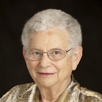 Elizabeth F. Neufeld (Elizabeth Neufeld)合作论文数Department of Biological Chemistry, David Geffen School of Medicine, University of California, Los Angeles3Helmut Ankel合作论文数3T.H. Chiu合作论文数3J. G. Schiller合作论文数3Bk Pugashetti合作论文数2Eugene E. Grebner合作论文数2L Vadas合作论文数2
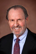 Kenneth A. Arndt合作论文数SkinCare Physicians2
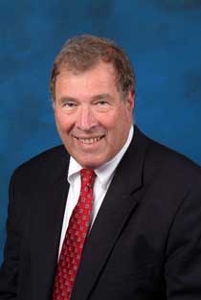 Sheldon Greenfield合作论文数School of Medicine, University of California, Irvine2Kuo Irene合作论文数2John Hedley-Whyte合作论文数Beth Israel Hospital, Harvard Medical School2W. Z. Hassid合作论文数2Gary C. Du Moulin合作论文数2Austin L. Vickery合作论文数2Inder J. Sud合作论文数2James M. Klick合作论文数2Amikam Cohen合作论文数2Shlomo Hestrin合作论文数2Julius S. Youngner合作论文数2Alan J. Eichler合作论文数2S RUDDY合作论文数1G Keleti合作论文数1Jr Lamontagne合作论文数1La Glasgow合作论文数1Cj Schleupner合作论文数1I JACOBSSON合作论文数1Hr Thacore合作论文数1Jn Goldman合作论文数1Patrick Campbell合作论文数1Ma Chirigos合作论文数1K.N. Rao合作论文数1Kf Austen合作论文数1Raymond Ishman合作论文数1Emilie Kovach合作论文数1Juris Zalitis合作论文数1Nancy L. Stagg合作论文数1




