Abstract The tumor-suppressor p53 prevents cancer development via initiating cell-cycle arrest, cell death, repair, or antiangiogenesis processes. Over 50% of human cancers harbor cancer-causing mutant p53. p53 mutations not only abrogate its tumor-suppressor function, but also endow mutant p53 with a gain of function (GOF), creating a proto-oncogene that contributes to tumorigenesis, tumor progression, and chemo- or radiotherapy resistance. Thus, targeting mutant p53 to restore a wild-type p53 signaling pathway provides an attractive strategy for cancer therapy. We demonstrate that small-molecule NSC59984 not only restores wild-type p53 signaling, but also depletes mutant p53 GOF. NSC59984 induces mutant p53 protein degradation via MDM2 and the ubiquitin–proteasome pathway. NSC59984 restores wild-type p53 signaling via p73 activation, specifically in mutant p53-expressing colorectal cancer cells. At therapeutic doses, NSC59984 induces p73-dependent cell death in cancer cells with minimal genotoxicity and without evident toxicity toward normal cells. NSC59984 synergizes with CPT11 to induce cell death in mutant p53-expressing colorectal cancer cells and inhibits mutant p53-associated colon tumor xenograft growth in a p73-dependent manner in vivo. We hypothesize that specific targeting of mutant p53 may be essential for anticancer strategies that involve the stimulation of p73 in order to efficiently restore tumor suppression. Taken together, our data identify NSC59984 as a promising lead compound for anticancer therapy that acts by targeting GOF-mutant p53 and stimulates p73 to restore the p53 pathway signaling. Cancer Res; 75(18); 3842–52. ©2015 AACR.
p53-dependent apoptosis is a major determinant of its tumor suppressor activity and can be triggered by hypoxia. No p53 target is known to be induced by p53 or to mediate p53-dependent apoptosis during hypoxia. We report that p53 can directly upregulate expression of Bnip3L, a cell death inducer. During hypoxia, Bnip3L is highly induced in wild-type p53-expressing cells, in part due to increased recruitment of p53 and CBP to Bnip3L. Apoptosis is reduced in hypoxia-exposed cells with functional p53 following Bnip3L knockdown. In vivo, Bnip3L knockdown promotes tumorigenicity of wild-type versus mutant p53-expressing tumors. Thus, Bnip3L, capable of attenuating tumorigenicity, mediates p53-dependent apoptosis under hypoxia, which provides a novel understanding of p53 in tumor suppression.
BACKGROUND:We previously reported the identification of ONC201/TIC10, a novel small molecule inducer of the human TRAIL gene that improves efficacy-limiting properties of recombinant TRAIL and is in clinical trials in advanced cancers based on its promising safety and antitumor efficacy in several preclinical models. METHODS:We performed a high throughput luciferase reporter screen using the NCI Diversity Set II to identify TRAIL-inducing compounds. RESULTS:Small molecule-mediated induction of TRAIL reporter activity was relatively modest and the majority of the hit compounds induced low levels of TRAIL upregulation. Among the candidate TRAIL-inducing compounds, TIC9 and ONC201/TIC10 induced sustained TRAIL upregulation and apoptosis in tumor cells in vitro and in vivo. However, ONC201/TIC10 potentiated tumor cell death while sparing normal cells, unlike TIC9, and lacked genotoxicity in normal fibroblasts. Investigating the effects of TRAIL-inducing compounds on cell signaling pathways revealed that TIC9 and ONC201/TIC10, which are the most potent inducers of cell death, exclusively activate Foxo3a through inactivation of Akt/ERK to upregulate TRAIL and its pro-apoptotic death receptor DR5. CONCLUSION:These studies reveal the selective activity of ONC201/TIC10 that led to its selection as a lead compound for this novel class of antitumor agents and suggest that ONC201/TIC10 is a unique inducer of the TRAIL pathway through its concomitant regulation of the TRAIL ligand and its death receptor DR5.
Supplementary Figures S1-S12. Validation of kinase regulators of ONC201 sensitivity (S1); KSR1 shifts the ONC201, but not lapatinib, dose-response curve downward (S2); KSR1 does not regulate ONC201-induced TRAIL or DR5 (S3); KSR1 does not affect ONC201-mediated inhibition of MAPK or Akt inhibition (S4); Distribution of ONC201 synergy instances by drug (S5); Validation of ONC201 combinatorial activity with approved anti-cancer small molecules (S6); ONC201 synergizes with sorafenib in HepG2 cells (S7); ONC201 and sorafenib cooperatively induce apoptosis, TRAIL, and DR5 in vivo (S8); ONC201 and sorafenib cooperatively induce apoptosis in HepG2 cells (S9); ONC201 and sorafenib combination therapy is well tolerated and effective in vivo (S10); FLIP, Mcl-1, and KSR-1 knockdown enhances ONC201 response in HCT116 cells (S11); Model of ONC201-induced TRAIL signaling and sensitivity factors (S12).
PDF file, 84K, Excess soluble DR4, but not DR5, blocks the cytotoxic effect of agonist DR4 Atrimer complexes. Atrimer complex 1C9 was pre-incubated with or without soluble DR4-Fc or DR5-Fc for 1 hour before addition to Colo205 cells. Cell viability was measured at 48 hours using the ViaLight Plus kit.
Supplementary Figure 1. UPR genes in DU145 cells. Supplementary Figure 2. ONC201 does not inhibit translocation of AR-GFP to the nucleus upon DHT treatment and does not induce AR degradation. Supplementary Figure 3. ONC201 partly synergizes with docetaxol. Supplementary Figure 4. ONC201 partly synergizes with crizotinib. Supplementary Figure 5. Biweekly measurements of mouse weights after the indicated treatments in different prostate cancer xenograft models.
PDF file - 338KB, Figure S1. COX-2 is overexpressed in MDA-MB-231 cells. Figure S2. Celecoxib inhibits the invasion and migration capacity of breast cancer cells in vitro. Figure S3. Celecoxib inhibits the migration of breast cancer cells into the cerebrospinal fluid. Figure S4. Enumerating human breast cancer cells in mouse blood using the CellSearch system. Figure S5. Changes in CTC and CSFTC counts as a function of changes in neurological or systemic clinical symptoms of disease. Table S1. Receptor status of enrolled breast cancer patients.
PDF file, 121K, Screening for cytotoxicity of SLM3 in cancer cell lines derived from multiple tumor types.
PDF file, 63K, Combining DR4 Atrimer complexes with TRAIL. Sub-G1 content in HCT116 cells treated with 1G2 (50 ng/mL) or TRAIL (25 ng/mL) for 24 hours (n=2).
<p>ONC201/TIC10-mediated CSC depletion involves inhibition of Akt, ERK and activation of Foxo3a.</p>
 Wafik S. El-Deiry合作论文数Department of Pathology and Laboratory Medicine, Warren Alpert Medical School, Brown University;Cancer Center, Brown University;Lifespan Cancer Institute, Rhode Island Hospital224
 Joshua E Allen合作论文数Chimerix, Inc.100
 Varun Vijay Prabhu合作论文数Chimerix48
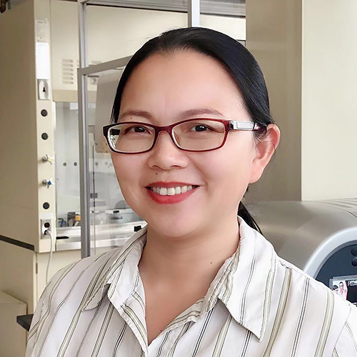 Lanlan Zhou合作论文数Department of Pathology and Laboratory Medicine, Brown University38
 Lulla Amriti合作论文数Penn State Hershey Cancer Institute, Penn State College of Medicine38
 Nathan G. Dolloff合作论文数Milton S. Hershey Medical Center, Hershey Medical Center32Avital Lev合作论文数28
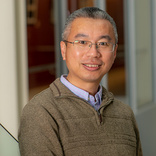 Shengliang Zhang合作论文数Department of Pathology and Laboratory Medicine, Brown University26Wagner Jessica合作论文数19Hong Bo合作论文数17
 Das Avisnata合作论文数a Hematology/Oncology Division;Penn State Hershey Cancer Institute;Her15Mayes Patrick A合作论文数13A. P. J. Van Den Heuvel合作论文数13Christina Kline合作论文数13
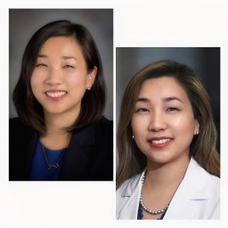 Bora Lim合作论文数Department of Breast Medical Oncology, The University of Texas MD Anderson Cancer Center12
 Talekar Mala合作论文数Penn State Hershey Cancer Institute, Penn State College of Medicine12Lori S. Hart合作论文数12Elise Chen合作论文数11Elizabeth M Matthew合作论文数11Oltean Daniela I合作论文数11Mark W. Renshaw合作论文数11Batzer Glenda合作论文数11Ferrini Roger合作论文数11Bing Lin合作论文数11
 Arunasalam Navaraj合作论文数Department of Biochemistry, Indian Institute of Science11Anke Kretz-Rommel合作论文数11Charles D. Smith合作论文数Apogee Biotechnology Corporation11Wafik El-Deiry合作论文数10Shuai Zhao合作论文数Brown University, Brown University Providence10Liz Hernandez Borrero合作论文数9Marie D. Ralff合作论文数9k peters合作论文数9J. M. Sheehan合作论文数9Malysz Jozef合作论文数9
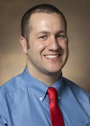 Gallant Jean-Nicolas合作论文数Penn State Hershey Medical Center, Penn State Hershey Cancer Institute9Akshal S. Patel合作论文数9
 Jussuf T Kaifi合作论文数Department of General, Visceral and Thoracic Surgery, University Medical Center Hamburg-Eppendorf9
 Michael Glantz合作论文数Berkshire Medical Center, University of Massachusetts9
 Giampaolo Talamo合作论文数Milton S. Hershey Medical Center, Penn State College of Medicine9Nicole Aqui合作论文数8Humphreys Robin C合作论文数8Zhaohai Yang合作论文数8Miriam Kunkel合作论文数8Levy Kopelovich合作论文数Division of Cancer Prevention, NCI8
 Junaid Abdulghani合作论文数Department of Cancer Biology, Kimmel Cancer Center8
 Dan T. Vogl合作论文数Division of Hematology/Oncology, University of Pennsylvania School of Medicine8Yingqiu Y. Liu合作论文数7Marie D. Baumeister合作论文数7
 Jay Fitzgerald Dorsey合作论文数Department of Radiation Oncology, Perelman School of Medicine, University of Pennsylvania7




