In this retrospective review, a head-mounted augmented reality (AR) display was investigated to evaluate its clinical usability and ergonomic benefits across multiple musculoskeletal image-guided procedures. Data were collected through an electronic survey with 5-point Likert scale (n = 9) and single-user motion analysis. Nine interventional radiologists rated their experience in procedures including drainage, biopsy, endovascular procedure, ablation, and cement augmentation. Kinematic data were obtained from 14 needle placement procedures (10 kyphoplasties and 4 spinal biopsies), comparing sessions with AR enabled (n = 7) versus without AR (n = 7). User feedback indicated high comfort (median Likert score, 5), ease of use (median Likert score, 4), and positive ergonomic impact (median Likert score, 5), with 66% reporting improved ergonomics. Motion analysis revealed procedures without AR required more frequent (P = .003) and greater magnitude (P = .0002) head turns during image-guided needle advancement. AR demonstrated improvements in ergonomic efficiency and user satisfaction for musculoskeletal interventional procedures, although image quality refinement could enhance clinical adoption.
Interventional radiology (IR) procedures traditionally rely on two-dimensional imaging for guidance. Augmented reality (AR) offers a transformative approach by overlaying two-dimensional radiological images or three-dimensional digital reconstructions onto the real-world environment, potentially improving procedural precision and operator ergonomics. This narrative literature review examines AR systems, procedural workflow, operator ergonomics, current clinical applications, and outcomes. Inclusion criteria include studies investigating AR in IR, excluding articles unrelated to IR procedures. Current literature suggests improved ergonomics, mainly enhanced depth perception, decreased eye movement/fatigue, and decreased cybersickness with overall operator tolerability. Clinical studies demonstrated reductions in radiation exposure and procedural times, with comparable accuracy to standard techniques. Limitations include the lack of large-scale clinical data and standardized evaluation metrics.
To evaluate the feasibility of using generative artificial intelligence (GenAI) in determining entry angles for lung needle biopsies. This retrospective IRB-approved study analyzed de-identified computed tomography (CT) images from 30 lung biopsy procedures using GPT-4 (OpenAI, San Francisco, CA) to identify biopsy paths that minimize tissue disruption and avoid critical structures. A radial grid was overlaid on axial CT images to facilitate identification of entry angles (EA). The proposed GenAI EA were compared with the biopsy trajectories selected by clinicians during manual procedures. The median number of iterations required to identify the GenAI EA was 2 (interquartile range: 1–3). Independent reviewers deemed 28 of 30 (93.3
Sinistral portal hypertension, also known as left-sided portal hypertension, is a rare cause of gastric variceal bleeding which occurs secondary to occlusion of the splenic vein. We present a case of venous occlusion and sinistral portal hypertension secondary to distal pancreatic cancer requiring treatment of gastric variceal bleeding. After failing conservative management, transvenous intervention was attempted, but a venous communication with the gastric varices was unable to be identified on multiple venograms. A percutaneous trans-splenic approach using a 21-G needle and ultrasound guidance was successful in directly accessing an intraparenchymal vein feeding the gastric varices, and glue embolization was performed directly through the access needle with excellent results.
The purpose of this study was to assess characteristics and outcomes of patients with breast cancer with liver metastasis who have and have not undergone radioembolization. Medical records of patients with breast cancer with liver metastases (4,332 patients who had not undergone radioembolization, 166 patients who had) seen between January 1, 2009 and October 31, 2021 were assessed. Propensity Score Matching was performed. Radioembolization patients were significantly younger (47.7 ± 10.1 vs. 52.3 ± 12.5, p <0.001) and underwent more systemic treatment lines (8.6 ± 4.1 vs 5.2 ± 3.2, p < 0.001). Median survival was longer in unmatched (67.1 vs 40.7 months, p=0.001) and matched (67.1 vs. 38.0 months, p=0.001) cohorts. Radioembolization favorably affected survival (p=0.002). In conclusion, patients with breast cancer with liver metastases that undergo radioembolization are significantly younger and undergo more treatment lines. Radioembolization was associated with longer median survival.
PURPOSE:To compare the effectiveness of percutaneous lung biopsy using a patient-mounted needle-driving robotic system with that using a manual insertion of needles under computed tomography (CT) fluoroscopy guidance. MATERIALS AND METHODS:In this institutional review board approved study, the cohort consisted of a series of patients who underwent lung biopsies following the intention-to-treat protocol from September 2022 to September 2023 using robot (n = 15) or manual insertion under single-rotation CT fluoroscopy (n = 66). Patient and procedure characteristics were recorded as well as outcomes. RESULTS:Although age, body mass index, and skin-to-target distance were not statistically different, target size varied (median, 8 mm [interquartile range, 6.5-9.5 mm] for robot vs 12 mm [8-18 mm] for single-rotation CT fluoroscopy; P = .001). No statistical differences were observed in technical success (86.7% [13/15] vs 89.4% [59/66], P = .673), Grade 3 adverse event (AE) (6.7% [1/15] vs 12.1% [8/66], P = .298), procedural time (28 minutes [22-32 minutes] vs 19 minutes [14.3-30.5 minutes], P = .086), and patient radiation dose (3.9 mSv [3.2-5.6 mSv] vs 4.6 mSv [3.3-7.5 mSv], P = .398). In robot-assisted cases, the median angle out of gantry plane was 10° (6.5°-16°), although it was null (0°-5°) for single-rotation CT fluoroscopy (P = .001). CONCLUSIONS:Robot-assisted and single-rotation CT fluoroscopy-guided percutaneous lung biopsies were similar in terms of technical success, diagnostic yield, procedural time, AEs, and radiation dose, although robot allowed for out-of-gantry plane navigation along the needle axis.
Objectives: This systematic review aims to assess existing research concerning the use of robotic systems to execute percutaneous lung biopsy. Methods: A systematic review was performed and identified 4 studies involving robotic systems used for lung biopsy. Outcomes assessed were operation time, radiation dose to patients and operators, technical success rate, diagnostic yield, and complication rate. Results: One hundred and thirteen robot-guided percutaneous lung biopsies were included. Technical success and diagnostic yield were close to 100%, comparable to manual procedures. Technical accuracy, illustrated by needle positioning, showed less frequent needle adjustments in robotic guidance than in manual guidance ( P < .001): 2.7 ± 2.6 (range 1-4) versus 6 ± 4 (range 2-12). Procedure time ranged from comparable to reduced by 35% on average (20.1 ± 11.3 minutes vs 31.4 ± 10.2 minutes, P = .001) compared to manual procedures. Patient irradiation ranged from comparable to reduced by an average of 40% (324 ± 114.5 mGy vs 541.2 ± 446.8 mGy, P = .001). There was no significant difference in reported complications between manual biopsy and biopsies that utilized robotic guidance. Conclusion: Robotic systems demonstrate promising results for percutaneous lung biopsy. These devices provide adequate accuracy in probe placement and could both reduce procedural duration and mitigate radiation exposure to patients and practitioners. However, this review underscores the need for larger, controlled trials to validate and extend these findings.
Aim: To investigate how the sequence of checkpoint immunotherapy (CPI) and transarterial embolization (TAE) affects overall survival (OS) of patients with metastatic melanoma. Materials & methods: This retrospective cohort study included 65 patients with metastatic melanoma who underwent both TAE and CPI between September 2011 and January 2022. Results: Significantly higher OS was seen in patients who received CPI before and after embolization (22 months, 95% CI 14-NR, p < 0.001) compared with only before embolization (4.5 months 95% CI, 14-NR). <= 3 hepatic metastasis (p < 0.01), more TAE procedures (p < 0.001) and CPI sequence (before and after embolization) (p < 0.001) were independent predictors of survival. Conclusion: Metastatic melanoma patients who underwent TAE have longer survival when CPI was sequenced both before and after embolization. This study looked at how the order of two treatments, called checkpoint immunotherapy (CPI) and transarterial embolization (TAE), affects how long people with metastatic melanoma live. Sixty-five patients who had both treatments between September 2011 and January 2022 took part in the study. Patients with fewer than three liver metastases, cancer in just one part of the liver, and who had more TAE treatments tended to live longer. Patients who got CPI both before and after TAE lived longer compared with those who only got CPI before TAE.
Background Lower extremity ischemia due to extrinsic arterial compression by venous stent placement is a rare but increasingly recognized occurrence. Given the rise of complex venous interventions, awareness of this entity is becoming increasingly important to avoid serious complications. Case Presentation A 26-year-old with progressively enlarging pelvic sarcoma despite chemoradiation developed recurrent symptomatic right lower extremity deep venous thrombosis due to worsening mass effect on a previously placed right common iliac vein stent. This was treated with thrombectomy and stent revision, with extension of the right common iliac vein stent to the external iliac vein. During the immediate post-procedure period the patient developed symptoms of acute right lower extremity arterial ischemia including diminished pulses, pain, and motorsensory loss. Imaging confirmed extrinsic compression of the external iliac artery by the newly placed adjacent venous stent. The patient underwent stenting of the compressed artery with complete resolution of ischemic symptoms. Conclusions Awareness and early recognition of arterial ischemia following venous stent placement is important to prevent serious complication. Potential risk factors include patients with active pelvis malignancy, prior radiation, or scarring from surgery or other inflammatory processes. In cases of threatened limb, prompt treatment with arterial stenting is recommended. Further study is warranted to optimize detection and management of this complication.
This retrospective study evaluated the feasibility and safety of percutaneous computed tomography (CT)-guided bone biopsies in patients with cancer using a patient-mounted robotic system with steering capabilities. The study included 39 patients (17 women, 22 men; median age, 65.5 years; interquartile range [IQR], 54.8–71.0 years). Forty biopsies were performed in the pelvis, spine, ribs, shoulder, femur, and sternum. The technical success rate was 100%, and the median trajectory length was 55.9 mm (IQR, 47.1–73.6 mm). Intermediate checkpoints were used in 8 biopsies. Median time from the first to final scan was 21 minutes (IQR, 17–37 minutes). The overall procedure time was 30 minutes (IQR, 24–36 minutes). The median dose length product and effective dose were 536.6 mGy⋅cm (IQR, 396.2–837.7 mGy∗cm) and 7.1 mSv (IQR, 4.7–10.8 mSv), respectively. No adverse events occurred. The diagnostic yield for cancer was 72.5%. Percutaneous robotic-assisted bone biopsies demonstrated high technical success, adequate diagnostic yield, and favorable safety profile.
To assess the prognostic value of total lesion glycolysis (TLG) and to assess for a relationship between tumor absorbed dose (TAD) and change in total lesion glycolysis (ΔTLG) in patients with breast cancer liver metastases undergoing radioembolization. This is an IRB-approved retrospective study. From January 2015 to April 2018 a total of 15 female patients with breast cancer liver metastases (ages 37-72 years, mean 54.8) underwent ecanal-90 radioembolization using glass microspheres. Pre-treatment and post-treatment PET/CT were evaluated to determine metabolic tumor volume (MTV), SUVmean, SUVmax, and TLG using Aquarius iNtuition. ΔTLG was subsequently calculated. Treatment response was assessed using PERCIST. Post 90Y bremsstrahlung SPECT/CT was evaluated to determine TAD with MIRD partition model using HERMES. The relationship between TAD and ΔTLG was analyzed using Spearman correlation coefficients. Univariable analysis of Overall Survival (OS) was performed through Kaplan-Meier survival curves and log-rank tests. Median OS was 18.8 months. 1, 2 and 3year survival was 75%, 26.7% and 13.3% respectively. Patients with low TLG (TLG< 1000) compared with those with high TLG had significantly improved survival (46.7 months vs 14.8 months P = 0.014). There was a statistically significant difference in OS when TAD in the target lesion was >200 Gy (38.9 months vs 12.5 months P < 0.05). ΔTLG greater than 70% was associated with statistically significant improved survival (42.1 months vs 11.4 months P< 0.05). Finally, there is a positive correlation between the TAD and ΔTLG (P< 0.05). Pretreatment TLG appears to be a prognostic factor influencing OS in patients with metastatic breast cancer. Higher tumor absorbed doses lead to improved outcomes with higher ΔTLG and improved OS. ΔTLG may be an effective method to quantify and monitor post-treatment response for radioembolization.
To assess the long-term outcomes of patients with breast cancer liver metastases treated with PET-guided microwave ablation and analyze variables affecting survival. This is an IRB-approved retrospective review. From March 2012 to October 2017 a total of 20 patients (ages 39-83, mean 53.9years) with breast cancer liver metastases underwent FDG-18-PET-guided microwave ablation using split dose technique. The number of lesions ranged from 1 to 3. Lesions ranged in size from 1.2-4.6 cm. Margin assessment at time of ablation was performed using PET overlay. Independent variables assessed included number of lesions, lesion size > 3 cm, hormone receptor status, presence of margins >10 mm, and presence of extrahepatic disease. Univariate analysis of overall survival (OS) was performed through Kaplan-Meier survival curves and log-rank tests. Technical efficacy was 95% with complete metabolic response in 19 of 20 patients. Median OS was 46.3 months. 1, 3 and 5 year survival was 93%, 56% and 48% respectively. There was a statistically significant increase in OS when ablation margins were >10 mm (63.9 months vs 23.8 months, P < 0.05). Other factors which significantly decreased OS were multiplicity of lesions (P = 0.05) and presence of extrahepatic disease (P = 0.028). Lesion size >3 cm and hormone receptor status did not significantly impact OS. PET-guided microwave ablation of breast cancer liver metastases is most effective with a margin >10 mm in patients with a solitary lesion and no extrahepatic disease. PET guidance may be an effective method for treating larger lesions >3 cm compared with conventional image guided ablation techniques.
Objective Robotic-guided interventions are emerging techniques that are gradually becoming a common tool for performing biopsies and tumor ablations in liver. This systematic review aims to evaluate their advancements, challenges, and outcomes. Methods A systematic review was conducted using the PubMed database to identify relevant articles published between January 2000 and February 2023. Inclusion criteria focused on studies that assessed robotic systems for percutaneous liver biopsies and tumor ablations. Data extraction was performed to collect information on study characteristics; robotic systems; components and software; imaging modality; degree of freedom; and needle insertion methods. The outcome measures analyzed were procedure time, radiation dose, and accuracy. Results 10 studies met the inclusion criteria. The robotic devices used included MAXIO, EPIONE, ROBIO-EX, AcuBot, and ACE robotic systems. The data set consisted of 429 percutaneous thermal ablations and 57 biopsies, both robot-guided. On average, the mean deviation of probes was reduced by 30% (from 1.6 vs 3.3 mm to 2.4 vs 3.9 mm (p < 0.001)), and 40% (p < 0.05) fewer readjustments were required during the robotic-assisted interventions. Moreover, robotic systems contributed to a reduction in operating time, ranging from 15% (18.3 vs 21.7 min, p < 0.001) to 25% (63.5 vs 87.4 min, p < 0.001). Finally, the radiation dose delivered to both the patient and the operator was decreased by an average of 50% (p < 0.05) compared to manual procedures. Advances in knowledge Robotic systems could provide precise navigation and guidance during liver biopsies and percutaneous ablations.
To examine the outcomes of patients treated with combination therapy transarterial ethanol and Lipiodol embolization and percutaneous microwave ablation of unresectable intermediate to large-sized hepatocellular carcinoma. This an IRB-approved retrospective study. From January 2018- October 2018, 20 patients (16M, 4 F, ages 51-85, Mean age 67) with hepatocellular carcinoma (HCC) with solitary lesions less than 6 cm (3.1–6.0 cm, median 4.1 cm) without extrahepatic disease were treated with combination therapy with trans-arterial ethanol Lipiodol embolization followed by percutaneous image-guided microwave ablation. Embolization was performed selectively utilizing an emulsion with 1:2 concentration of ethanol and Lipiodol. Image-guided microwave ablation was performed 1 week post embolization. Univariable analysis of overall survival (OS) and progression-free survival (PFS) was performed through Kaplan-Meier survival curves and log-rank tests. Technical success of TAELE followed by MW ablation was 100%. Median OS was not reached as 16 of 20 patients were still living at greater than 36 months. 1,2,3 year survival is 100%, 90% and 80% respectively. Median PFS was not reached as 12 of 20 patients demonstrated complete response with no progression. PFS at 1,2,3 year was 80%, 70%, and 60% respectively. There was a statistically significant increase in OS for lesions < 5 cm (P< 0.0001). Hepatitis B was associated with lower PFS (P< 0.05). Location of lesion and presence of cirrhosis did not significantly impact PFS or OS. Combination therapy with trans-arterial ethanol and Lipiodol embolization is an effective method for treating intermediate to large unresectable HCC. This combination therapy is most effective when lesion size is less than 5 cm.
 Francois H. Cornelis合作论文数11
 Stephen Solomon合作论文数7Petre Elena N合作论文数5
 Bodard Sylvain合作论文数Laboratoire d'Imagerie Biomédicale, Sorbonne Université4D. Igonkin合作论文数3
 Brett Marinelli合作论文数3Etay Ziv合作论文数3Chenyang Zhan合作论文数Center for Synchrotron Biosciences, Albert Einstein College of Medicine3Constantinos T. Sofocleous合作论文数3G. Carbonella合作论文数2S. Sobolevsky合作论文数2Y. Bryce合作论文数2
 Erica S Alexander合作论文数2Ken Zhao合作论文数2Sylvain Guinebert合作论文数2
 Molly Hoy合作论文数2h yarmohammadi合作论文数2Yolanda Bryce合作论文数2
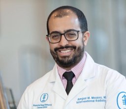 Amgad M Moussa合作论文数Department of Radiology, Memorial Sloan Kettering Cancer Center2Erinjeri Joseph P合作论文数2
 Amy Deipolyi合作论文数Intervent Radiol Serv, Mem Sloan Kettering Canc Ctr2
 Majid Maybody合作论文数2
 Alexander N. Shoushtari合作论文数David H. Koch Center for Cancer Care, Memorial Sloan Kettering Cancer Center2Gongidi Preetam合作论文数2Eric Lis合作论文数2K. Zhao合作论文数1V. Sotirchos合作论文数1Mohamed M. Soliman合作论文数1Eric Behling合作论文数1E. Petre合作论文数1E. Santos合作论文数1Adie Friedman合作论文数1D. Mobley合作论文数1A. Birney合作论文数1Thomas Presenza合作论文数1Javin Schefflein合作论文数1Emily Scattergood合作论文数1Matthias Barral合作论文数1
 Leila Haghani合作论文数Chan Medical School, University of Massachusetts1Rishav Adhikari合作论文数1d tsai合作论文数1
 Mohammad Elsayed合作论文数Department of Radiology, Memorial Sloan Kettering Cancer Center1
 Hooman Yarmohammadi合作论文数1
 Philipp Berning合作论文数Univ Hosp Munster, Dept Med Hematol Oncol & Pneumol A, Albert Schweitzer Campus 1, D-48149 Munster, Germany1S. Honig合作论文数1Omar Dzaye合作论文数Johns Hopkins Ciccarone Center for the Prevention of Cardiovascular Disease1
 Alexander C Razavi合作论文数Department of Medicine, Emory University1
 Frank Annie合作论文数Charleston Area Med Ctr CAMC1Jacqueline Bromberg合作论文数Memorial Sloan Kettering Cancer Center1




