Radial sorting of axons is a critical process in nerve development, ensuring proper segregation of axons to form myelinated and unmyelinated Schwann cell-axon units. This process is regulated by signals mediating communication between Schwann cells and the extracellular matrix, with laminin-211 as a key component. However, the molecular signals involved in directing Schwann cell-axon interactions are less understood, highlighting the need to identify additional molecules that mediate axon recognition and segregation. Gaining a deeper understanding of these mechanisms may shed light on the pathogenesis of genetic neuropathies. In this study, we utilized a mouse model of either sex with defects in axonal sorting, resulting from the conditional inactivation of the COP9 signalosome component Csn5 (Jab1) in Schwann cells. Transcriptome analysis was performed to identify adhesion molecules dysregulated during nerve development. Notably, we discovered that the repulsive molecule Slit2 was significantly overexpressed in Jab1-KO nerves and was particularly abundant in axon bundles with improper sorting. We demonstrated that while Slit2 is highly expressed in embryonic nerves, its expression must be precisely regulated in mature Schwann cells. Gain- and loss-of-function mutants for Slit2 further confirmed the role of Slit2 in nerve development. Transgenic mice overexpressing Slit2 displayed defects in radial sorting and hypomyelination, while Slit2 loss led to hypermyelination and misalignment of unmyelinated axons in Remak bundles. Additionally, Slit2 dysregulation interfered with nerve regeneration following cut injury. Our findings suggest that Slit2 plays a significant role in multiple stages of nerve development and some aspects of nerve regeneration.
Objectives:The AFG3L2 gene encodes a mitochondrial AAA-protease involved in inner mitochondrial membrane (IMM) proteostasis. Heterozygous variants in the AFG3L2 proteolytic domain cause Spinocerebellar Ataxia type 28, heterozygous variants in the AFG3L2 ATPase domain cause Optic Atrophy type 12, while biallelic variants lead to recessive Spastic Ataxia type 5. In this study, we aimed to investigate the link between AFG3L2 haploinsufficiency and Charcot-Marie-Tooth (CMT) phenotypes. Methods:We performed clinical evaluation, genetic analyses, electrophysiology, and functional studies in 1 patient's fibroblasts. Results:In this study, we report a patient presenting with progressive symmetric distal muscle atrophy, weakness, pes cavus, and sensory deficits at lower limbs, in the absence of cerebellar or pyramidal signs. Electrophysiologic studies confirmed axonal sensorimotor neuropathy. After excluding common CMT-related genes, clinical exome sequencing revealed a heterozygous truncating variant in AFG3L2 [NM_006793.3:c.121C > T; p.(Arg41*)]. In patient's fibroblasts, we observed ∼50% reduction in AFG3L2 protein levels, which is sufficient to hyperactivate the stress-sensitive IMM protease OMA1. Consequently, we detected increased OPA1 processing, mitochondrial shortening, and activation of the integrated stress response. Discussion:These findings suggest that AFG3L2 haploinsufficiency can underlie axonal CMT, expanding the clinical spectrum of AFG3L2-related diseases and emphasizing its potential inclusion in CMT diagnostic panels.
BACKGROUND:Spinal cord stimulation is emerging as a novel approach to restore movement in patients affected by spinal cord injury. Recent advances have shown restoration of locomotion in patients with complete motor lesions using sophisticated new systems including a brain machine interface. Despite the great potential, this approach is hindered by the inherent complexity and absence of approvals for clinical use. METHODS:Here, we tested a novel strategy to restore locomotion in patients with motor-complete spinal cord injury using voluntary trunk movements to modulate the motor output produced by a commercially available epidural spinal cord stimulator. FINDINGS:Four participants with thoracic AIS A-B injuries gained volitional modulation of stimulation-enabled movement without external interfaces or brain decoding, achieving standing and walking over four months of rehabilitation. The approach transformed passive stimulation into self-initiated movement, enabling functional mobility. CONCLUSIONS:This simple and potentially scalable method successfully leverages clinically available spinal cord stimulators to allow locomotion even in patients unable to produce voluntary contractions below the lesion, advancing accessible neuromodulation for motor-complete spinal cord injury. FUNDING:This work was funded by Università Vita-Salute San Raffaele, Boston Scientific Spa, Fondazione Cariplo, Bertarelli Foundation, #NEXTGENERATIONEU (NGEU), and the Ministry of University and Research (MUR).
Charcot-Marie-Tooth (CMT) neuropathies represent a broad and very heterogeneous group of disorders for which no therapies are yet available. Due to the huge genetic heterogeneity, therapeutical approaches that can benefit several forms independently of the unique pathogenetic mechanism have been sought. Niacin, nicotinic acid, is a vitamin used for many decades as anti-dyslipidaemic and anti-cholesterol drug product under the commercial name of Niaspan (R), the extended-release formulation of niacin. Of note, niacin can have other effects depending on the dose, formulation and physiology and it has been used to reduce inflammation, to promote angiogenesis and to protect neurons, muscle and axons by boosting nicotinamide adenine dinucleotide (NAD+) levels. Niacin also activates TNF-alpha convertase enzyme (TACE) secretase, which negatively regulates Neuregulin type I-mediated signalling in the peripheral nervous system and myelination. We previously postulated that niacin-mediated TACE activation can be effective in reducing aberrant excessive myelin associated with different CMT forms. Here, we explored efficacy of this strategy by performing a long-term preclinical trial and we provided evidence that a novel niacin-based long-lasting formulation ameliorates neurophysiology and reduces fibre degeneration in a model of Charcot-Marie-Tooth type 4B1 (CMT4B1) neuropathy, characterized by aberrant myelin. We also sought to determine whether this strategy might interfere with nerve regeneration, which is dependent on Neuregulin type I signalling. Surprisingly, we found that the Mtmr2 knockout mice, a model of CMT4B1, have a defect in nerve regeneration and that niacin-based treatment is not detrimental to nerve regeneration. Cipriani et al. report that a novel long-lasting formulation of niacin (vitamin B3) ameliorates neurophysiology and reduces fibres degeneration in a mouse model of Charcot-Marie-Tooth (CMT) 4B1, a severe demyelinating neuropathy with aberrant myelin. These findings are clinically relevant as fibre degeneration and loss correlate with clinical disability in patients.
This study evaluated the efficacy of atidarsagene autotemcel (arsa-cel) gene therapy in mitigating the severity and progression of peripheral neuropathy as assessed by nerve conduction velocity (NCV) in individuals affected by late-infantile metachromatic leukodystrophy (LI-MLD). A post hoc analysis was conducted on pre-symptomatic patients affected by LI-MLD treated with ex vivo autologous haematopoietic stem cell gene therapy (arsa-cel) in the context of prospective open-label, single-arm, interventional trials and expanded access programmes. All patients were followed longitudinally with nerve conduction studies (NCSs) of peripheral motor (ulnar, deep peroneal) and sensory (median, sural) nerves. These results were compared with those from a control group of untreated patients (NHx) studied with the same standardized protocol. We then analysed the effects of baseline characteristics (age at treatment, severity of neuropathy pre-treatment expressed as age-matched NCV Z-scores) and arylsulphatase A (ARSA) enzyme activity (measured in peripheral blood myeloid CD15+ cells post-treatment) on NCVs of treated patients. The primary end point of this post hoc analysis was NCV, reflecting the severity of demyelinating neuropathy. Changes in dermal nerve histopathology in skin biopsies were used as an exploratory outcome. Fifteen treated and 16 NHx patients were included in the analyses, with a median age (interquartile range) at treatment of 13 (9.1-14.5) months. At 36 months of age, treated patients showed higher estimated NCVs in all nerves compared with age-matched controls (∼15 m/s difference in motor nerves). Peripheral neuropathy was observed in the majority of treated patients at their pre-treatment examination (age range 7.3-17.4 months). Severity of pre-treatment neuropathy in treated patients did not have an effect on NCV values at 2 years post-gene therapy or on the rate of NCV-slowing afterwards. A younger age at treatment was associated with higher NCVs of motor ulnar nerve and sensory medial nerve 2 years post-gene therapy. Overall, ARSA levels in CD15+ cells correlated with NCVs of motor deep peroneal nerve at 2 years post-gene therapy, and ARSA levels were associated with a slower decrease or a slight increase in NCVs of the deep peroneal, ulnar and medial nerves afterwards. In summary, peripheral neuropathy assessed by NCV is significantly ameliorated in LI patients treated with arsa-cel compared with untreated patients of similar age. In addition to the potential role of early age at treatment in the preservation of myelin, supraphysiological ARSA levels may slow demyelination of the deep peroneal and other peripheral nerves. Arsa-cel may exert a stronger effect on NCV than allogeneic haematopoietic stem cell transplantation due to its greater ARSA expression.
BackgroundWhile there is strong evidence for botulinum toxin-A (BoNT-A) in post-stroke spasticity, there is a paucity of data in multiple sclerosis and other conditions in real-world settings.ObjectiveDocument the use of BoNT-A in the management of spasticity, with focus on the treatment of spasticity due to stroke and multiple sclerosis.MethodsThis noninterventional, retrospective study included all adults treated for upper and/or lower limb spasticity (any etiology) at three centers in Italy who received ≥3 BoNT-A injection cycles between 2008 and 2018.ResultsInjection data from 149 patients were analyzed (n = 67 post-stroke, n = 47 MS, n = 35 other etiologies). The median treatment duration was 54.9 months in the post-stroke population and 41.9 months in the MS population. Total doses for the commercially available BoNT-A formulations were typically lower than approved for use in spasticity; we also observed clinically relevant differences in the muscle patterns treated between the MS and post-stroke indications. Regardless of etiology, most patients were satisfied with treatment.ConclusionsThis retrospective study provides a snapshot of spasticity management for patients referred for BoNT-A treatment. Most patients were satisfied with their treatment over several cycles and the data support the effectiveness of BoNT-A for focal spasticity regardless of etiology.
Mutations in myelin protein zero (MPZ) are generally associated with Charcot-Marie-Tooth type 1B (CMT1B) disease, one of the most common forms of demyelinating neuropathy. Pathogenesis of some MPZ mutants, such as S63del and R98C, involves the misfolding and retention of MPZ in the endoplasmic reticulum (ER) of myelinating Schwann cells. To cope with proteotoxic ER-stress, Schwann cells mount an unfolded protein response (UPR) characterized by activation of the PERK, ATF6 and IRE1α/XBP1 pathways. Previous results showed that targeting the PERK UPR pathway mitigates neuropathy in mouse models of CMT1B; however, the contributions of other UPR pathways in disease pathogenesis remains poorly understood. Here, we probe the importance of the IRE1α/XBP1 signalling during normal myelination and in CMT1B. In response to ER stress, IRE1α is activated to stimulate the non-canonical splicing of Xbp1 mRNA to generate spliced Xbp1 (Xbp1s). This results in the increased expression of the adaptive transcription factor XBP1s, which regulates the expression of genes involved in diverse pathways including ER proteostasis. We generated mouse models where Xbp1 is deleted specifically in Schwann cells, preventing XBP1s activation in these cells. We observed that Xbp1 is dispensable for normal developmental myelination, myelin maintenance and remyelination after injury. However, Xbp1 deletion dramatically worsens the hypomyelination and the electrophysiological and locomotor parameters observed in young and adult CMT1B neuropathic animals. RNAseq analysis suggested that XBP1s exerts its adaptive function in CMT1B mouse models in large part via the induction of ER proteostasis genes. Accordingly, the exacerbation of the neuropathy in Xbp1 deficient mice was accompanied by upregulation of ER-stress pathways and of IRE1-mediated RIDD signaling in Schwann cells, suggesting that the activation of XBP1s via IRE1 plays a critical role in limiting mutant protein toxicity and that this toxicity cannot be compensated by other stress responses. Schwann cell specific overexpression of XBP1s partially re-established Schwann cell proteostasis and attenuated CMT1B severity in both the S63del and R98C mouse models. In addition, the selective, pharmacologic activation of IRE1α/XBP1 signaling ameliorated myelination in S63del dorsal root ganglia explants. Collectively, these data show that XBP1 has an essential adaptive role in different models of proteotoxic CMT1B neuropathy and suggest that activation of the IRE1α/XBP1 pathway may represent a therapeutic avenue in CMT1B and possibly for other neuropathies characterized by UPR activation.
BACKGROUND:Emerging research increasingly supports that epidural spinal cord electrical stimulation (EES) combined with neurorehabilitation can improve motor recovery in spinal cord injury (SCI) subjects. Patients with lesions involving the medullary cone may be challenging to treat with this approach, probably due to potential peripheral nervous system damage, leaving the open question of whether this large population may benefit from EES. METHODS:A T11-T12 SCI patient, with medullary cone involvement, underwent EES implant in a clinical trial (NCT05926843). During three months of testing, we determined optimal stimulation protocols for improving isolated movements and integrated them to reinstate independent walking with a walker. FINDINGS:EES substantially boosted hip flexor, spinal erector, and abdominal muscle contraction, improving the patient's performance in isolated movements. Over three months of combining continuous subthreshold EES with personalized rehabilitation, the patient progressed from being unable to walk to overground ambulation using a two-wheeled walker and bilateral knee and foot orthoses. At the time of hospital discharge, the patient managed to cover 58 m in the 6-min walking test and completed the 10-meter walking test in 40.29 s. Six months after EES implant, the patient was able to walk independently for 1 km with a walker. CONCLUSIONS:These results underscore the potential of neurorehabilitation protocols integrating EES also for patients with medullary cone lesions and pave the way for new rehabilitation prospects. FUNDING:This work was funded by Università Vita-Salute San Raffaele, Boston Scientific Spa, Fondazione Cariplo, Bertarelli Foundation, and the Ministry of University and Research (MUR).
Spinal cord injury (SCI) causes severe motor and sensory deficits, and there are currently no approved treatments for recovery. Nearly 70% of patients with SCI experience pathological muscle cocontraction and spasticity, accompanied by clinical signs such as patellar hyperreflexia and ankle clonus. The integration of epidural electrical stimulation (EES) of the spinal cord with rehabilitation has substantial potential to improve recovery of motor functions; however, abnormal muscle cocontraction and spasticity may limit the benefit of these interventions and hinder the effectiveness of EES in promoting functional movements. High-frequency excitation block introduced in peripheral nerve stimulation could reduce abnormal activity and lead to more physiological activation patterns. Here, we evaluated the application of high-frequency EES (HF-EES) in alleviating undesired muscular cocontraction and spasticity in two patients with motor incomplete SCI implanted with a commercial 32-channel EES paddle commonly used for pain therapy. To design custom HF-EES protocols, we first mapped the muscles targeted by different EES configurations. Our results showed that HF-EES substantially reduced patellar reflex in one participant and eliminated both patellar reflex and ankle clonus in the other participant. By combining HF-EES and low-frequency EES (LF-EES) to enhance functional movements with intensive rehabilitation, we observed notable improvements in lower limb kinematics, muscle strength, and clinical lower limb motor assessments over the trial period. This study suggests that HF-EES could be an important supplementary tool in SCI treatment, emphasizing the importance of personalized rehabilitation approaches and advanced tools to optimize EES treatments and offering hope for individuals with SCI-related motor deficits.
Background:Spinal nerve sheath tumors (NSTs), including schwannomas and neurofibromas, are slow-growing lesions often arising from dorsal sensory roots. Surgical resection is the gold-standard treatment, but the necessity of sacrificing the affected nerve root to achieve gross total resection remains controversial due to concerns about postoperative deficits. Methods:A comprehensive review of the literature was conducted to evaluate neurological outcomes following nerve root sacrifice and the functional reorganization by adjacent roots during the resection of large spinal schwannomas and neurofibromas. In addition, we report a representative case of a patient with a 20-year history of a large left L5 schwannoma, showing preoperative and intraoperative electrophysiological evidence of chronic radicular impairment. The patient underwent microsurgical tumor excision under intraoperative neurophysiological monitoring, with sacrifice of the affected root to allow the resection of the lesion tightly adherent to adjacent structures. Results:The literature consistently demonstrates that chronically compressed nerve roots in large, slow-growing NSTs are often nonfunctional or poorly functional, with compensatory reorganization by adjacent cranial and caudal roots. Reported rates of new postoperative motor deficits are low, even after root sacrifice. In our case, postoperative clinical and electromyographic follow-up confirmed the absence of new neurological deficits and the persistence of chronic, compensated impairment of the sacrificed root. Conclusion:Chronic impairment of spinal nerve roots in patients with large neoplasms, as documented by preoperative and intraoperative electromyographic data, allows for the safe resection of adherent NSTs with the possibility of sacrificing nonfunctional nerve roots. Functional compensation by adjacent roots preserves motor function, thereby minimizing the risk of postoperative deficits following root sacrifice.
INTRODUCTION:Evaluating the neural correlates of sensorimotor control deficits in cervical dystonia (CD) is fundamental to plan the best treatment. This study aims to assess kinematic and resting-state functional connectivity (RS-FC) characteristics in CD patients relative to healthy controls. METHODS:Seventeen CD patients and 14 age-/sex-matched healthy controls were recruited. Electromagnetic sensors were used to evaluate dystonic pattern, mean/maximal cervical movement amplitude and joint position error with eyes open and closed, and movement quality during target reaching with the head. RS-fMRI was acquired to compare the FC of brain sensorimotor regions between patients and controls. In patients, correlations between motion analysis and FC data were assessed. RESULTS:CD patients relative to controls showed reduced mean and maximal cervical range of motion (RoM) in rotation both towards and against dystonia pattern and reduced total RoM in rotation both with eyes open and closed. They had less severe dystonia pattern with eyes open vs eyes closed. CD patients showed an altered movement quality and sensorimotor control during target reaching and a higher joint position error. Compared to controls, CD patients showed reduced FC between supplementary motor area (SMA), occipital and cerebellar areas, which correlated with lower cervical RoM in rotation both with eyes open and closed and with worse movement quality during target reaching. CONCLUSIONS:FC alterations between SMA and occipital and cerebellar areas may represent the neural basis of cervical sensorimotor control deficits in CD patients. Electromagnetic sensors and RS-fMRI might be promising tools to monitor CD and assess the efficacy of rehabilitative interventions.
 Comi Giancarlo合作论文数Department of Neurology, Scientific Institute San Raffaele;Institute Experimental Neurology, Scientific Institute San Raffaele;Vita-Salute San Raffaele University35
 Francesca Bianchi合作论文数33Angelo Quattrini合作论文数27Stefano Amadio合作论文数24
 Maurizio D'Antonio合作论文数DIBIT, San Raffaele Scientific Institute21
 Filippi Massimo合作论文数San Raffaele Scientific Institute, Vita-Salute San Raffaele University17Cinzia Ferri合作论文数17Letizia Leocani合作论文数16Calogera Butera合作论文数14Lawrence Wrabetz合作论文数DIBIT, San Raffaele Scientific Institute14Feltri M Laura合作论文数Dept Genet & Cell Biol, Ist Sci San Raffaele12Alessandra Biffi合作论文数12
 Vittorio Martinelli合作论文数Università Vita-Salute San Raffaele;Multiple Sclerosis Centre, IRCCS Ospedale San Raffaele;Institute of Experimental Neurology, IRCCS Ospedale San Raffaele11Nilo Riva合作论文数11S. Previtali合作论文数10
 Luigi Naldini合作论文数Vita-Salute San Raffaele University;San Raffaele Telethon Institute for Gene Therapy10R. Fazio合作论文数10Lucia Moiola合作论文数9
 Marco Cursi合作论文数9Luca Bosco合作论文数8
 John Svaren合作论文数Department of Comparative Biosciences, School of Veterinary Medicine, University of Wisconsin-Madison8Stefania Medaglini合作论文数8Serena Valenzano合作论文数7Anke Claessens合作论文数7Phu Duong合作论文数7Rosa Mastrangelo合作论文数7Francesca Veneri合作论文数7
 Maria Sessa合作论文数Ospedale Niguarda7
 Roberto Santangelo合作论文数7Giovanni Di Maggio合作论文数7Giorgia Dina合作论文数7Daniela Ungaro合作论文数6Maria Grazia Natali Sora合作论文数6Francesca Fumagalli合作论文数6Baldoli Cristina合作论文数6Bolino A合作论文数6
 Claudio Bordignon合作论文数Università Vita-Salute San Raffaele;MolMed S.p.A.;San Raffaele Institute6
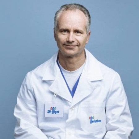 Alessandro Aiuti合作论文数Università Vita-Salute San Raffaele;Pediatric Immunohematology Unit, IRCCS Ospedale San Raffaele;Pediatric Clinical Research Unit, SR-Tiget6Roberta Guerriero合作论文数6Carla Taveggia合作论文数6
 Clelia di Serio合作论文数CUSSB, Univ Vita Salute San Raffaele6
 Cristina Scapin合作论文数6Federica Cerri合作论文数6
 Pietro Mortini合作论文数5Sabrina Canale合作论文数5Marco Pisa合作论文数5Yuri Falzone合作论文数5
 Thierry Touvier合作论文数5
 Emanuela Porrello合作论文数5




