Conjunctival impression cytology was performed on 236 Indonesian preschool children, half of whom had mild xerophthalmia and half of whom were age-matched controls. We devised an applicator that applies a paper disk of fixed area to the conjunctiva with even pressure. The disk applicator was used to collect impression cytology specimens from the temporal bulbar conjunctiva of one eye while the original strip technique was used on the other eye. Mean (+/- SD) serum retinol values for children with normal and abnormal discs were 22.0 +/- 8.6 micrograms/dL and 18.0 +/- 7.2 micrograms/dL P less than .0001). Mean serum values for normal and abnormal strips were 21.7 +/- 8.6 micrograms/dL and 19.0 +/- 7.7 micrograms/dL (P less than .03). Specimens obtained with the new disk applicator corresponded more closely with serum vitamin A levels and therefore vitamin A status than those obtained with the traditional strip technique.
Vitamin A status was assessed by conjunctival impression cytology (CIC-A) and clinical signs in 60 systematically sampled 36–83 month old children attending a hospital outpatient department on Moen, Truk. Seventeen percent had xerophthalmia (12% with XN, 5% with XN+X1B). More than half (57%) were CIC-A abnormal reflecting mild subclinical vitamin A deficiency. CIC-A abnormal children were more anemic (by hematocrit, p<0.05) and were at greater risk of middle ear infection (OR=3.5, 95% CL: 0.8, 14.5). Vitamin A deficiency appears to be a previously unrecognized nutritional problem among Micronesian children, the causes for which are speculative at present.
 Keith P. West合作论文数Division of Human Nutrition, Department of International Health, Bloomberg School of Public Health, Johns Hopkins University3
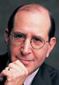 Alfred Sommer合作论文数Department of Epidemiology, Bloomberg School of Public Health, Johns Hopkins University;Department of International Health, Bloomberg School of Public Health, Johns Hopkins University;School of Medicine, Johns Hopkins University;Center for Global Health, Johns Hopkins University2
 Frank Mahoney合作论文数1Joan Mahoney合作论文数1Pn Manson合作论文数1JH HUMPHREY合作论文数1Muhilal合作论文数1Merbs Shannath L合作论文数1
 Richard Semba合作论文数Center for a Livable Future, Johns Hopkins Bloomberg School of Public Health, Johns Hopkins University;School of Medicine, Johns Hopkins University1Wirasasmita S合作论文数1
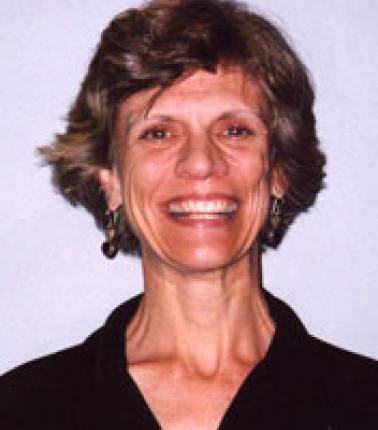 Jean Humphrey合作论文数Center for Human Nutrition, Bloomberg School of Public Health, Johns Hopkins University1
 Dean Eliott合作论文数Harvard Medical School, Harvard University1
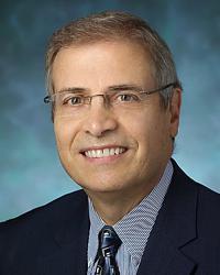 Peter A. Campochiaro合作论文数The Wilmer Eye Institute, Johns Hopkins University School of Medicine1Nicholas T. Iliff合作论文数1
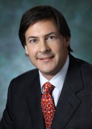 Michael P. Grant合作论文数Departments of ' Pharmacology;Case Western Reserve University;School of Medicine;School of Medicine, Case Western Reserve University1Michelle Lloyd-Puryear合作论文数1G Natadisastra合作论文数1Kiosi Aniol合作论文数1




