Objective.Future motor brain-computer interfaces (BCIs) are expected to benefit from integrating neural signals from multiple motor-related brain regions. While decoding studies have largely focused on lateral sensorimotor cortex, the medial wall of the cerebral hemisphere remains relatively underexplored. Here, we investigate the contribution of medial wall regions to finger movement decoding using human electrocorticography (ECoG) recordings.Approach.We analyzed ECoG data from four subjects performing finger movements. Single- and multi-channel decoding analyses were applied to medial wall electrodes, examining the contribution of time-domain and frequency-domain features, including local motor potentials (LMP) and oscillatory power in the(8-12 Hz) and(12-34 Hz) bands. Decoding performance was assessed for movement detection and finger discrimination.Main results.Significantly above-chance finger movement detection was observed across multiple medial wall subregions. LMP and-band power contributed most strongly to decoding performance. Feature dynamics shared key properties with primary motor cortex, including pre-movement-desynchronization, while also exhibiting region-specific patterns such as anatomically dependent positive or negative LMP modulations. Although movement detection was the dominant outcome, medial wall channels in two subjects enabled significant differentiation between individual fingers. In one subject, both contralateral and ipsilateral finger movements could be decoded with some generalization across hands; however, this observation is based on a single case and should be interpreted as preliminary.Significance.These findings identify the medial wall as a viable source of motor-related signals for finger movement decoding, with potential for future invasive motor BCI applications, while underscoring the need for further studies to confirm generalizability across individuals.
The effects of bilateral subthalamic nucleus (STN) stimulation on spontaneous language production are poorly understood. Using a pre-post surgery design, semantic and morphosyntactic parameters were evaluated according to the “Analysis of Spontaneous Speech in Aphasia” in seven people with Parkinson's disease (PD). Relative to normative data, the number of nouns and percentage of correct sentences produced were most subjective to changes over time and effects of stimulation. Twelve months after surgery, three patients with predominant left hemispheric dopamine depletion showed a normalization of the number of nouns during STN stimulation, compared to results below the norm in the bilateral OFF condition. Moreover, the effect on the percentage of correct sentences was variable, but the results remained below the norm when pre-operative deficits were present. These outcomes suggest that motor symptom laterality and pre-operative language functioning are among the variables that mediate STN stimulation effects on spontaneous language production, and emphasize the relevance to include the number of nouns and percentage of correct sentences produced as parameters in future larger-scale research.
Objective.Speech brain-computer interfaces (BCIs) aim to provide an alternative means of communication for individuals who are not able to speak. Remarkable progress has been achieved to decode attempted speech in individuals with severe anarthria. In contrast, imagined speech remains challenging to decode. The underlying neural mechanisms and relations to other speech modes are still elusive.Approach.In this study, we collected low-density electrocorticography signals from ten participants during a word repetition task. Electrodes were implanted for presurgical epilepsy evaluation in participants with preserved speech abilities. Models were developed using linear discriminant analysis to classify five words in response to different speech modes. We compared models trained during speaking, listening, imagining speaking, mouthing and reading. The relations between speech modes were investigated by transferring and augmenting models across speech modes.Main results.As expected, performed speech achieved the highest word classification accuracy followed by listening, mouthing, imagining and reading. While the accuracies obtained were not high enough for practical application, model transfer and augmentation could be investigated across speech modes. Transferring or augmenting models from one speech mode to another mode could significantly improve model performance. In particular, patterns learned from performed and perceived speech could generalize to imagined speech, leading to significantly improved imagined speech performance in seven participants. For four participants, imagined speech could be decoded above chance exclusively when models were transferred or augmented with performed or perceived speech.Significance.Imagined speech is often preferred by speech BCI users over attempted speech, as it requires less effort and can be produced more quickly. Transferring models across speech modes has the potential to facilitate and boost the development of imagined speech decoders.
BACKGROUND:Drug-resistant epilepsy (DRE) affecting the paracentral region poses significant therapeutic challenges, given resective surgery is often infeasible owing to the proximity to eloquent cortex. Neurostimulation modalities such as vagus nerve stimulation, anterior nucleus thalamic deep brain stimulation, and responsive neurostimulation offer alternatives, but their efficacy in achieving seizure freedom remains limited. Chronic subthreshold cortical stimulation (CSCS) represents a novel approach, directly targeting the epileptogenic zone through continuous or cyclic, open-loop subthreshold stimulation, potentially offering superior outcomes in paracentral epilepsy. This systematic review evaluates the efficacy, safety, stimulation parameters, and neurologic outcomes of CSCS in patients with paracentral DRE. MATERIALS AND METHODS:A literature search of PubMed/MEDLINE and EMBASE up to October 2025 identified reports involving chronic electrical stimulation of the primary motor cortex or paracentral region for seizure reduction in human subjects. The inclusion criteria encompassed case reports, case series, and observational studies. Data extracted included seizure outcomes, stimulation parameters, imaging findings, adverse events, quality of life (QOL), and neurologic effects. The primary end points were ≥90% seizure reduction and complete seizure freedom. Risk of bias and quality of evidence were assessed using Oxford, Grading Recommendation Assessment Development and Education, and the criteria of Murad et al. RESULTS: Six studies involving 17 patients were included. The mean follow-up duration was 31.3 ± 22.3 months. Complete seizure freedom was achieved in 47% of patients (8/17), whereas an additional 35% (6/17) experienced ≥90% seizure reduction. Patients with lesional epilepsy indicated significantly higher rates of seizure freedom than did nonlesional cases (p = 0.007). No major surgical or stimulation-related adverse events were reported. Stimulation parameters varied widely (frequency 2-130 Hz; pulse width 90-450 μs; amplitudes up to 7V or 4 mA), and both continuous and cyclic modes were effective. Several patients showed neurologic improvements beyond seizure control, including enhanced motor function, mood, and cognitive performance. QOL improvements also were reported. CONCLUSIONS:CSCS appears to be a promising treatment modality for paracentral DRE, showing high rates of seizure freedom, favorable safety profiles, and potential neuromodulatory benefits beyond seizure suppression. Although preliminary evidence suggests superior efficacy in paracentral cases to other neurostimulation modalities, the present data are limited by publication bias and lack of randomized trials. Future prospective, controlled studies are essential to validate these findings, optimize stimulation protocols, and refine patient selection criteria.
Reward positivity (RewP) is an event-related brain potential component that emerges ∼250-350 ms after receiving reward-related feedback stimuli and is believed to be important for reinforcement learning and reward processing. Although numerous localization studies have indicated that the anterior cingulate cortex (ACC) is the neural generator of this component, other studies have identified sources outside of the ACC, fuelling a debate about its origin. Because the results of EEG and magnetoencephalography source-localization studies are severely limited by the inverse problem, we addressed this question by leveraging the high spatial and temporal resolution of intracranial EEG. We predicted that we would identify a neural generator of rthe RewP in the caudal ACC. We recorded intracranial EEG in 19 patients with refractory epilepsy who underwent invasive video-EEG monitoring at Ghent University Hospital, Belgium. Participants engaged in the virtual T-maze task, a trial-and-error task known to elicit a canonical RewP, while scalp and intracranial EEG were recorded simultaneously. The RewP was identified using a difference wave approach for both scalp and intracranial EEG. The data were aggregated across participants to create a virtual 'meta-participant' that contained all the recorded intracranial event-related brain potentials with respect to their intracranial contact locations. We used both hypothesis-driven (focused on ACC) and exploratory (whole-brain analysis) approaches to segment the brain into regions of interest. For each region of interest, we evaluated the degree to which the time course of the absolute current density (ACD) activity mirrored the time course of the RewP, and we confirmed the statistical significance of the results using permutation analysis. The grand average waveform of the scalp data revealed a RewP at 309 ms after reward feedback with a frontocentral scalp distribution, consistent with the identification of this component as the RewP. The meta-participant contained intracranial event-related brain potentials recorded from 582 intracranial contacts in total. The ACD activity of the aggregated intracranial event-related brain potentials was most similar to the RewP in the left caudal ACC, left dorsolateral prefrontal cortex, left frontomedial cortex and left white matter, with the highest score attributed to caudal ACC, as predicted. To our knowledge, this is the first study to use intracranial EEG aggregated across multiple human epilepsy patients and current source density analysis to identify the neural generator(s) of the RewP. These results provide direct evidence that the ACC is a neural generator of the RewP.
Nonpharmaceutical approaches based on gamma entrainment using sensory stimuli (GENUS) have shown promise in reducing Alzheimer's disease pathology in mouse models. While human studies remain limited, GENUS has been shown to alleviate aspects of neurodegeneration in patients with Alzheimer's disease. In this study, we analyze intracranial EEG data from 490 contacts across eleven patients with refractory epilepsy in response to three visual stimulation conditions. We find that 40 Hz visual stimulation successfully entrains neural activity beyond early visual areas, including the hippocampus and other cortical regions such as the temporal and frontal lobes. Additionally, we show that synchronization increases between the hippocampus and other cortical areas in response to the 40 Hz visual stimulation. Furthermore, combining stimulation with a simple visual oddball task alters the direction of information flow from frontal regions to the hippocampus and enhances both the strength and spatial extent of neural entrainment. These findings highlight the potential influence of cognitive engagement during sensory gamma stimulation and provide additional insights into the neurophysiological effects of 40 Hz visual stimulation.
OBJECTIVE:Resective epilepsy surgery is an evidence-based treatment option for patients with focal drug-resistant epilepsy (DRE). Seizure outcome after surgery is largely dependent on detection and delineation of an epileptogenic lesion on magnetic resonance imaging (MRI). However, detection fails in 30% of patients at 3 Tesla (T) MRI, thereby limiting surgical options. Diagnostic and therapeutic gain of ultra-high-field MRI in patients with 3T MRI-negative DRE is evaluated in the EpiUltraStudy. Here we report the diagnostic gain of structural 7T MRI. METHODS:Inclusion criteria were age ≥12 years and DRE with a suspected epileptogenic focus and negative conventional 3T MRI during pre-surgical workup. Images were evaluated independently by two neuroradiologists and a neurologist or neurosurgeon in two runs: blinded (Run 1) and with the results of additional clinical investigations (Run 2). RESULTS:Sixty patients underwent 7T MRI. No persistent adverse events were reported. Visual assessment of 7T MRI identified lesions in 9 cases (15%), undetected on prior 3T MRI. Possible positive scan rates increased from 17% (10/60) in the blinded run to 47% (28/60) in the informed run. However, after consensus review, many of these were reclassified as negative. Eight of nine positive 7T MRI scans were initially identified by only one or two assessors. After reassessment, a total of 56% (5/9) of 7T lesions were retrospectively identified on 3T. SIGNIFICANCE:Our data suggest a benefit of 7T MRI for the detection of subtle epileptogenic lesions in patients with DRE and negative 3T MRI. Although the detection rate may appear modest compared to other reports, we present a nuanced discussion of our methodology and patient population, contributing meaningful context to the current literature. The availability of multimodal information and consensus reviews enhanced diagnostic accuracy but with higher rates of false positives, underscoring the importance of multidisciplinary cooperation in the clinical care for patients with DRE. TRIAL REGISTRATION NUMBER:www.trialregister.nl: NTR7536.
Objective.Speech brain-computer interfaces (BCIs) aim to restore communication for individuals who have lost the ability to speak by interpreting their brain activity and decoding the intended speech. As an initial component of these decoders, speech detectors have been developed to distinguish between the intent to speak and silence. However, it is important that these detectors account for real-life scenarios in which users may engage language-related brain areas-such as during reading or listening-without any intention to speak.Approach.In this study, we analyze the interplay between different speech modes: speaking, listening, imagining speaking, reading and mouthing. We gathered a large dataset of 29 participants implanted with electrocorticography electrodes and developed a speech mode classifier. We also assessed how well classifiers trained on data from a specific participant transfer to other participants, both in the case of a single- and multi-electrode classifier.Main results.High accuracy was achieved using linear classifiers, for both single-electrode and multi-electrode configurations. Single-electrode classification reached 88.89% accuracy and multi-electrode classification 96.49% accuracy in distinguishing among three classes (speaking, listening, and silence). The best performing electrodes were located on the superior temporal gyrus and sensorimotor cortex. We found that single-electrode classifiers could be transferred across recording sites. For multi-electrode classifiers, we observed that transfer performance was higher for binary classifiers compared to multiclass classifiers, with the optimal source subject of the binary classifiers depending on the speech modes being classified.SignificanceAccurately detecting speech from brain signals is essential to prevent spurious outputs from a speech BCI and to advance its use beyond lab settings. To achieve this objective, the transfer between participants is particularly valuable as it can reduce training time, especially in cases where subject training is challenging.
The next generation of motor brain-computer interfaces (BCIs) will likely benefit from integrating recordings from multiple motor-related brain regions. Among these is the medial wall, yet it remains relatively understudied in the case of finger movement decoding. Using electrocorticographic (ECoG) recordings from a subject implanted both over medial and lateral cortical areas, we first assessed the medial wall's potential for multiclass classification (5 fingers + rest). We achieved a six-class accuracy of 0.46, significantly above chance, with rest trials classified most accurately, followed by thumb movement trials. Several frequency features contributed to decoding, with Local Motor Potentials (LMP) being the most influential one, with distinctive activity already prior to movement onset, and power in the α (8-12 Hz) band aiding in decoding rest trials over finger movement trials. Next, we explored whether combining the best medial wall channel with lateral cortical channels could improve decoding performance. We found a significant accuracy improvement for most lateral channels (from an average of 0.36 to 0.42), except for the channel closest to the finger primary motor region, whose accuracy was already high (0.77). These findings highlight the medial wall's potential for motor decoding and its value as a target region for future motor BCIs, especially for individuals with impaired hand motor areas.
Introduction:Epilepsy is a neurological disorder affecting over 50 million people globally, with around 30 % of them classified as having drug-resistant epilepsy (DRE). Temporal lobe epilepsy (TLE) is the most frequently encountered type of surgically treated epilepsy. The primary surgical approaches for TLE include anterior temporal lobectomy (ATL) and selective amygdalohippocampectomy (selAH). Research question:This study sought to gather expert European consensus on surgical strategies and complication rate for ATL and selAH, in both adult and pediatric patients. Materials and methods:A modified Delphi technique was employed, with 39 experienced epilepsy surgeons from 35 different European centers. A 22-item questionnaire addressed key surgical considerations, including mortality, morbidity, neurological deficits, infection rates, and potential psychiatric and cognitive complications. Results:The survey had a 43 % response rate. Mortality rates for both surgical approaches ranged between 0 and 1 %. Visual field deficits (VFDs) were more frequently observed after ATL (over 16 %) compared to selAH (2-10 %). Permanent motor deficits were rare (<2 %), while complications such as infections and hematomas were reported in 0-2 % and less than 5 % of cases, respectively for both procedures. While psychiatric and cognitive complications were acknowledged, no consensus was reached regarding their prevalence or screening methods. Discussion:The results underscore the value of advanced imaging, thorough preoperative evaluation, and intraoperative monitoring. Future research is needed to refine outcome optimization and standardize training protocols. Conclusions:Consensus was achieved on critical aspects of surgical planning and complication management, providing support for the development of standardized practices in temporal lobe epilepsy surgery.
Speech brain-computer interfaces aim to support communication-impaired patients by translating neural signals into speech. While impressive progress was achieved in decoding performed, perceived and attempted speech, imagined speech remains elusive, mainly due to the absence of behavioral output. Nevertheless, imagined speech is advantageous since it does not depend on any articulator movements that might become impaired or even lost throughout the stages of a neurodegenerative disease. In this study, we analyzed electrocortigraphy data recorded from 16 participants in response to 3 speech modes: performed, perceived (listening), and imagined speech. We used a linear model to detect speech events and examined the contributions of each frequency band, from delta to high gamma, given the speech mode and electrode location. For imagined speech detection, we observed a strong contribution of gamma bands in the motor cortex, whereas lower frequencies were more prominent in the temporal lobe, in particular of the left hemisphere. Based on the similarities in frequency patterns, we were able to transfer models between speech modes and participants with similar electrode locations. The authors analyze electrocortigraphy data to demonstrate a contribution of gamma oscillations and low frequency waves to imagined speech, developing a model for speech detection capable of generalizing across participants and speech modes.
Introduction: Open resective surgery remains the main treatment modality for refractory epilepsy, but is often considered a last resort option due to its invasiveness. Research question: This manuscript aims to provide an overview on traditional as well as minimally invasive surgical approaches in modern state of the art epilepsy surgery. Materials and methods: This narrative review addresses both historical and contemporary as well as minimal invasive surgical approaches in epilepsy surgery. Peer-reviewed published articles were retrieved from PubMed and Scopus. Only articles written in English were considered for this work. A range of traditional and minimally invasive surgical approaches in epilepsy surgery were examined, and their respective advantages and disadvantages have been summarized. Results: The following approaches and techniques are discussed: minimally invasive diagnostics in epilepsy surgery, anterior temporal lobectomy, functional temporal lobectomy, selective amygdalohippocampectomy through a transsylvian, transcortical, or subtemporal approach, insulo-opercular corticectomies compared to laser interstitial thermal therapy, radiofrequency thermocoagulation, stereotactic radiosurgery, neuromodulation, high intensity focused ultrasound, and disconnection surgery including callosotomy, hemispherotomy, and subpial transections. Discussion and conclusion: Understanding the benefits and disadvantages of different surgical approaches and strategies in traditional and minimal invasive epilepsy surgery might improve the surgical decision tree, as not all procedures are appropriate for all patients.
Chronic subthreshold cortical stimulation (CSCS) is a form of neurostimulation consisting of continuous or cyclic, open-loop, subthreshold electrical stimulation of a well-defined epileptogenic zone (EZ). CSCS has seen limited clinical use but could be a safe and effective long-term treatment of focal drug resistant epilepsy, in particular when the EZ is located in the motor cortex. We present a case of a 49-year-old woman suffering from debilitating focal motor seizures. Treatment with CSCS resulted in significant clinical improvement, enabling her to walk unaided for the first time in years.
Background and AimsGlioma growth in eloquent language areas induces adaptive activation changes in the language network. The present study aimed to (1) investigate the pre- to postoperative evolution of neural phoneme perception processes in glioma patients and (2) assess if event-related potentials (ERPs) reflecting phoneme perception can provide added value to the diagnostic approach in individuals undergoing awake surgery.Methods and ProceduresIn five persons undergoing an awake craniotomy for the resection of a glioma, pre- and postoperative behavioural language assessment (Aachen Aphasia Test, Comprehensive Aphasia Test and Boston Naming Test) and electrophysiological investigation of phoneme perception was performed. For the latter, ERPs were obtained through the administration of an inattentive (Mismatch Negativity; MMN) and attentive (P300) oddball paradigm containing a phonemic articulation place contrast during EEG recording.Outcomes and ResultsAberrant phoneme categorization processing was evidenced in all five participants preoperatively based on the MMN and P300 amplitude and latency. Moreover, mild behavioural impairments were found in four participants at this time. Postoperatively, three out of five participants reached behavioural ceiling effects, while four individuals displayed normalization of electrophysiological measures.ConclusionsWhile neural processing of phoneme contrasts was preoperatively affected by glioma-induced disturbances, a high potential for postsurgical plasticity was shown. As four participants presented with a high grade glioma, tumour grade might partially account for this pattern. Addition of electrophysiological tests to the language assessment could provide benefits in both the pre- and postoperative clinical diagnostic approach in glioma patients. These preliminary results need validation in a larger sample.
BACKGROUND AND OBJECTIVES:The efficacy of deep brain stimulation of the anterior nucleus of the thalamus (ANT DBS) in patients with drug-resistant epilepsy (DRE) was demonstrated in the double-blind Stimulation of the Anterior Nucleus of the Thalamus for Epilepsy randomized controlled trial. The Medtronic Registry for Epilepsy (MORE) aims to understand the safety and longer-term effectiveness of ANT DBS therapy in routine clinical practice. METHODS:MORE is an observational registry collecting prospective and retrospective clinical data. Participants were at least 18 years old, with focal DRE recruited across 25 centers from 13 countries. They were followed for at least 2 years in terms of seizure frequency (SF), responder rate (RR), health-related quality of life (Quality of Life in Epilepsy Inventory 31), depression, and safety outcomes. RESULTS:Of the 191 patients recruited, 170 (mean [SD] age of 35.6 [10.7] years, 43% female) were implanted with DBS therapy and met all eligibility criteria. At baseline, 38% of patients reported cognitive impairment. The median monthly SF decreased by 33.1% from 15.8 at baseline to 8.8 at 2 years (p < 0.0001) with 32.3% RR. In the subgroup of 47 patients who completed 5 years of follow-up, the median monthly SF decreased by 55.1% from 16 at baseline to 7.9 at 5 years (p < 0.0001) with 53.2% RR. High-volume centers (>10 implantations) had 42.8% reduction in median monthly SF by 2 years in comparison with 25.8% in low-volume center. In patients with cognitive impairment, the reduction in median monthly SF was 26.0% by 2 years compared with 36.1% in patients without cognitive impairment. The most frequently reported adverse events were changes (e.g., increased frequency/severity) in seizure (16%), memory impairment (patient-reported complaint, 15%), depressive mood (patient-reported complaint, 13%), and epilepsy (12%). One definite sudden unexpected death in epilepsy case was reported. DISCUSSION:The MORE registry supports the effectiveness and safety of ANT DBS therapy in a real-world setting in the 2 years following implantation. CLASSIFICATION OF EVIDENCE:This study provides Class IV evidence that ANT DBS reduces the frequency of seizures in patients with drug-resistant focal epilepsy. TRIAL REGISTRATION INFORMATION:MORE ClinicalTrials.gov Identifier: NCT01521754, first posted on January 31, 2012.
Introduction:Alzheimer's disease is one of the great challenges in the coming decades, and despite great efforts, a widely effective disease-modifying therapy in humans remains elusive. One particular promising non-pharmacological therapy that has received increased attention in recent years is based on the Gamma ENtrainment Using Sensory stimulation (GENUS), a high-frequency neural response elicited by a visual and/or auditory stimulus at 40 Hz. While this has shown to be effective in animal models, studies on human participants have reported varying success. The current work hypothesizes that the varying success in humans is due to differences in cognitive workload during the GENUS sessions.Methods:We recruited a cohort of 15 participants who underwent a scalp-EEG recording as well as one epilepsy patient who was implanted with 50 subdural surface electrodes over temporo-occipital and temporo-basal cortex and 14 depth contacts that targeted the hippocampus and insula. All participants completed several GENUS sessions, in each of which a different cognitive task was performed.Results:We found that the inclusion of a cognitive task during the GENUS session not only has a positive effect on the strength and extent of the gamma entrainment, but also promotes the propagation of gamma entrainment to additional neural areas including deep ones such as hippocampus which were not recruited when no cognitive task was required from the participants. The latter is of particular interest given that the hippocampal complex is considered to be one of the primary targets for AD therapies.Discussion:This work introduces a possible improvement strategy for GENUS therapy that might contribute to increasing the efficacy of the therapy or shortening the time needed for the positive outcome.
Intraoperative electrocorticography (ECoG) captures neural information from the surface of the cerebral cortex during surgeries such as resections for intractable epilepsy and tumors. Current clinical ECoG grids come in evenly spaced, millimeter-sized electrodes embedded in silicone rubber. Their mechanical rigidity and fixed electrode spatial resolution are common shortcomings reported by the surgical teams. Here, advances in soft neurotechnology are leveraged to manufacture conformable subdural, thin-film ECoG grids, and evaluate their suitability for translational research. Soft grids with 0.2 to 10 mm electrode pitch and diameter are embedded in 150 µm silicone membranes. The soft grids are compatible with surgical handling and can be folded to safely interface hidden cerebral surface such as the Sylvian fold in human cadaveric models. It is found that the thin-film conductor grids do not generate diagnostic-impeding imaging artefacts (<1 mm) nor adverse local heating within a standard 3T clinical magnetic resonance imaging scanner. Next, the ability of the soft grids to record subdural neural activity in minipigs acutely and two weeks postimplantation is validated. Taken together, these results suggest a promising future alternative to current stiff electrodes and may enable the future adoption of soft ECoG grids in translational research and ultimately in clinical settings.
Background and purpose The subventricular zone (SVZ) is an important niche for neural stem cells but probably also for brain tumor propagating cells, including the glioblastoma stem cell. The SVZ may become a target for radiation therapy in glioblastoma patients. However, reports studying the effect of irradiation of the SVZ on glioblastoma patient survival show conflicting results. We studied the correlation between incidental SVZ radiation dose and survival in a cohort of isocitrate dehydrogenase-wildtype (IDHwt) glioblastoma patients with inclusion of important survival prognosticators. Patients and methods In this retrospective analysis, only adult patients with supratentorial IDHwt glioblastoma were included who were treated with temozolomide-based chemoradiotherapy after surgery. The SVZ was contoured on the radiotherapy planning imaging. Cox proportional regression overall survival (OS) analysis was used to study the correlation between SVZ dose and survival. Age, Karnofsky Performance Score, extent of resection and O-6-methylguanine-methyl-DNA-transferase gene promoter (MGMTp) methylation were used as covariates in multivariate analysis. Results In total, 137 patients were included. Median OS was 13.3 months. The MGMTp methylation was present in 40% of cases. Ipsilateral SVZ (iSVZ) mean dose was 44.4 Gy and 27.2 Gy for the contralateral SVZ (cSVZ). Univariate survival analysis showed an inverse relationship between cSVZ mean dose and OS (HR 1.029 (1.003-1.057); p= .032). However, there was no correlation between cSVZ mean dose and OS in multivariate analysis. iSVZ dose did not correlate with survival. Conclusion In this cohort of 137 IDHwt glioblastoma patients, iSVZ did not correlate with OS. Higher cSVZ dose was inversely correlated with OS in univariate survival analysis but lost its significance in multivariate analysis, including MGMTp-methylation. Hence, the correlation between SVZ radiation and glioblastoma patient survival remains unclear. Carefully designed prospective studies are needed to provide unequivocal results on this controversial topic.
 Paul Boon合作论文数54
 Kristl Vonck合作论文数42Alfred Meurs合作论文数39
 Evelien Carrette合作论文数35Robrecht Raedt合作论文数27
 Johannes Schramm合作论文数Department of Neurosurgery, Medical School, University of Bonn21Frank Dewaele合作论文数18Baert Edward合作论文数17
 Veerle De Herdt合作论文数Reference Center for Refractory Epilepsy, Ghent University Hospital17Carlo Schaller合作论文数17Marc M. Van Hulle合作论文数Campus Gasthuisberg;Laboratorium voor Neurofysiologie16Caemaert Jacques合作论文数15
 Christian Elger合作论文数Center for Economics and Neuroscience, University of Bonn15
 Pieter Van Mierlo合作论文数Ghent University15Giorgio Hallaert合作论文数14
 Benjamin Wittevrongel合作论文数Katholieke Univ Leuven, Dept Neurosci, Lab Neuro & Psychophysiol, Leuven, Belgium12
 De Taeye Leen合作论文数Department of Neurology, Ghent University12
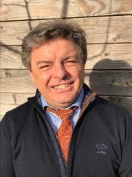 Patrick Santens合作论文数Department of Neurology, Ghent University Hospital12
 Ine Dauwe合作论文数12Jean-Pierre Kalala Okito合作论文数Ghent University Hospital12Elvira Khachatryan合作论文数Lab Neuro & Psychophysiol, Katholieke Univ Leuven11Annelies Van Dycke合作论文数11Eric Achten合作论文数10
 Miet de letter合作论文数Department of Oto-Rhino-Laryngology- Logopaedic and Audiologic Sciences, Ghent University10Martina Deckert合作论文数10Serge Goldman合作论文数Université libre de Bruxelles9Thomas Grunwald合作论文数9
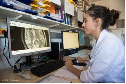 Riëm El Tahry合作论文数Reference Center for Refractory Epilepsy, Ghent University Hospital9Manuel Montesinos-Rongen合作论文数9
 Klaus Lehnertz合作论文数University of Bonn7N Pezer合作论文数7Wytse J. Wadman合作论文数7
 Mansoureh Fahimi Hnazaee合作论文数Katholieke Univ Leuven, Lab Neuro & Psychophysiol, Dept Neurosci, Leuven, Belgium6
 Dimitri Hemelsoet合作论文数Ghent University;Ghent University Hospital6
 Katja Batens合作论文数6
 Strobbe Gregor合作论文数6
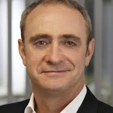 Bernhard Meyer合作论文数School of Medicine and Health, Technical University of Munich;Neurosurgical Clinic and Policlinic, Technical University of Munich6Martin Kurthen合作论文数6
 Xavier De Tiege合作论文数5Od Wiestler合作论文数5Vandersteene Jelle合作论文数5
 Sarah Vanhoutte合作论文数5Philippe David合作论文数Department of Neuroradiology, Hôpital ERASME Université Libre de Bruxelles5Karel Deblaere合作论文数5Stefanie Dedeurwaerdere合作论文数Heymans Institute, Ghent University Hospital5
 Luc Defreyne合作论文数5
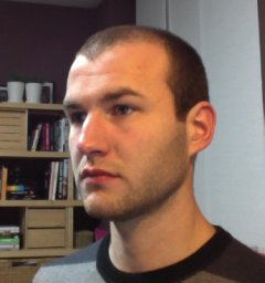 Mathieu Bourguignon合作论文数Centre for Research in Cognition & Neurosciences, Faculté des Sciences Psychologiques et de l'Education, Université Libre de Bruxelles5Olaf E. M. G. Schijns合作论文数5
 Benjamin Legros合作论文数Reference Center for Refractory Epilepsy, ULB-Hôpital Erasme5




